. 2021 Feb 1;218(2):e20200844.
doi: 10.1084/jem.20200844.

STING agonist promotes CAR T cell trafficking and persistence in breast cancer

Affiliations

STING agonist promotes CAR T cell trafficking and persistence in breast cancer

Nuo Xu et al. J Exp Med. .

Abstract

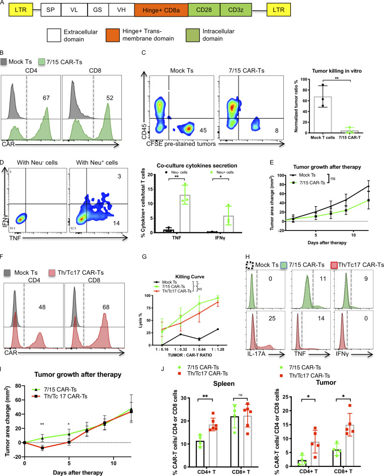
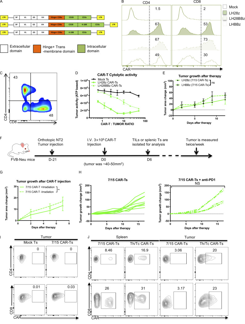
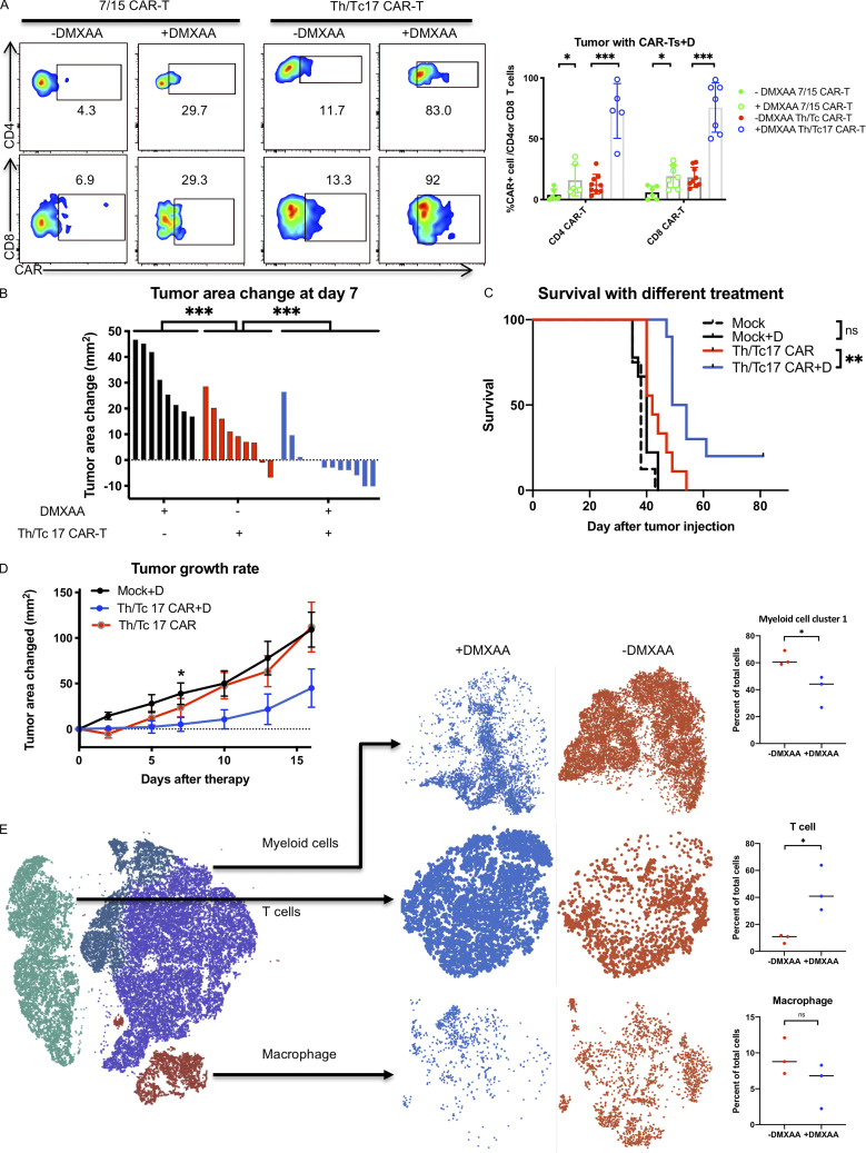
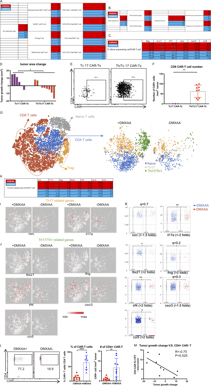
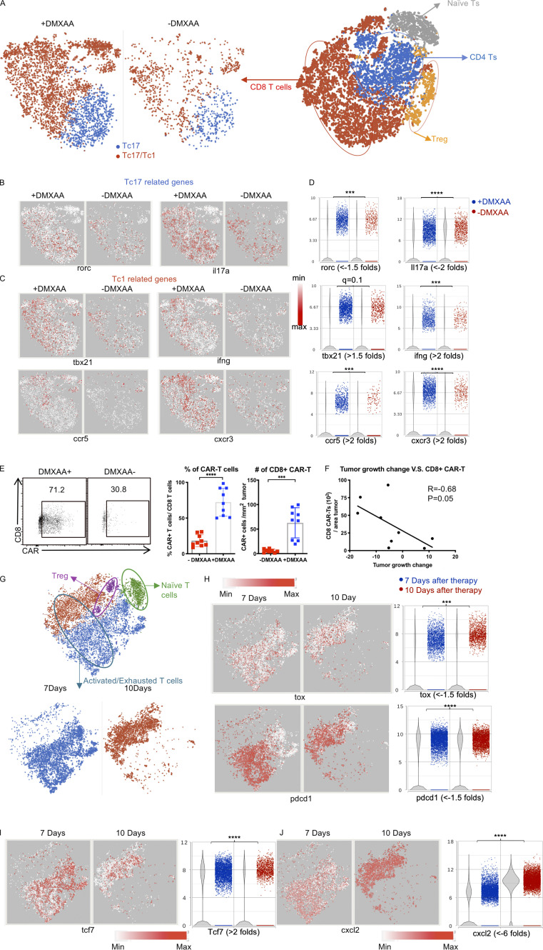
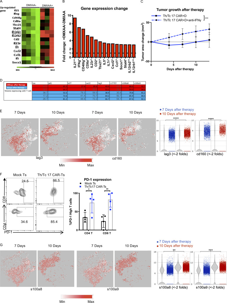
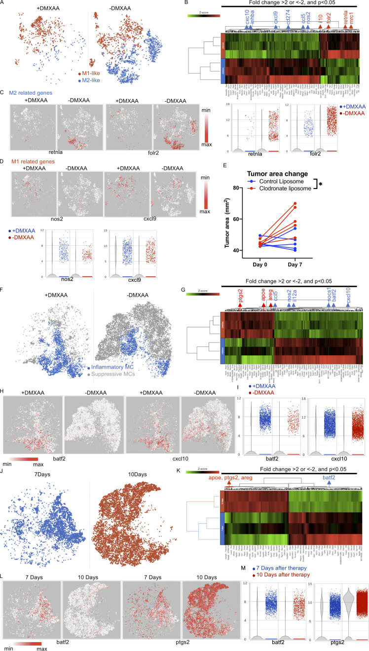
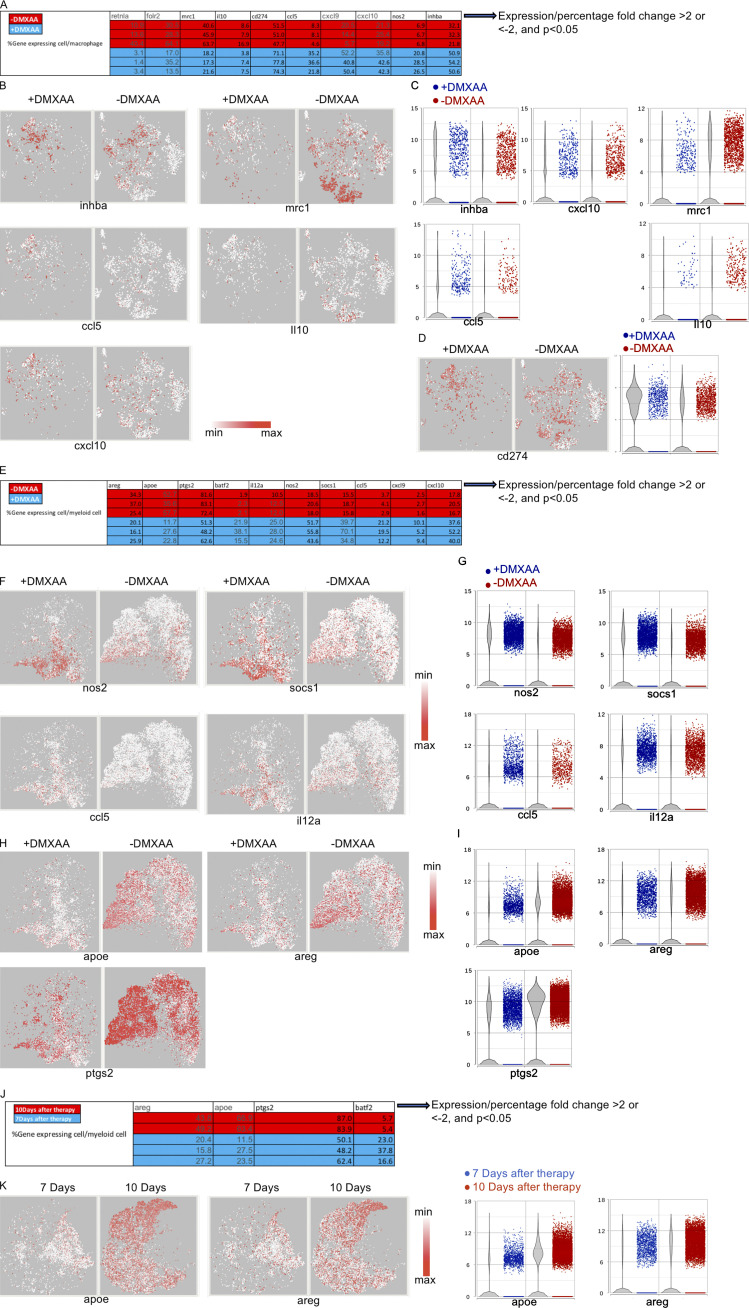
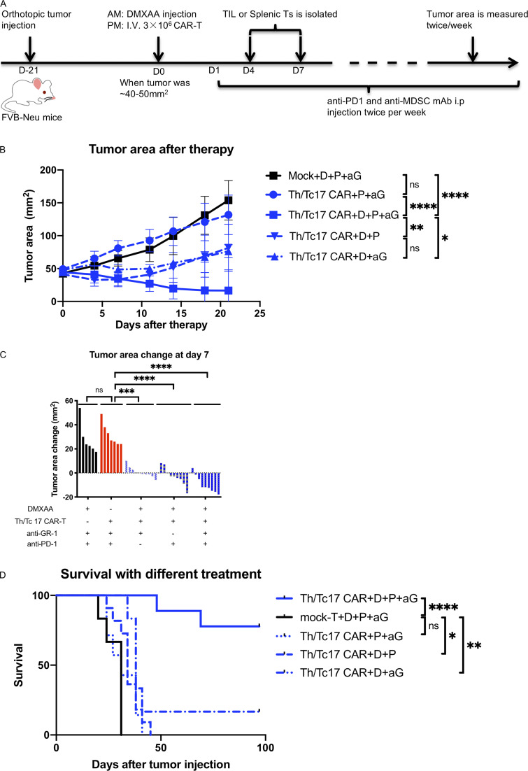
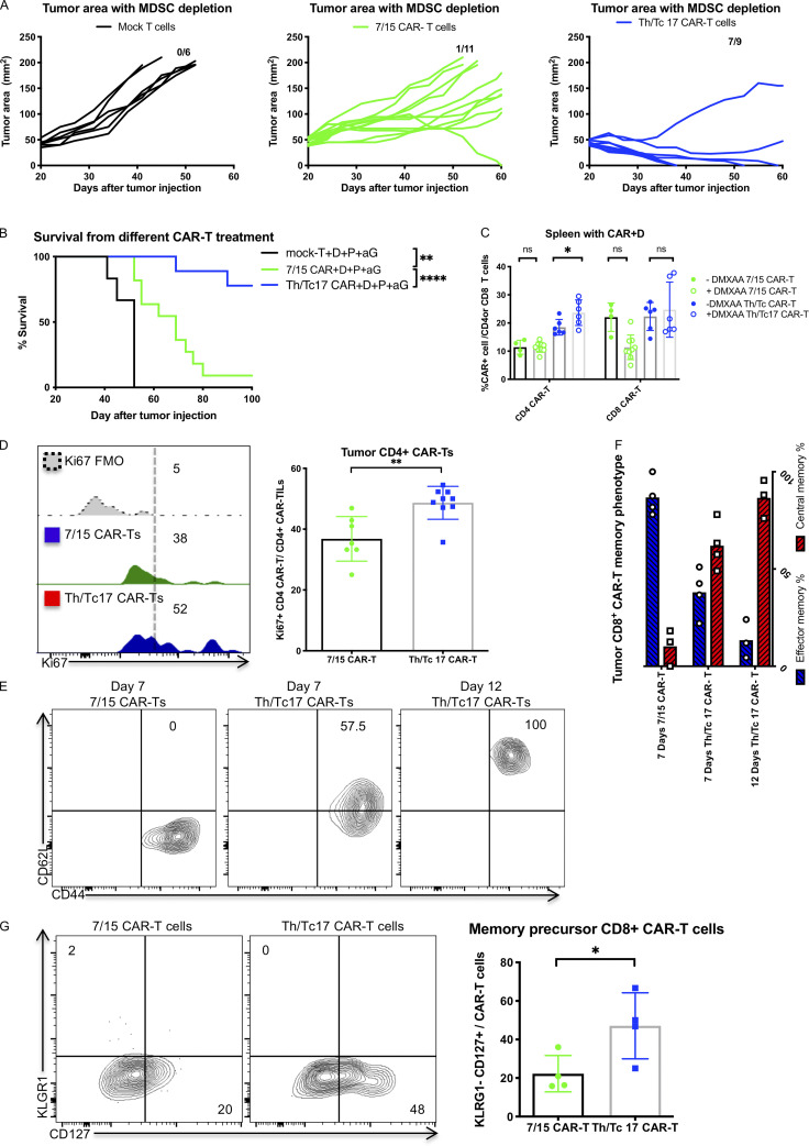
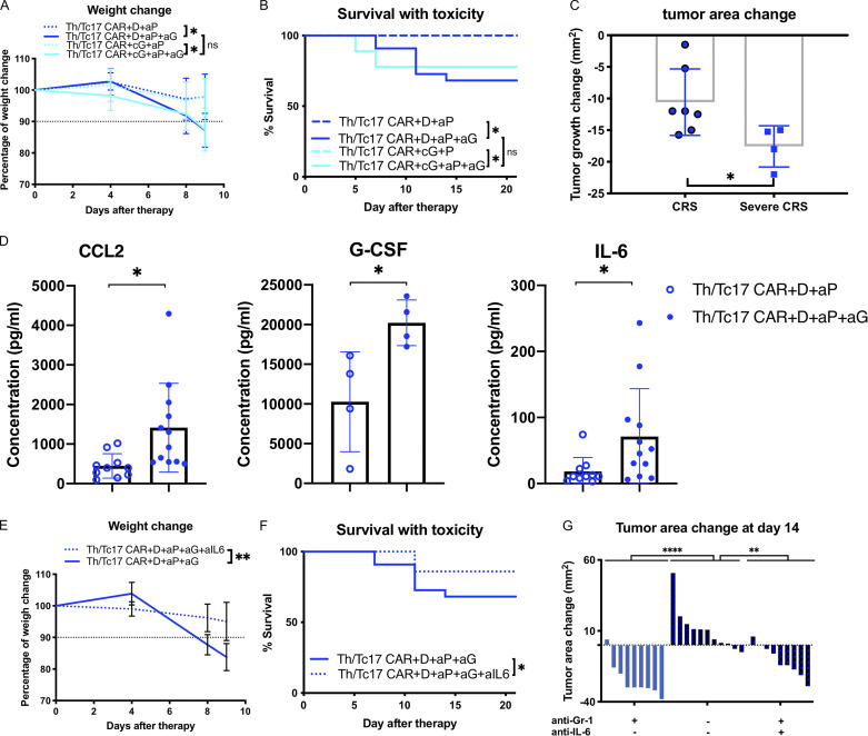
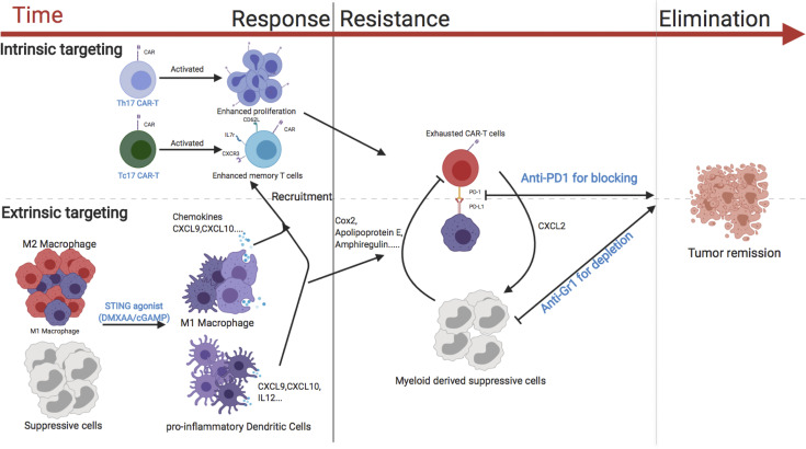
CAR T therapy targeting solid tumors is restrained by limited infiltration and persistence of those cells in the tumor microenvironment (TME). Here, we developed approaches to enhance the activity of CAR T cells using an orthotopic model of locally advanced breast cancer. CAR T cells generated from Th/Tc17 cells given with the STING agonists DMXAA or cGAMP greatly enhanced tumor control, which was associated with enhanced CAR T cell persistence in the TME. Using single-cell RNA sequencing, we demonstrate that DMXAA promoted CAR T cell trafficking and persistence, supported by the generation of a chemokine milieu that promoted CAR T cell recruitment and modulation of the immunosuppressive TME through alterations in the balance of immune-stimulatory and suppressive myeloid cells. However, sustained tumor regression was accomplished only with the addition of anti-PD-1 and anti-GR-1 mAb to Th/Tc17 CAR T cell therapy given with STING agonists. This study provides new approaches to enhance adoptive T cell therapy in solid tumors.


Conflict of interest statement

Disclosures: N. Xu reported a patent to STING on CAR T therapy against solid tumor pending. A.C. Robeson reported personal fees from Precision BioSciences, Inc. outside the submitted work. B.G. Vincent reported "other" from GeneCentric Therapeutics outside the submitted work. N.P. Restifo reported a patent to UNC/NCI pending. J.S. Serody reported grants from Merck Inc. and Glaxo Smith Kline outside the submitted work; in addition, J.S. had a patent for the use of STING agonist to enhance CAR T cell function pending. No other disclosures were reported.

Figures

None
Graphical abstract
Figure 1.
Figure 1.
Th/Tc17 CAR T cells exhibit enhanced early control of tumor growth over 7/15 CAR T cells owing to enhanced persistence in the tumor. (A) Schematic of the LH28z CAR cassette encoding the scFv (7.16.4), hinge, and transmembrane domain from CD8, and intracellular domains from CD28 and CD3ζ. (B) Expression of CAR T receptor on transduced murine 7/15 CD4+ (left) and CD8+ (right) T cells. (C) Representative flow cytometry histograms depicting viability of Neu+ NT2 tumor cells in vitro after 3 d of coculture with 7/15 CAR T cells. Tumor cells were prelabeled with CFSE and plated before the addition of CAR T cells at a 1:1 ratio with tumor cells. (D) Intracellular staining illustrating IFN-γ and TNF production by 7/15 CAR T cells after coculture with Neu+ cells or Neu 3T3 cells at a 2:1 ratio. (E) Tumor area change (tumor area before therapy subtracted from area after therapy) was determined and compared in FVB-neu mice that received 7/15 CAR T cells or mock-transduced T cells (Mock Ts). (F) Expression of CAR T receptor on transduced Th17 (left) or Tc17 (right) cells. (G) In vitro killing of NT2 tumor cells by 7/15 CAR T and Th/Tc17 CAR T cells after overnight culture. (H) Histogram flow plots of IL-17A, TNF, and IFN-γ secretion by Th/Tc17 CAR T and 7/15 CAR T compared with mock T cells after coculture with Neu+ cells at 3:1 ratio for 6 h. (I) Tumor area change was calculated and compared in FVB-neu mice that received Th/Tc17 CAR T cells or 7/15 CAR T cells. (J) Detection of CD4+ and CD8+ CAR T cells in spleen or tumor by flow cytometry 5 d after injection of 7/15 CAR T cells or Th/Tc17 CAR T cells. Data are shown as mean ± SD; *, P < 0.05; **, P < 0.01; significance was determined by Student’s t test or two-way ANOVA. n ≥ 5 mice per group with data from at least two independent experiments with the presented data pooled.
Figure S1.
Figure S1.
IL-7/15 cultured CAR T cells fail to control in vitro or in vivo tumor growth control compared with Th/Tc17 CAR T cells. (A) Schematic of the different CAR cassettes encoding the scFv (7.16.4), hinge and transmembrane domain from CD8 and intracellular domains from CD3ζ, 4-1BB, and/or CD28. (B) Expression of CAR T receptor on IL-7/15 cultured LH28BBz or LHBBz CAR T cells compared with LH28z CAR T cells. (C) Ratio of CD4+:CD8+ CAR T cells after 6 d of expansion with IL-7 and IL-15. (D) NT2 cells were cocultured with different ratios of indicated CAR T cells for 6 h, and tumor cell ATP activity was measured. Tumor ATP level indicating death of tumor cells was characterized at different ratios of mock T cells, LH28z, or LH28BBz CAR T cells. (E) Tumor area change was calculated following the administration of IL-7/15 cultured LHBBz or LH28z CAR T cells (3 × 10⁶ CAR T cells intravenously at 1:1 CD4+:CD8+ ratio) into tumor-bearing mice when the tumor size reached 50 mm2. (F) Schematic of the tumor model in which 5 × 10⁴ NT2 tumor cells were injected orthotopically into the mammary fat pad at day −21. Mice received 3 × 10⁶ CAR T cells intravenously at 1:1 CD4+:CD8+ ratio at day 0 when the tumor size reached 50 mm2. (G) Tumor area change was calculated following injection of 7/15 CAR T before lymphopenia induced by 5 Gy total body irradiation. (H) Tumor area change was calculated following the administration of anti–PD-1 (200 µg/mouse) twice a week following 7/15 CAR T injection. (I) Representative flow cytometry histograms show detection of CAR T cells within the tumor 5 d after 7/15 CAR T infusion. (J) Representative flow plots showing detection of CAR T cells within the tumor or spleen 5 d after 7/15 CAR T or Th/Tc17 CAR T infusion. Data are shown as mean ± SD; ns, not significant; significance was determined by Student’s t test or two-way ANOVA. Mouse studies used a minimum of five mice per group and represent at least two independent experiments. Data were shown as either representative batch or pooled.
Figure 2.
Figure 2.
Tumor control by Th/Tc17 CAR T cells is enhanced by DMXAA injection owing to altered composition of tumor infiltrating immune cells. NT2 tumors were orthotopically injected into murine mammary pads. 21 d later, 500 µg of DMXAA was injected at a site distal to the tumor site, followed by injection of 3 × 10⁶ mock T cells, 7/15 CAR T cells, or Th/Tc17 CAR T cells (CD4+:CD8+ at ∼1:1 ratio). (A) Representative flow cytometry histograms of CAR T detection in TME (left) and summary of accumulation of CAR T cells (right). (B) Change in tumor area was measured 7 d after treatment. (C) Kaplan–Meier survival curve for the treatment cohorts. End point criteria for sacrifice was a tumor area of ≥200 mm2. (D) Tumor area change 14 d after indicated therapy. (E) t-SNE analysis of pooled single-cell sequencing data generated from tumor-infiltrating CD45+ immune cells 7 d after receiving Th/Tc17 CAR T cells in the presence or absence of DMXAA. Data were analyzed by unsupervised clustering, and populations were determined by expression of key markers, including Cd3e (T cells), Adgre1 (macrophages), and Itgam and/or Itgax (myeloid cells). Right: Further classification of subpopulation between DMXAA-treated (+DMXAA) and non–DMXAA-treated (−DMXAA) mice and summary data comparing the frequency of indicated cell populations between +DMXAA and −DMXAA animals. Gating of flow cytometry data used fluorescence minus one controls for each individual experiment. Data are shown as mean ± SD; ns, not significant; *, P < 0.05; **, P < 0.01; ***, P < 0.001; significance was determined by Student’s t test or log-rank Mantel–Cox test when comparing survival. n ≥ 5 mice per group with data from at least two independent experiments (except for scRNA-seq), with the presented data pooled.
Figure S2.
Figure S2.
Th17 CAR T cells are indispensable for DMXAA therapeutic efficacy, which enhances the number and cytotoxicity of Th17 CAR T cells in the TME. (A and B) Tables for the percentage of T cell subsets (A) and macrophage and myeloid cell subsets (B) calculated from scRNA-seq data. (C) Table for the percentage of CD8+ T cells expressing indicated genes calculated from scRNA-seq data. (D–F) Assessment of CAR expression on tumor-infiltrating T cells isolated 7 d after therapy by flow cytometry. (D) Determination of change in tumor area 7 d after injection of Tc17 or Th/Tc17 CAR T cells. (E) Assessment of intratumoral CD8+ CAR T cells was performed 7 d after therapy. (F) Absolute number of CD8+ CAR T cells/mm2 tumor 7 d after therapy. Data in G–M data were derived from the characterization of T cells expressing CD4 from scRNA-seq result. (G) CD4+ T cells were subdivided based on absence of Cd44 (naive T cells), expression of Cd4 and Cd44 (activated CD4 T cells), Cd4 and Foxp3 (T reg cells). CD4+ T cells were compared between the +DMXAA and −DMXAA treatment groups, and each subpopulation was examined further as described below. (H) Table for the percentage of CD4+ T cells expressing the indicated genes from the scRNA-seq data. (I) t-SNE plot of CD4+ T cells for Th17-related genes. (J) t-SNE plot of CD4+ T cells for Th17/Th1-related genes. (K) Violin plot shows the distribution and change of indicated gene expression in B and C. (L) Validation of single-cell data using flow cytometry to detect CD4+ CAR T cells. (M) Analysis of the correlation between tumor growth change and absolute cell number of CAR-expressing CD4 T cells/cm2 tumor. Data (excluding scRNA-seq) represent one of two independent experiments (n ≥ 5 mice per group). Single-cell sequencing data represent three mice/treatment group, with statistical significance determined by differential GSA with the Partek flow workstation. *, q < 0.05; **, q < 0.01, in the change of gene expression level. Fold change is +DMXAA versus −DMXAA. For percentage/number change, please refer to Fig. S2 H. Flow cytometry data were pooled from two independent experiments and shown as mean ± SD. Statistics analysis for cytometric analysis is determined by Student’s t test; *, P < 0.05; **, P < 0.01; ****, P < 0.0001.
Figure 3.
Figure 3.
DMXAA treatment enhances the number and cytotoxicity of Tc17 CAR T cells in the TME, although they eventually become exhausted. (A) T cell populations were subdivided based on expression of Cd8 and Cd44 (activated CD8 T cells), Cd44 (naive T cells), and Cd4 and Foxp3 (T reg cells). CD8+ T cells were compared between the +DMXAA and −DMXAA treatment groups, and each subpopulation was examined further as described below. (B) t-SNE plots of CD8+ T cells for Tc17-related genes. (C) t-SNE plots of CD8+ T cells for Tc1-related genes. (D) Violin plots depicting the distribution and change in expression of genes identified in B and C. (E) Validation of single-cell sequencing data using flow cytometry to detect CD8+ CAR T cells. Representative flow plots (left) and frequency (middle) and number (right) of CD8+ CAR T cells. (F) Analysis of the correlation between tumor growth and absolute cell number of CAR-expressing CD8+ T cells/cm2 tumor. (G) Activated or exhausted T cells were selected by unsupervised clustering result of tumor-infiltrating immune cells from 7 and 10 d after Th/Tc17 CAR+DMXAA therapy and shown as a t-SNE plot. (H) t-SNE plot (left) and violin plot (right) of T cells assessing genes associated with T cell exhaustion. (I) t-SNE plot (left) and violin plot (right) of T cells for genes associated with T cell effector function. (J) t-SNE plot (left) and violin plot (right) of T cells for genes associated with chemokine secretion. Data (excluding scRNA-seq) represent one of two independent experiments (n ≥ 5 mice per group). Single-cell sequencing data represent two to three mice/treatment group, and statistical significance was determined by differential GSA from Partek flow workstation. *, q < 0.05; **, q < 0.01; ***, q < 0.001; ****, q < 0.0001 in the change of gene expression level. Fold change is +DMXAA versus −DMXAA or 7 versus 10 d. For percentage/number change, please refer to Fig. S2 C. Flow-cytometric analysis was pooled from two independent experiments and is shown as mean ± SD. Statistical analysis for cytometric analysis was determined by Student’s t test; ***, P < 0.001; ****, P < 0.0001.
Figure S3.
Figure S3.
DMXAA treatment enhances IFN-γ–dependent antitumor function of Th/Tc17 CAR T cells, but T cell exhaustion limits the cumulative beneficial effect of DMXAA on Th/Tc17 CAR T therapy. Mice received Th/Tc17 CAR T cells in the presence or absence of DMXAA therapy. 7 d later, RNA from tumors was isolated and analyzed by microarray for Th17/Th1 response. (A) Heatmap depicts genes where fold-change was significant, with a threefold increase. Each column represents an individual mouse. (B) Quantification of significant changes (P < 0.05) in TME for Th17 or Th1 response following DMXAA treatment. (C) Mice were treated i.p. with anti–IFN-γ (250 µg/mouse) twice a week after Th/Tc17 CAR T therapy. Significance was determined by Student’s t test or two-way ANOVA. Studies involved at least four mice per independent experiment. Data from C represent two independent experiments with four mice per group. (D–G) T cell cluster selected based on expression of Cd3e by unsupervised clustering of tumor-infiltrating immune cells from 7 and 10 d after Th/Tc17 CAR + D therapy. (D) Table shows the percentage of T cells expressing the indicated genes calculated from scRNA-seq data. (E) t-SNE plot (left) and violin plot (right) of T cells for genes associated with T cell exhaustion. (F) Validation of PD-1 expression using flow-based method to detect high levels of PD-1 expression on T cells from mice receiving Th/Tc17 CAR T or mock T cell therapy. Representative flow cytometry histograms (left); percentage of CAR T (right) within the CD4 or CD8 T cell group (n = 4/group). (G) t-SNE plot (left) and violin plot (right) of T cells for genes associated with T cell apoptosis. Data (excluding scRNA-seq) represent one of two independent experiments (n ≥ 5 mice per group). Single-cell sequencing data represent two to three mice/treatment group. Statistical significance was determined by differential GSA with the Partek flow workstation. *, q < 0.05; **, q < 0.01; ***, q < 0.001; ****, q < 0.0001 in the change of gene expression level. Fold change is 7 versus 10 d. For percentage/number change, please refer to Fig. S3 D. Flow cytometric analysis is shown as mean ± SD. Statistics analysis for cytometric analysis was determined by Student’s t test; *, P < 0.05; **, P < 0.01; ***, P < 0.001; ****, P < 0.0001.
Figure 4.
Figure 4.
DMXAA reduces the accumulation of suppressive macrophages and enhances the trafficking of T cells in the TME, but this effect is eventually lost due to the return of immunosuppressive cells. (A) t-SNE plot of macrophage populations identified by unsupervised clustering (Fig. 2), selected and classified as M1- or M2-like, and compared between mice receiving therapy with or without DMXAA treatment. (B) Heatmap of genes identified in A exhibiting a greater than twofold change in expression following DMXAA treatment, with a significant difference of P < 0.05 (determined by GSA). (C) t-SNE plot (left) and violin plots (right) showing expression of M2-related genes. (D) t-SNE plot (left) and violin plots (right) showing expression of M1-related genes. (E) Tumor area before and after 7 d of treatment with Th/Tc17 CAR T and DMXAA therapy after injection of clodronate or control liposomes. (F) t-SNE plot of myeloid populations identified by unsupervised clustering (Fig. 2), selected and classified as inflammatory myeloid cells or suppressive myeloid cells, and compared between mice in the presence or absence of DMXAA treatment. (G) Heatmap of genes identified in F exhibiting a greater than twofold change in expression following DMXAA treatment, with a significant difference of P < 0.05 (determined by GSA). (H) t-SNE plot depicting genes preferentially expressed in inflammatory myeloid cells. (I) Violin plots showing distribution and change in expression of genes highlighted in H. Myeloid cells from unsupervised clustering 7 and 10 d after therapy were selected. (J) t-SNE plot of myeloid cells between 7 and 10 d after therapy. (K) Heatmap of proinflammatory or suppressive genes that exhibited a greater than twofold change in expression following DMXAA treatment, with a significant difference of P < 0.05. (L) t-SNE plot of myeloid cells illustrating genes associated with the inflammatory and suppressive functions of myeloid cells. (M) Violin plots showing distribution and change of indicated gene expression in L. *, P < 0.05; significance was determined by Student’s t test. Single-cell sequencing data represent two to three mice/treatment group. Macrophage depletion assay was done independently twice with representative result shown.
Figure S4.
Figure S4.
M2 macrophages and myeloid cells are reduced upon DMXAA treatment, but later replaced by suppressive myeloid cells. Macrophages and myeloid cells from day 7 are selected as in Fig. 3 F. (A) Table showing the percentage of macrophages expressing the indicated genes calculated from scRNA-seq data. (B) t-SNE plot on M1-like (left) and M2-like (right) related genes. (C) Violin plot shows the distribution and change of indicated gene expression in B. (D) t-SNE and violin plot of cd274 (PD-L1). (E) Table showing the percentage of myeloid cells expressing the indicated gene calculated from scRNA-seq data. (F) t-SNE plot on genes expressed in iMCs. (G) Violin plot shows the distribution and change of indicated gene expression in F. (H) t-SNE plot of genes expressed in myeloid-like suppressor cells. (I) Violin plot shows the distribution and change of indicated gene expression in H. Myeloid cells from days 7 and 10 are selected as in Fig. 3 J. (J) Table showing the percentage of myeloid cells expressing the indicated genes calculated from scRNA-seq data. (K) t-SNE plot (left) and violin plot (right) of myeloid cells for genes associated with myeloid-like suppressor cells. Single-cell sequencing data represent two to three mice/treatment group.
Figure 5.
Figure 5.
DMXAA is required for tumor remission after Th/Tc17 CAR T, DMXAA, anti–PD-1, and anti-Gr1 treatment. Animals received injections of anti–PD-1 (200 µg/mouse) and anti-Gr1 (300 µg/mouse) twice weekly, beginning a day after CAR T injection in additional to therapy described in Fig. 2 (Th/Tc17 CAR + D + aP + aG). (A) Schematic describing therapy schedule. (B) Summary of tumor growth (in area) in the first 3 wk after CAR T therapy. (C) Change in tumor area was assessed 7 d after administration of Th/Tc CAR + triple therapy or in the absence of DMXAA, anti–PD-1, anti–Gr-1, or CAR T cells (mock T cells to model the absence of CAR T cells). (D) Kaplan–Meier survival curve indicating mortality over 100 d for the four treatment groups. Data are shown as mean ± SD; ns, not significant; *, P < 0.05; **, P < 0.01; ***, P < 0.001; ****, P < 0.0001; significance was determined by Student’s t test or two-way ANOVA when comparing tumor growth change between groups or log-rank Mantel–Cox test when comparing survival. n ≥ 5 mice per group with the data from at least two independent experiments, with the presented data pooled.
Figure S5.
Figure S5.
Th/Tc17 cells function better than IL-7/15 CAR T cells when combined with a STING agonist, anti–PD-1, and anti–GR-1 mAb therapy. Mice were treated with anti–GR-1, anti–PD-1, DMXAA, and Th/Tc17 CAR T cells, 7/15 CAR T cells, or mock T cells as described in Fig. S5. (A) Tumor growth in individual mice receiving therapy. (B) Kaplan–Meier survival curve. (C) Summary of CAR T cell accumulation in the spleen. (D–G) Mice received 7/15 CAR + D + aP or Th/Tc17 CAR + D + aP treatment. T cells were isolated from the tumor 7 d after therapy and characterized by flow cytometry. (D) Representative plot (left) and summary graph (right) of Ki67 expression from CD4+ CAR T cells with six to nine mice per group. (E and F) Assessment of the memory phenotype of CD8+ CAR T cells from the tumor at 7 or 12 d after 7/15 CAR + D + aP or Th/Tc17 CAR + D + aP treatment. (G) Comparison of memory precursor effector cells of CD8+ CAR T cells within the tumor 7 d after 7/15 CAR + D + aP or Th/Tc17 CAR + D + aP treatment. Data shown as mean ± SD; ns, not significant; *, P < 0.05; **, P < 0.01; ***, P < 0.001; ****, P < 0.0001, significance was determined by Student’s t test or log-rank Mantel–Cox test comparing survival. Mouse studies used a minimum of five mice per group and represent at least two independent experiments where data are shown as either representative or pooled.
Figure 6.
Figure 6.
Tumor control by Th/Tc17 CAR T cells is enhanced by human STING agonist 2,3-cGAMP. NT2 tumors were orthotopically injected into murine mammary pads. 21 d later, 25 µg of cGAMP was injected at a site distal to the tumor site, followed by injection of 3 × 10⁶ mock T cells, 7/15 CAR T cells, or Th/Tc17 CAR T cells (CD4+:CD8+ at ∼1:1 ratio). (A) Representative flow cytometry histograms of CAR T cells detection at in the TME (left) and summary of accumulation of CAR T cells (right). (B) Change in tumor area measured over 7 d after treatment. (C) Tumor area change 21 d after indicated therapy. (D) Kaplan–Meier survival curve for the treatment cohorts. Endpoint criteria for sacrifice was tumor area of ≥200 mm2. Gating of flow cytometry data used fluorescence minus one controls for each individual experiment. Data are shown as mean ± SD; ns, not significant; *, P < 0.05; **, P < 0.01; ***, P < 0.001; significance was determined by Student’s t test or log-rank Mantel–Cox test when comparing survival. n ≥ 5 mice per group with the data from at least two independent experiments, with the presented data pooled.
Figure 7.
Figure 7.
Depletion of myeloid cells with anti-Gr1 during Th/Tc17 CAR + D/cG + aP regimen leads to cytokine release-like syndrome. Mice received Th/Tc17 CAR + D + P with or without twice-weekly anti–Gr-1 injections after CAR T therapy. (A) Quantification of weight loss as percentage of total change in body weight following Th/Tc17 CAR + D/cG + P or Th/Tc17 CAR + D/cG + aP + aG. (B) Kaplan–Meier survival curve indicating mortality over 20 d in the presence or absence of anti–Gr-1 treatment. (C) Change in tumor area 7 d after Th/Tc17 CAR + D + P or Th/Tc17 CAR + D + aP + aG treatment in mice that survived but lost >10% of their initial weight (CRS-like) compared with mice that eventually died from CRS-like symptoms (severe CRS-like). (D) Serum cytokine levels of CCL2, mG-CSF, and IL-6 measured 5 d after therapy. (E) Quantification of weight loss as percentage of total change in body weight following Th/Tc17 CAR + D + aP + aG + aIL-6 or Th/Tc17 CAR + D + aP + aG treatment. (F) Kaplan–Meier survival curve indicating mortality over 20 d in the presence or absence of anti–IL-6 mAb treatment. (G) Tumor growth change at 14 d after the therapy in the presence or absence of anti–Gr-1 and/or anti–IL-6 mAb. Data are shown as mean ± SD; ns, not significant; *, P < 0.05; **, P < 0.01; ****, P < 0.0001; significance was determined by Student’s t test and log-rank Mantel–Cox test when comparing survival. n ≥ 5 mice per group with the data from at least two independent experiments with the presented data pooled.
Figure 8.
Figure 8.
Model of the activity of CAR T cells against breast cancer. The use of Th/Tc17 CAR T cells with enhanced proliferation and memory status (intrinsic modification) along with DMXAA/cGAMP to enhance trafficking and reduce immunosuppression in the TME (extrinsic modification) is critical for the effective antitumor response of CAR T cells against breast cancer (left). CAR T exhaustion (intrinsic resistance) and reversion back to an immunosuppressive TME state (extrinsic resistance) are associated with loss of CAR T cell function (middle). The addition of anti–PD-1 to overcome exhaustion (intrinsic modification) and the depletion of suppressive myeloid cells with anti–Gr-1 (extrinsic modification) leads to sustained tumor remission (right).

References

    1. Adachi, K., Kano Y., Nagai T., Okuyama N., Sakoda Y., and Tamada K.. 2018. IL-7 and CCL19 expression in CAR-T cells improves immune cell infiltration and CAR-T cell survival in the tumor. Nat. Biotechnol. 36:346–351. 10.1038/nbt.4086 - DOI - PubMed
    1. Beatty, G.L., O’Hara M.H., Lacey S.F., Torigian D.A., Nazimuddin F., Chen F., Kulikovskaya I.M., Soulen M.C., McGarvey M., Nelson A.M., et al. . 2018. Activity of mesothelin-specific chimeric antigen receptor T cells against pancreatic carcinoma metastases in a phase 1 trial. Gastroenterology. 155:29–32. 10.1053/j.gastro.2018.03.029 - DOI - PMC - PubMed
    1. Boni, A., Muranski P., Cassard L., Wrzesinski C., Paulos C.M., Palmer D.C., Gattinoni L., Hinrichs C.S., Chan C.C., Rosenberg S.A., and Restifo N.P.. 2008. Adoptive transfer of allogeneic tumor-specific T cells mediates effective regression of large tumors across major histocompatibility barriers. Blood. 112:4746–4754. 10.1182/blood-2008-07-169797 - DOI - PMC - PubMed
    1. Buonocore, S., Ahern P.P., Uhlig H.H., Ivanov I.I., Littman D.R., Maloy K.J., and Powrie F.. 2010. Innate lymphoid cells drive interleukin-23-dependent innate intestinal pathology. Nature. 464:1371–1375. 10.1038/nature08949 - DOI - PMC - PubMed
    1. Burga, R.A., Thorn M., Point G.R., Guha P., Nguyen C.T., Licata L.A., DeMatteo R.P., Ayala A., Joseph Espat N., Junghans R.P., and Katz S.C.. 2015. Liver myeloid-derived suppressor cells expand in response to liver metastases in mice and inhibit the anti-tumor efficacy of anti-CEA CAR-T. Cancer Immunol. Immunother. 64:817–829. 10.1007/s00262-015-1692-6 - DOI - PMC - PubMed

Publication types

LinkOut - more resources