

Piperine Inhibits Cell Proliferation and Induces Apoptosis of Human Gastric Cancer Cells by Downregulating Phosphatidylinositol 3-Kinase (PI3K)/Akt Pathway
- PMID: 33382670
- PMCID: PMC7784594
- DOI: 10.12659/MSM.928403
Piperine Inhibits Cell Proliferation and Induces Apoptosis of Human Gastric Cancer Cells by Downregulating Phosphatidylinositol 3-Kinase (PI3K)/Akt Pathway
Abstract
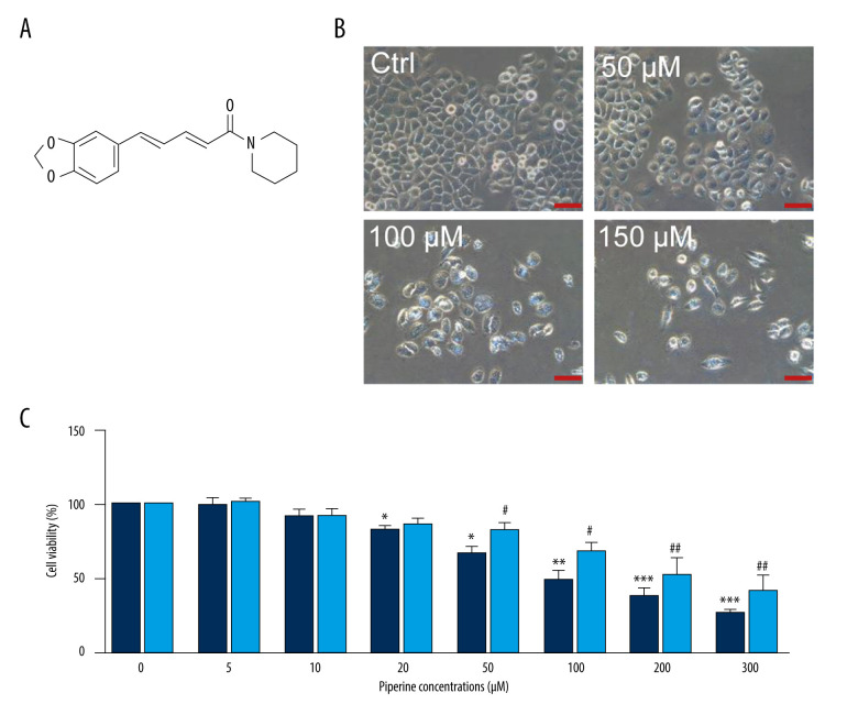
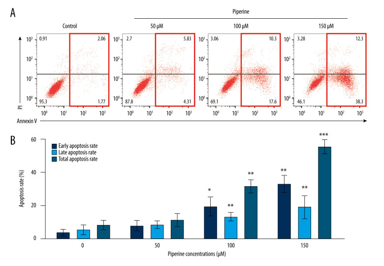
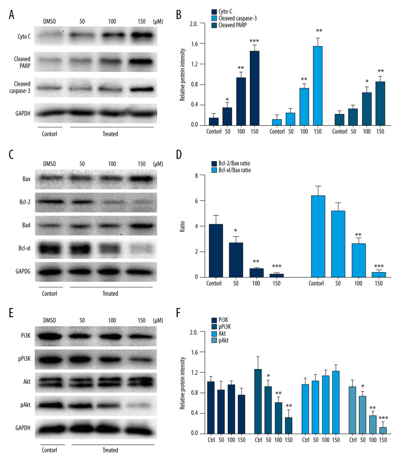
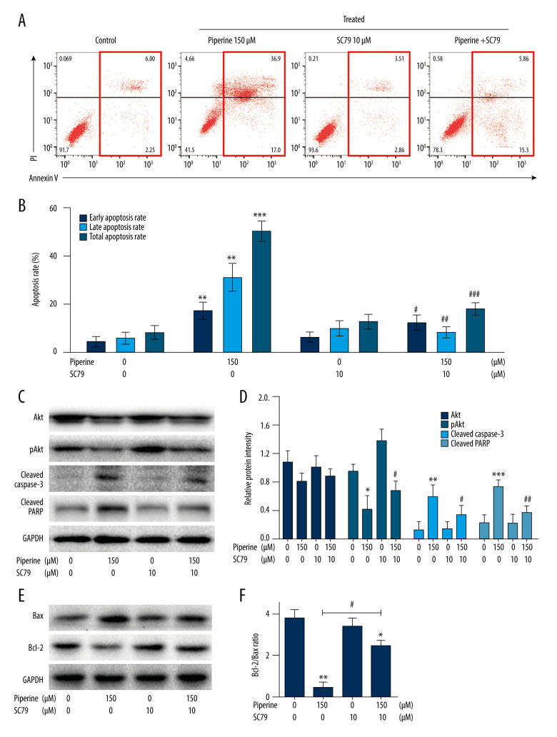
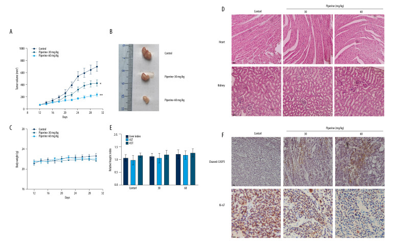
BACKGROUND Piperine has been reported to inhibit proliferation and induce apoptosis in various cancer cells. This study aimed to explore the efficacy and underlying mechanism of piperine in human gastric cancer. MATERIAL AND METHODS MTT assay was performed to examine the effect of piperine (concentrations of 0-300 μM) on the proliferation of human gastric cancer SNU-16 cells and normal human gastric epithelial GES-1 cells. Flow cytometry and Western blot were used to determine cell apoptosis and the expression level of protein (Cyto C, cleaved PARP, cleaved caspase-3, Bax, Bcl-2, Bad, Bcl-xl, PI3K, pPI3K, Akt, and pAkt), respectively. To further investigate the anti-tumor mechanism of piperine in SNU-16 cells, we used a small-molecule Akt activator SC79 in this study. The in vivo mechanism of piperine against gastric cancer was evaluated using a xenograft tumor model. RESULTS The results showed that piperine inhibited proliferation and induced apoptosis of SNU-16 cells. Piperine upregulated the protein expression of Bax, Bad, Cyto C, cleaved PARP, and cleaved caspase-3, but downregulated the protein expression of Bcl-2, Bcl-xl, pPI3k, and pAkt. However, SC79 reversed the function of piperine on the apoptosis-related proteins. An in vivo study revealed that, compared with the control group, the tumor volume of mice treated with piperine was significantly reduced. Piperine enhanced cleaved caspase-3 expression but decreased Ki-67 expression in a dose-dependent manner. Moreover, the nontoxicity effect of piperine was confirmed by H&E staining analysis in kidney and heart tissues of mice. CONCLUSIONS Our findings suggest that piperine inhibits proliferation and induces apoptosis of human gastric cancer cells through inhibition of the PI3K/Akt signaling pathway.
Conflict of interest statement
None.
Figures
Similar articles
-
Synergistic effect of piperine and paclitaxel on cell fate via cyt-c, Bax/Bcl-2-caspase-3 pathway in ovarian adenocarcinomas SKOV-3 cells.Eur J Pharmacol. 2016 Nov 15;791:751-762. doi: 10.1016/j.ejphar.2016.10.019. Epub 2016 Oct 15. Eur J Pharmacol. 2016. PMID: 27756602
-
Antitumor and Apoptosis-inducing Effects of Piperine on Human Melanoma Cells.Anticancer Res. 2019 Apr;39(4):1883-1892. doi: 10.21873/anticanres.13296. Anticancer Res. 2019. PMID: 30952729
-
The natural secolignan peperomin E induces apoptosis of human gastric carcinoma cells via the mitochondrial and PI3K/Akt signaling pathways in vitro and in vivo.Phytomedicine. 2016 Jul 15;23(8):818-27. doi: 10.1016/j.phymed.2016.04.001. Epub 2016 May 11. Phytomedicine. 2016. PMID: 27288917
-
Piperine and its nanoformulations: A mechanistic review of their anti-cancer activities.Biomed Pharmacother. 2025 Jun;187:118075. doi: 10.1016/j.biopha.2025.118075. Epub 2025 Apr 23. Biomed Pharmacother. 2025. PMID: 40273688 Review.
-
Piperine: an emerging biofactor with anticancer efficacy and therapeutic potential.Biofactors. 2025 Jan-Feb;51(1):e2134. doi: 10.1002/biof.2134. Epub 2024 Oct 28. Biofactors. 2025. PMID: 39467259 Review.
Cited by
-
Piperine enhances doxorubicin sensitivity in triple-negative breast cancer by targeting the PI3K/Akt/mTOR pathway and cancer stem cells.Sci Rep. 2024 Aug 6;14(1):18181. doi: 10.1038/s41598-024-65508-0. Sci Rep. 2024. PMID: 39107323 Free PMC article.
-
Interactions between circRNAs and miR-141 in Cancer: From Pathogenesis to Diagnosis and Therapy.Int J Mol Sci. 2023 Jul 24;24(14):11861. doi: 10.3390/ijms241411861. Int J Mol Sci. 2023. PMID: 37511619 Free PMC article. Review.
-
The Hidden Power of Black Pepper: Exploring Piperine's Role in Cancer.Plant Foods Hum Nutr. 2025 May 29;80(3):129. doi: 10.1007/s11130-025-01374-z. Plant Foods Hum Nutr. 2025. PMID: 40439931 Free PMC article. Review.
-
Piperine Induces Apoptosis and Autophagy in HSC-3 Human Oral Cancer Cells by Regulating PI3K Signaling Pathway.Int J Mol Sci. 2023 Sep 11;24(18):13949. doi: 10.3390/ijms241813949. Int J Mol Sci. 2023. PMID: 37762259 Free PMC article.
-
An Overview of the Spices Used for the Prevention and Potential Treatment of Gastric Cancer.Cancers (Basel). 2024 Apr 22;16(8):1611. doi: 10.3390/cancers16081611. Cancers (Basel). 2024. PMID: 38672692 Free PMC article. Review.
References
-
- Liang X, Zhu J, Li Y, et al. Treatment strategies for metastatic gastric cancer: Chemotherapy, palliative surgery or radiotherapy? Future Oncol. 2020;16(5):91–102. - PubMed
-
- Shi Y, Luo X, Li P, et al. miR-7-5p suppresses cell proliferation and induces apoptosis of breast cancer cells mainly by targeting REGγ. Cancer Lett. 2015;358(1):27–36. - PubMed
MeSH terms
Substances
LinkOut - more resources
Full Text Sources
Medical
Research Materials
