. 2020 Dec 31;15(12):e0236495.
doi: 10.1371/journal.pone.0236495. eCollection 2020.

An unbiased template of the Drosophila brain and ventral nerve cord

Affiliations

An unbiased template of the Drosophila brain and ventral nerve cord

John A Bogovic et al. PLoS One. .

Abstract

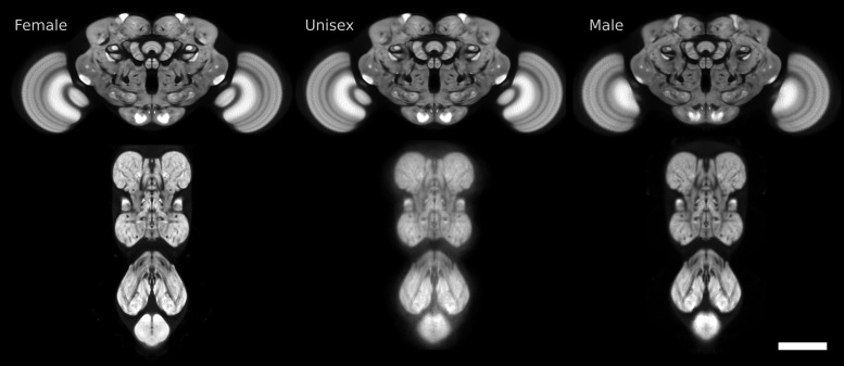
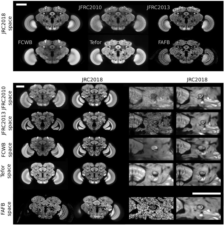
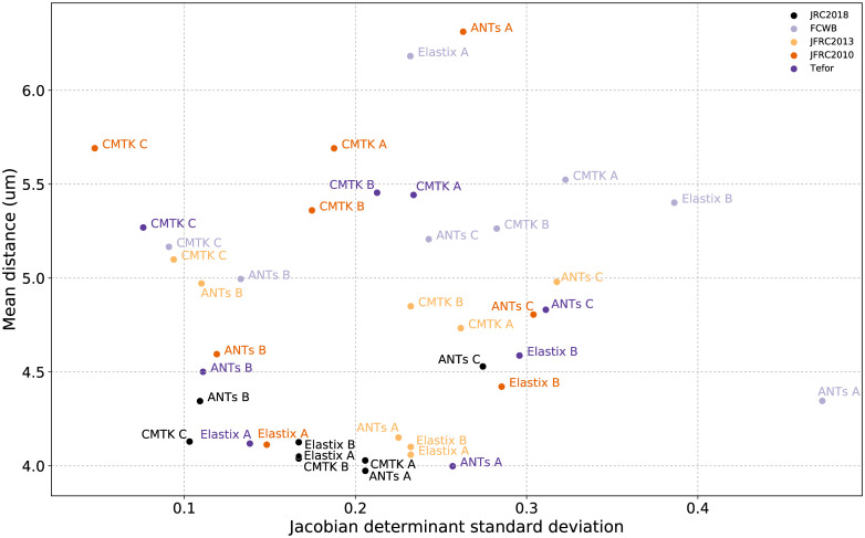
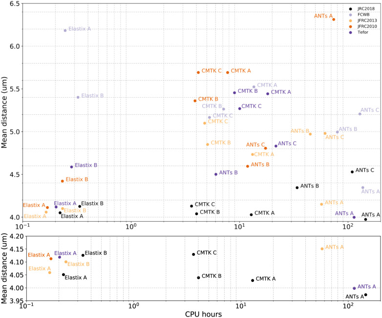
The fruit fly Drosophila melanogaster is an important model organism for neuroscience with a wide array of genetic tools that enable the mapping of individual neurons and neural subtypes. Brain templates are essential for comparative biological studies because they enable analyzing many individuals in a common reference space. Several central brain templates exist for Drosophila, but every one is either biased, uses sub-optimal tissue preparation, is imaged at low resolution, or does not account for artifacts. No publicly available Drosophila ventral nerve cord template currently exists. In this work, we created high-resolution templates of the Drosophila brain and ventral nerve cord using the best-available technologies for imaging, artifact correction, stitching, and template construction using groupwise registration. We evaluated our central brain template against the four most competitive, publicly available brain templates and demonstrate that ours enables more accurate registration with fewer local deformations in shorter time.


Conflict of interest statement

The authors have declared that no competing interests exist.

Figures

Fig 1
Fig 1. Slices of the six templates we created for female, unisex, and male Drosophila central brains and ventral nerve cords.
Scale bar 100 μm.
Fig 2
Fig 2. Maximum intensity projections of individuals from four of the GAL4 driver lines used to evaluate registration accuracy (magenta nc82, green GAL4).
Notice the broad arborization spanning most of the central brain and optic lobes. Scale bar 100 μm.
Fig 3
Fig 3. Visual comparison of Drosophila brain templates and bridging transformations.
The top two rows show four existing templates registered to our JRC 2018 female template, as well as synaptic cleft predictions derived from the FAFB EM volume, transformed into the space of JRC 2018F. The middle four rows show JRC 2018F (second and fourth columns) registered to each of the three templates, along with a close-up around the fan-shaped body and the pedunculus of the mushroom body. The bottom row shows JRC 2018F transformed into the space of FAFB. Scale bars 100 μm.
Fig 4
Fig 4. Horizontal (xz-slice) visualization of five brain templates and FAFB in physical coordinates.
Note that the lower z-resolution is appreciable for individual templates (JFRC 2010 and JFRC 2013). Furthermore, observe the significant differences in physical sizes across these brain templates. Scale bar 100 μm.
Fig 5
Fig 5. Scatterplot showing mean skeleton distance and standard deviation of the Jacobian determinant for all template-algorithm pairs.
Fig 6
Fig 6. Scatterplot showing mean skeleton distance and the mean computation time in CPU-hours for all template-algorithm pairs (above), and the best performing pairs (below).
Fig 7
Fig 7. A visualization of female vs male morphological differences.
(A) the maximum intensity projection (MIP) of the difference between male and female Jacobian determinant maps where values greater than 1 (yellow) indicate the male template is locally larger. (B) the MIP of the Jacobian determinant map overlayed with a MIP of the unisex template. Arrows indicate an artifact of this analysis in regions where the templates have no contrast, but the registration algorithm applies a strong deformation. Scale bar 100 μm.

References

    1. Talairach J, Tournoux P. Co-planar stereotaxic atlas of the human brain. Thieme, New York; 1988.
    1. Lancaster JL, Wolodorff MG, Parsons LM, Liotti M, Freitas CS, Rainey L, et al. Automated Talairach Atlas Labels for Functional Brain Mapping. Human Brain Mapping. 2000;10:120–131. 10.1002/1097-0193(200007)10:3<120::AID-HBM30>3.0.CO;2-8 - DOI - PMC - PubMed
    1. Allen Institute for Brain Science. Techincal White Paper: Allen Mouse Common Coordinate Framework; 2015. v.1. Available from: http//:help.brain-map.org/download/attachments/2818171/MouseCCF.pdf.
    1. Lein ES, Hawrylycz MJ, Jones AR, et al. Genome-wide atlas of gene expression in the adult mouse brain. Nature. 2007;445(7124):168–176. 10.1038/nature05453 - DOI - PubMed
    1. Long F, Peng H, Liu X, Kim SK, Myers E. A 3D digital atlas of C. elegans and its application to single-cell analyses. Nature Methods. 2009;6:667–672. 10.1038/nmeth.1366 - DOI - PMC - PubMed

Publication types

MeSH terms

LinkOut - more resources