. 2021 Jan 11;56(1):36-51.e5.
doi: 10.1016/j.devcel.2020.12.006. Epub 2020 Dec 30.

Chromosome Segregation and Peptidoglycan Remodeling Are Coordinated at a Highly Stabilized Septal Pore to Maintain Bacterial Spore Development

Affiliations

Chromosome Segregation and Peptidoglycan Remodeling Are Coordinated at a Highly Stabilized Septal Pore to Maintain Bacterial Spore Development

Ahmed M T Mohamed et al. Dev Cell. .

Abstract

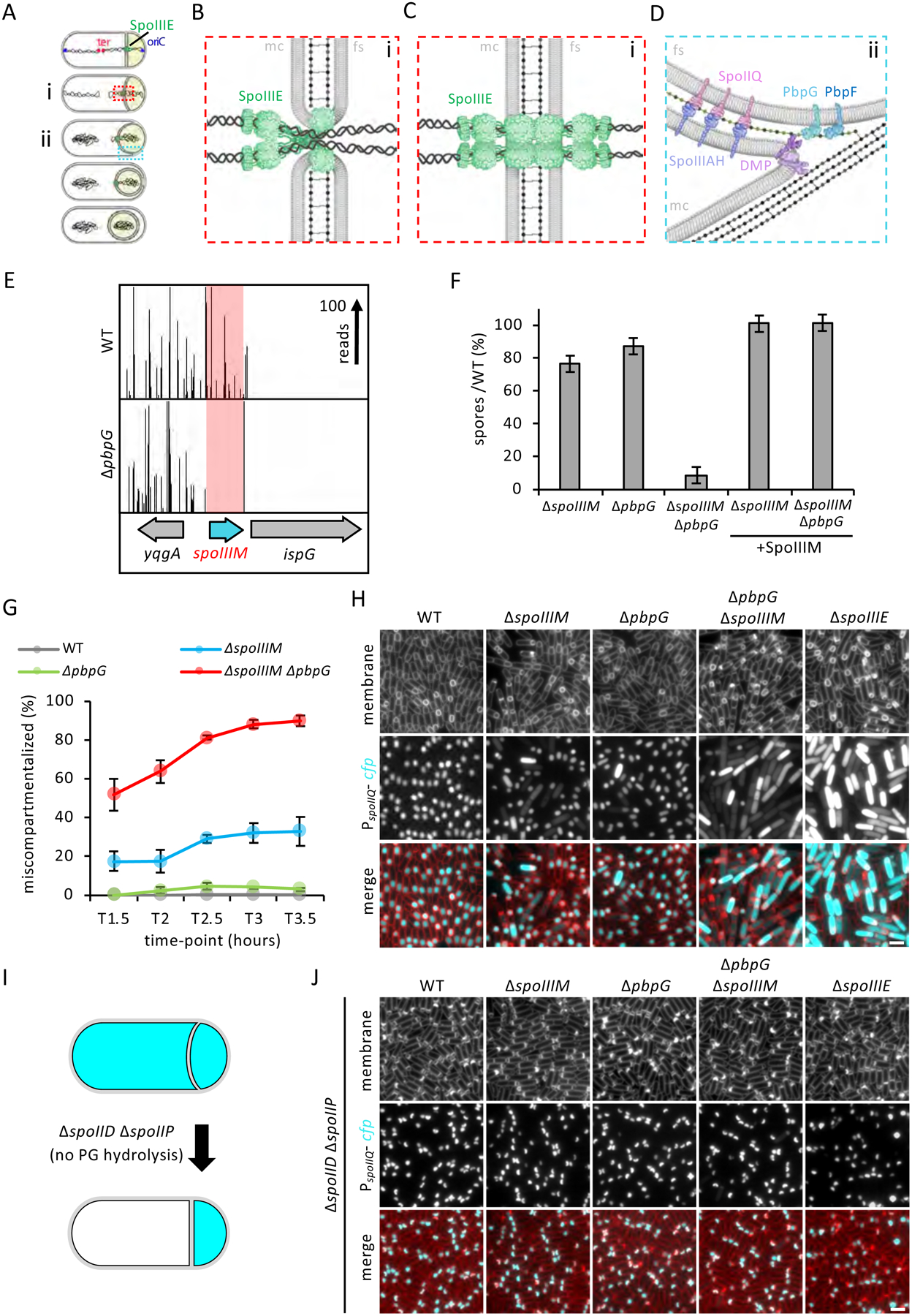
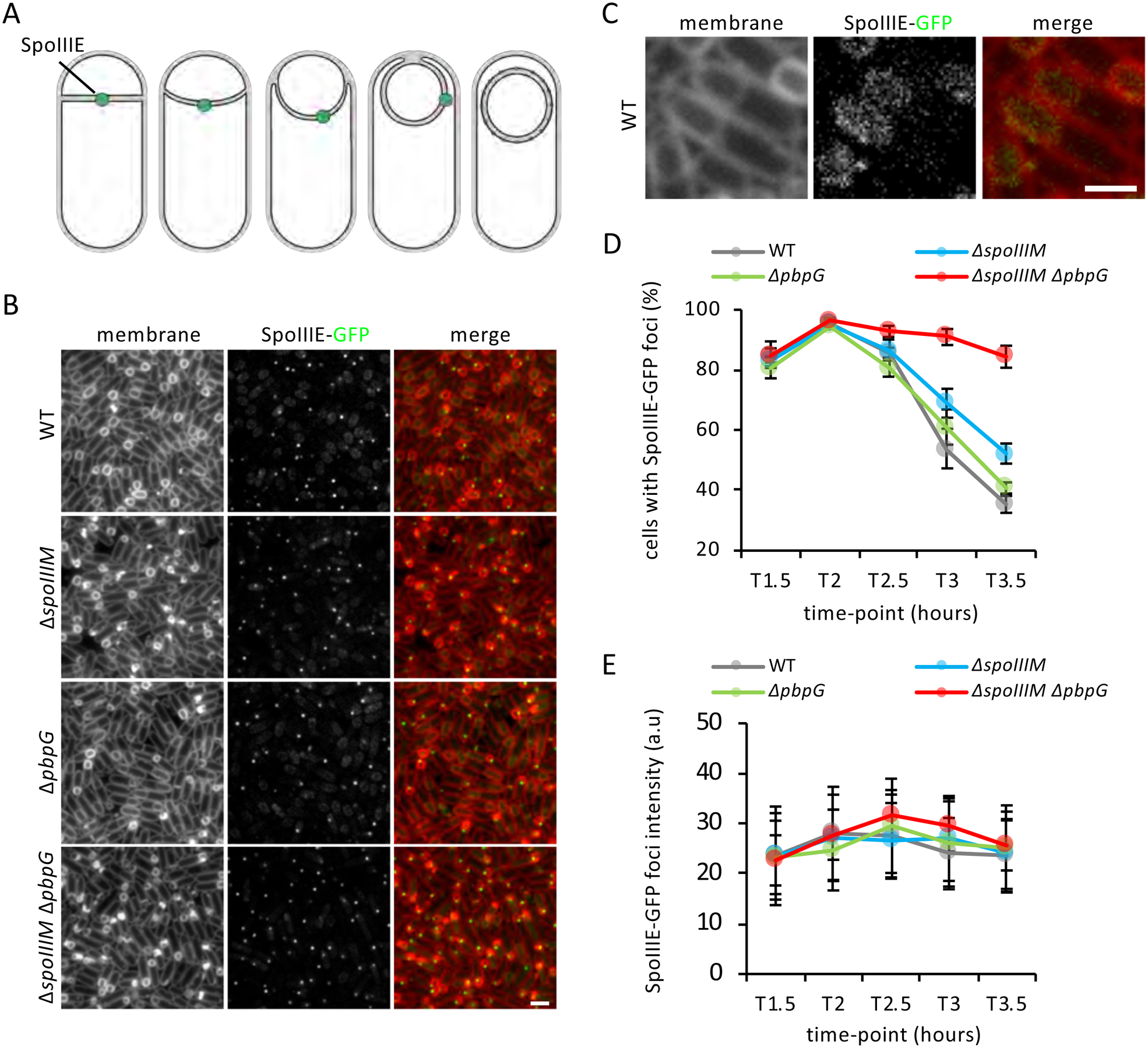
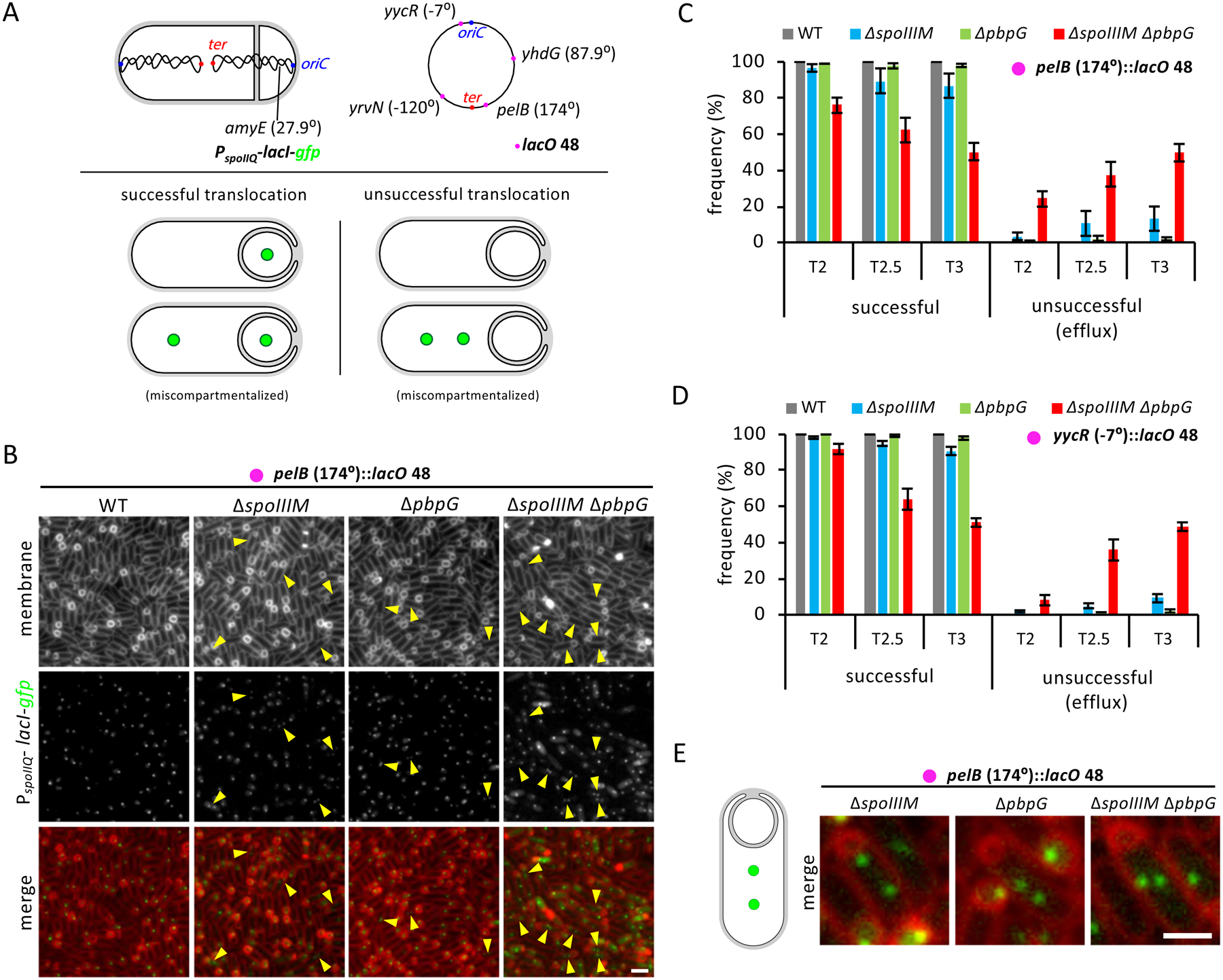
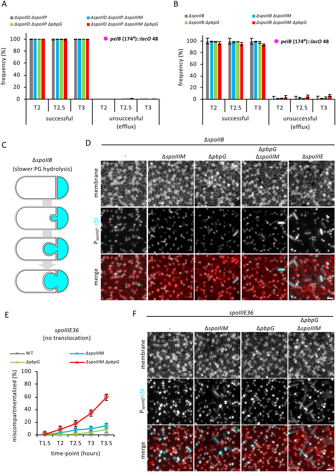
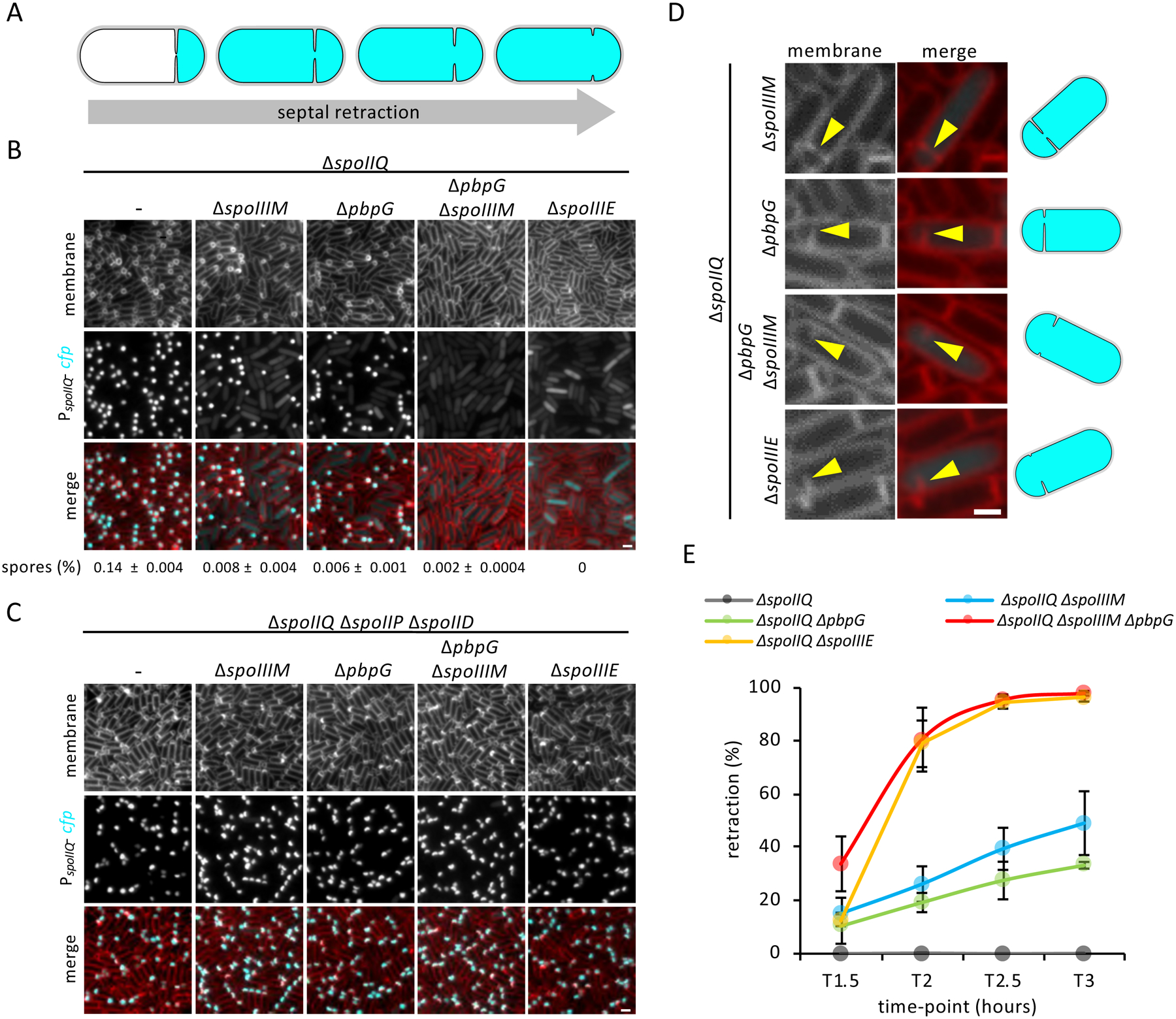
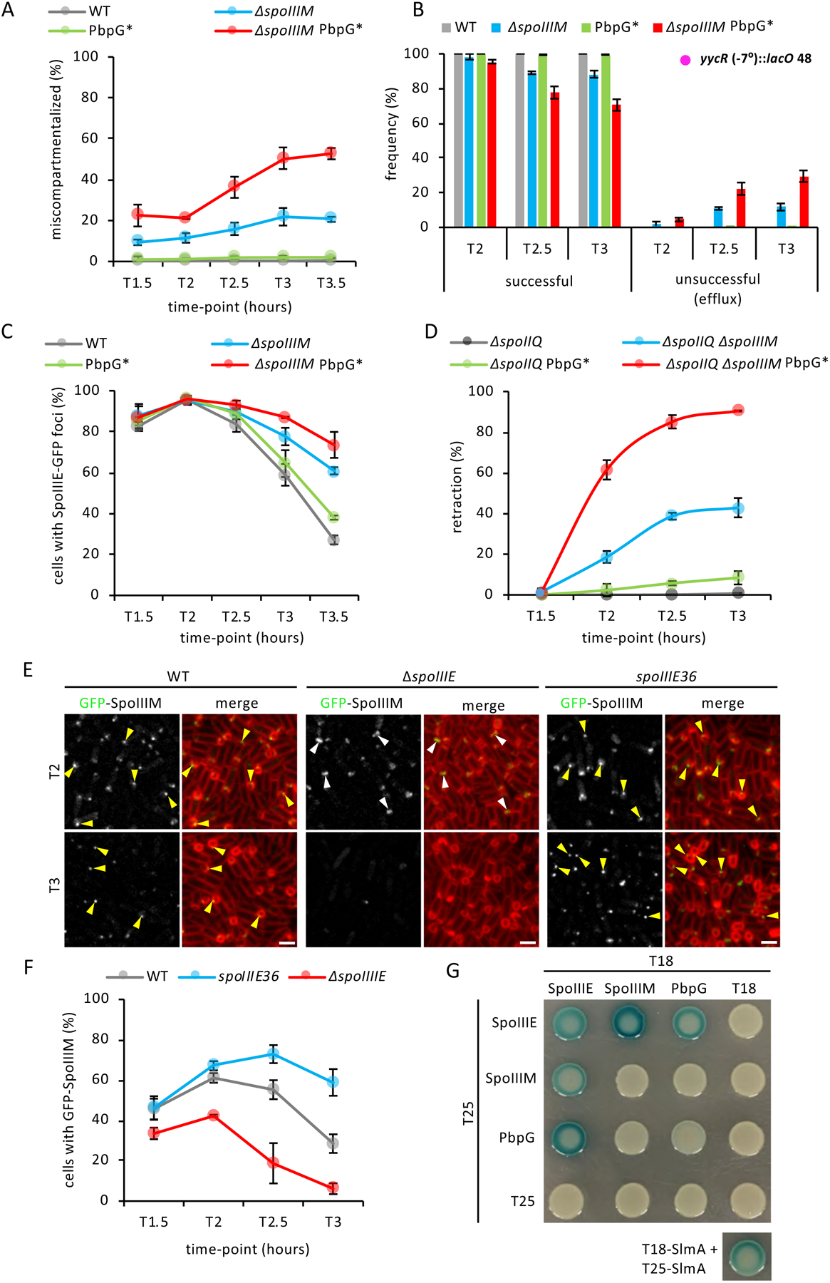
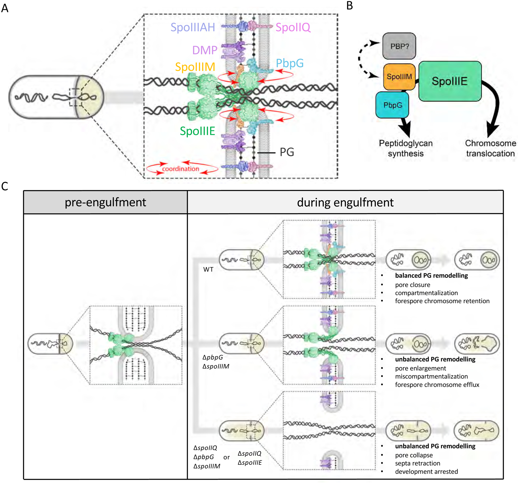
Asymmetric division, a hallmark of endospore development, generates two cells, a larger mother cell and a smaller forespore. Approximately 75% of the forespore chromosome must be translocated across the division septum into the forespore by the DNA translocase SpoIIIE. Asymmetric division also triggers cell-specific transcription, which initiates septal peptidoglycan remodeling involving synthetic and hydrolytic enzymes. How these processes are coordinated has remained a mystery. Using Bacillus subtilis, we identified factors that revealed the link between chromosome translocation and peptidoglycan remodeling. In cells lacking these factors, the asymmetric septum retracts, resulting in forespore cytoplasmic leakage and loss of DNA translocation. Importantly, these phenotypes depend on septal peptidoglycan hydrolysis. Our data support a model in which SpoIIIE is anchored at the edge of a septal pore, stabilized by newly synthesized peptidoglycan and protein-protein interactions across the septum. Together, these factors ensure coordination between chromosome translocation and septal peptidoglycan remodeling to maintain spore development.

Keywords: SpoIIIE; cell wall; chromosome segregation; chromosome translocation; development; endospores; peptidoglycan; spores; sporulation.


Conflict of interest statement

Declaration of Interests The authors declare no competing interests.

Figures

Figure 1:
Figure 1:. Identification of SpoIIIM and its requirement, together with PbpG, in maintaining compartmentalization.
(A) Diagram of chromosome translocation and the different stages of engulfment, showing membranes (black), PG (grey), chromosome (black squiggles), SpoIIIE (green), origin (oriC, blue) and terminus (ter, red). The forespore cytoplasm is shown in beige. (B) Diagram of Aqueous Pore Model (zoomed-in from red box in Ai), showing SpoIIIE on the mother cell (mc) side and not on the forespore (fs) side, localized within unfused septal membranes that form a pore in the membrane. (C) Diagram of Channel Model (zoomed-in from red box in Ai), showing SpoIIIE on both sides of the fused septal membranes. (D) Diagram of the leading edge of the engulfing membranes (zoomed-in from blue box in Aii), showing the SpoIIIAH-SpoIIQ interaction across the engulfing membranes, PbpG (light teal) and PbpF (dark teal) in the forespore membrane, the DMP complex (purple), new PG (green dots and lines) and PG in the lateral wall of the sporangium (grey dots and lines). (E) Tn-seq profiles in WT and ΔpbpG after 24 hours of growth and sporulation in exhaustion medium. The height of each line reflects the number of sequencing reads at this position. Red box highlights the spoIIIM (yqfZ) locus, which is depleted for transposon insertions in the ΔpbpG library compared to WT. (F) Average sporulation efficiency (% ± SD, n=3) of ΔspoIIIM, ΔpbpG, ΔspoIIIM ΔpbpG and the respective spoIIIM complementation strains, ΔspoIIIM and ΔpbpG ΔspoIIIM in DSM medium. (G) Average frequency (± SD of 3 biological replicates) of miscompartmentalized cells during a sporulation time-course in WT, ΔspoIIIM, ΔpbpG and ΔspoIIIM ΔpbpG (n>900 per time-course, per strain, per replicate). (H) Representative images of miscompartmentalization in WT, ΔspoIIIM, ΔpbpG, ΔspoIIIM ΔpbpG and ΔspoIIIE at T3.5 (related to Figure 1G). Scale bar, 2 μM. (I) Diagram showing the engulfment block occurring in the ΔspoIID ΔspoIIP and its effect on compartmentalization by preventing PG hydrolysis. (J) Representative images (from one biological replicate, out of 3 examined) of miscompartmentalization in ΔspoIID ΔspoIIP at T3.5, in WT, ΔspoIIIM, ΔpbpG, ΔspoIIIM ΔpbpG and ΔspoIIIE backgrounds. Scale bar, 2 μM.
Figure 2:
Figure 2:. SpoIIIE foci persist in the absence of SpoIIIM and PbpG.
(A) Diagram showing the dynamic localization of SpoIIIE during engulfment. (B) Representative images of the localization of SpoIIIE-GFP in WT, ΔspoIIIM, ΔpbpG and ΔspoIIIM ΔpbpG at T3.5. Scale bar, 2 μM. (C) Representative image of the diffused localization of SpoIIIE in WT cells that have completed engulfment. Scale bar, 1 μM. (D) Average frequency (± SD of of 3 biological replicates) of cells with SpoIIIE-GFP foci in WT, ΔspoIIIM, ΔpbpG and ΔspoIIIM ΔpbpG during a sporulation time-course (n>1000 per time-course, per strain, per replicate). (E) Average fluorescence (± STDEVP, n>400, per time-point, per strain) intensity of SpoIIIE-GFP foci in WT, ΔspoIIIM, ΔpbpG and ΔspoIIIM ΔpbpG during a sporulation time-course.
Figure 3:
Figure 3:. Efficient chromosome translocation requires SpoIIIM and PbpG.
(A) Diagram explaining experimental rationale of the LacI-lacO system for visualizing chromosome translocation. LacI-GFP, expressed in the forespore (PspoIIQ) from the amyE locus, binds to lacO48 sites inserted at the yhdG, pelB, yrvN or yycR chromosomal locus. The origin (oriC) and terminus (ter) loci are shown in blue and red, respectively. Successful chromosome translocation is indicated by a LacI-GFP focus in the forespore, with an additional focus in the mother cell compartment in the event of miscompartmentalization. Unsuccessful chromosome translocation is indicated by an absence of LacI-GFP foci, or by two LacI-GFP foci in the mother cell compartment in the event of miscompartmentalization. (B) Representative images of LacI-GFP foci at T3 in WT, ΔspoIIIM, ΔpbpG and ΔspoIIIM ΔpbpG strains containing lacO48 inserted at the pelB locus (174°). Yellow arrowheads point to cells with two mother cell foci. Scale bar, 2 μM. (C) Average frequency (± SD of 3 biological replicates) of cells with a LacI-GFP focus in the forespore (successful translocation) or with no or two LacI-GFP foci in the mother cell (unsuccessful translocation, efflux) during a sporulation time-course, with lacO48 integrated at the pelB locus (174°) and (D) at the yycR locus (−7°) in WT, ΔspoIIIM, ΔpbpG and ΔspoIIIM ΔpbpG (n>650 per time-course, per strain, per replicate). (E) Representative images of cells at T3 with two LacI-GFP foci in the mother cell of ΔspoIIIM, ΔpbpG and ΔspoIIIM ΔpbpG sporulating cells containing lacO48 inserted at the pelB locus (174°).
Figure 4:
Figure 4:. Blocking or impairing engulfment restores efficient chromosome translocation and compartmentalization in the absence of SpoIIIM and PbpG.
(A) Average frequency (± SD of 3 biological replicates) of cells with a LacI-GFP focus in the forespore or with two LacI-GFP foci in the mother cell during a sporulation time-course, with lacO 48 integrated at the pelB locus (174°) in the ΔspoIID ΔspoIIP alone or combined with the ΔspoIIIM, ΔpbpG and ΔpbpG ΔspoIIIM (n>600 per time-course, per strain, per replicate). (B) Average frequency (± SD of 3 biological replicates) of cells with a LacI-GFP focus in the forespore or with two LacI-GFP foci in the mother cell during a sporulation time-course, with lacO 48 integrated at the pelB locus (174°) in the ΔspoIIB alone or combined with the ΔspoIIIM, ΔpbpG and ΔspoIIIM ΔpbpG (n>600 per time-course, per strain, per replicate). (C) Schematic representation of the slower engulfment and membrane bulging phenotype observed in the ΔspoIIB. Cyan represents forespore-specific CFP. (D) Representative images of sporulating cells at T3 showing compartmentalization of forespore gene expression in ΔspoIIB mutant alone, or combined with the ΔspoIIIM, ΔpbpG, ΔspoIIIM ΔpbpG and ΔspoIIIE. Scale bar, 2 μM. (E) Average frequency (± SD of 3 biological replicates) of miscompartmentalized cells during a sporulation time-course in the spoIIIE36 mutant in otherwise WT (grey), ΔspoIIIM (blue), ΔpbpG (green) and ΔspoIIIM ΔpbpG (red) strains (n>750 per time-course, per strain). (F) Representative images of miscompartmentalization in the spoIIIE36 mutant in otherwise WT, ΔspoIIIM, ΔpbpG and ΔspoIIIM ΔpbpG strains at T3.5. Scale bar, 2 μM.
Figure 5:
Figure 5:. Septa retraction and its dependency on PG hydrolysis.
(A) Schematic representation of septal retraction, illustrating that as septal retraction progresses, CFP fluorescence (cyan) leaks from the forespore to fill the entire cell. (B) Representative images of septal retraction in WT, ΔspoIIIM, ΔpbpG, ΔspoIIIM ΔpbpG and ΔspoIIIE in a ΔspoIIQ background at T3. Scale bar,2 μM. Sporulation efficiency (%, average ± SD, n=3) is shown below the respective strains. (C) Representative images of septal retraction suppression in cells blocked for engulfment in WT, ΔspoIIIM, ΔpbpG, ΔspoIIIM ΔpbpG and ΔspoIIIE strains at T3. Scale bar, 2 μM. (D) Representative zoomed-in examples of septal retraction in ΔspoIIIM, ΔpbpG, ΔspoIIIM ΔpbpG and ΔspoIIIE at T3. Yellow arrowheads point to retracting septa. Scale bar,1 μM. Schematic representations of cells are shown on the right. (E) Average frequency (± SD of of 3 biological replicates) of cells exhibiting septal retraction in WT, ΔspoIIIM, ΔpbpG, ΔspoIIIM ΔpbpG and ΔspoIIIE during a sporulation time-course (n>600 per time-point, per strain, per replicate).
Figure 6:
Figure 6:. Peptidoglycan synthesis by PbpG is required for developmental characteristics and evidence that SpoIIIE, SpoIIIM and PbpG form a complex.
(A) Average frequency (± SD of 3 biological replicates) miscompartmentalized cells during a sporulation time-course in WT, ΔspoIIIM, pbpG* and ΔspoIIIM pbpG* strains (n>900 per time-course, per strain, per replicate). (B) Average frequency (± SD of of 3 biological replicates) of cells with a LacI-GFP focus in the forespore (successful translocation) or with no or two LacI-GFP foci in the mother cell (unsuccessful translocation, efflux) during a sporulation time-course, with lacO48 integrated at the yycR locus (−7°), in WT, ΔspoIIIM, PbpG* and ΔspoIIIM PbpG* (n>600 per time-course, per strain, per replicate). (C) Average frequency (± SD of 3 biological replicates) of cells with SpoIIIE-GFP foci during a sporulation time-course in WT (grey), ΔspoIIIM, PbpG* and ΔspoIIIM PbpG* (n>950 per time-course, per strain, per replicate). (D) Average frequency (± SD of of 3 biological replicates) of sporulating cells exhibiting septal retraction during a sporulation time-course in WT ΔspoIIIM, PbpG* and ΔspoIIIM PbpG* (n>700 per time-course, per strain, per replicate). (D) Representative images of GFP-SpoIIIM localization in WT, ΔspoIIIE and spoIIIE36 strains at T2 and T3. Yellow arrowheads indicate GFP-SpoIIIM fluorescence that appears to disperse, disappear or remain as a stable focus as engulfment nears completion in WT, ΔspoIIIE and spoIIIE36 mutant. Scale bar,2 μM. (F) Average frequency (± SD of 3 biological replicates) of cells with GFP-SpoIIIM fluorescence in WT, ΔspoIIIE and spoIIIE36 during sporulation time-course (n>750 per time-course, per strain, per replicate). (G) Bacterial two-hybrid assay of T18 and T25 fusions to SpoIIIE, SpoIIIM and PbpG. T18-SlmA and T25-SlmA interaction is included as a positive control (Cho et al., 2011).
Figure 7:
Figure 7:. Model illustrating coordination of PG remodelling and chromosome translocation at a highly-stabilized septal pore.
(A) Schematic illustration showing coordination (red arrows) of chromosome translocation and PG remodelling by SpoIIIM and PbpG via SpoIIIE, at the septal pore. (B) Interaction network of SpoIIIE, highlighting its role in chromosome translocation and its role in PG synthesis via interactions with SpoIIIM and PbpG. We hypothesize that a putative unidentified PG synthase (PBP) may also be involved in PG remodelling via interaction with SpoIIIM. (C) Schematic representation of the septal membrane, before and after initiation of engulfment and how different molecular components contribute to ensuring developmental compartmentalization and chromosome translocation upon initiation of engulfment. Note that in the ΔspoIIQ ΔspoIIIM ΔpbpG triple mutant, SpoIIIE is still present as focus in retracted septa (Fig. S4) but for simplicity this is not shown in the bottom panel.

References

    1. BARRE FX 2007. FtsK and SpoIIIE: the tale of the conserved tails. Mol Microbiol, 66, 1051–1055. - PubMed
    1. BEN-YEHUDA S, RUDNER DZ & LOSICK R 2003. Assembly of the SpoIIIE DNA translocase depends on chromosome trapping in Bacillus subtilis. Curr Biol, 13, 2196–2200. - PubMed
    1. BESPROZVANNAYA M, PIVORUNAS VL & BURTON BM 2014. Mechanistic study of classical translocation-dead SpoIIIE36 reveals the functional importance of the hinge within the SpoIIIE motor. Journal of Bacteriology, 196, 2481–2490. - PMC - PubMed
    1. BESPROZVANNAYA M, PIVORUNAS VL, FELDMAN Z & BURTON BM 2013. SpoIIIE protein achieves directional DNA translocation through allosteric regulation of ATPase activity by an accessory domain. Journal of Biological Chemistry, 288, 28962–28974. - PMC - PubMed
    1. BLAYLOCK B, JIANG X, RUBIO A, MORAN CP JR. & POGLIANO K 2004. Zipper-like interaction between proteins in adjacent daughter cells mediates protein localization. Genes Dev, 18, 2916–2928. - PMC - PubMed

Publication types

MeSH terms

LinkOut - more resources