Comparative Study
. 2021 Mar:194:111425.
doi: 10.1016/j.mad.2020.111425. Epub 2020 Dec 28.

Cellular hallmarks of aging emerge in the ovary prior to primordial follicle depletion

Affiliations
Comparative Study

Cellular hallmarks of aging emerge in the ovary prior to primordial follicle depletion

Victor A Ansere et al. Mech Ageing Dev. 2021 Mar.

Abstract

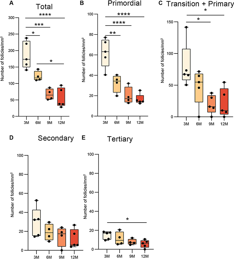
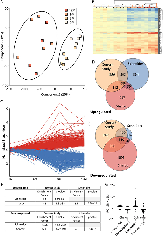
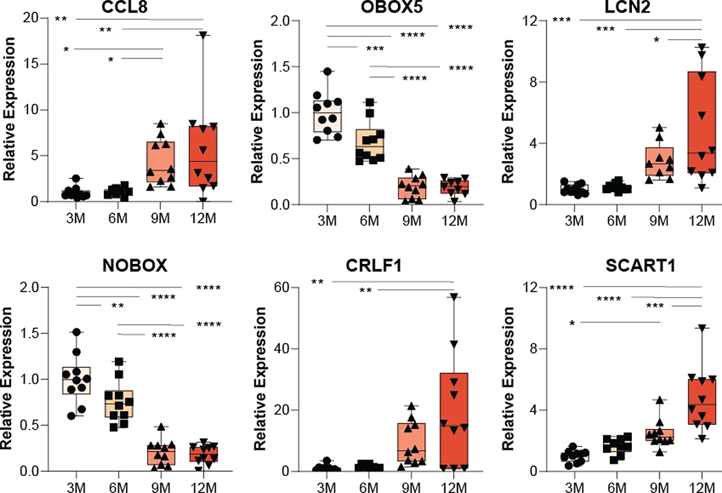
Decline in ovarian reserve with advancing age is associated with reduced fertility and the emergence of metabolic disturbances, osteoporosis, and neurodegeneration. Recent studies have provided insight into connections between ovarian insufficiency and systemic aging, although the basic mechanisms that promote ovarian reserve depletion remain unknown. Here, we sought to determine if chronological age is linked to changes in ovarian cellular senescence, transcriptomic, and epigenetic mechanisms in a mouse model. Histological assessments and transcriptional analyses revealed the accumulation of lipofuscin aggresomes and senescence-related transcripts (Cdkn1a, Cdkn2a, Pai-1 and Hmgb1) significantly increased with advancing age. Transcriptomic profiling and pathway analyses following RNA sequencing, revealed an upregulation of genes related to pro-inflammatory stress and cell-cycle inhibition, whereas genes involved in cell-cycle progression were downregulated; which could be indicative of senescent cell accumulation. The emergence of these senescence-related markers preceded the dramatic decline in primordial follicle reserve observed. Whole Genome Oxidative Bisulfite Sequencing (WGoxBS) found no genome-wide or genomic context-specific DNA methylation and hydroxymethylation changes with advancing age. These findings suggest that cellular senescence may contribute to ovarian aging, and thus, declines in ovarian follicular reserve. Cell-type-specific analyses across the reproductive lifespan are needed to fully elucidate the mechanisms that promote ovarian insufficiency.

Keywords: Aging; Cellular senescence; DNA methylation; Epigenetics; Ovary.


Conflict of interest statement

DECLARATION OF INTEREST

The authors have no conflicts of interest to declare.

Figures

Figure 1:
Figure 1:. Follicular density decreases with age.
Box plots demonstrates age-related decreases in follicular density represented as the mean of 5 ovarian sections per mouse (n = 5 mice/ group; *p<0.05, **p<0.01, ***p<0.005 and ****p<0.001; one-way ANOVA with Tukey post hoc test). For each section the follicular density was calculated as the number of follicles in the section divided by the area of the section. (A) Total, all follicles with clearly visible oocyte nucleus (B) Primordial follicles representing follicles surrounded by a single layer of flattened granulosa cells (C) Transition + Primary representing follicles surrounded by single layer of cuboidal cells or both flattened and cuboidal cells (D) Secondary follicles are surrounded by more than one layer of granulosa cuboidal cells with no visible antrum (E) Tertiary represents follicles with well-defined antral space and a layer of cumulus granulosa cells around the oocyte.
Figure 2:
Figure 2:. Lipofuscin aggresomes accumulate with age.
(A) Sudan black staining of ovarian sections for lipofuscin aggresomes with age. Images acquired at 10X magnification with dark blue areas representing lipofuscin (B) Quantification of lipofuscin positive area as a percentage of the total area of section evaluated expressed as mean ± SEM (n=3 mice/group; *p<0.05, **p<0.01; one-way ANOVA with Tukey post hoc test). (C) Correlation of lipofuscin staining to number of primordial follicles.
Figure 3 -
Figure 3 -. Aging causes senescence-related markers in the mouse ovary to increase.
qPCR analysis of ovarian Cdkn1a (p21), Cdkn2a (p16), PAI-1 and HMGB1 reveal age-related increases in all markers by 9 and/or 12 months of age as compared to younger animals (3 and/or 6 months of age) (n=10/group, one-way ANOVA, *p<0.05, **p<0.01, ***p<0.001 SNK post hoc).
Figure 4 –
Figure 4 –. Age-related ovarian transcriptomic patterns.
A) Using the full complement of genes detected as expressed in the study a principle component analysis was performed and a segregation of 3 and 6 month from 9 and 12 month was evident. B) 2,550 genes (Up/down) were differentially expressed with age and hierarchical clustering separated 3- and 6 -month from 9- and 12 -month groups. C) When examined for their trajectory over ages differentially expressed genes generally demonstrated a linear change (increase/decrease) across the time points. D&E) Comparison of statistically significant changes to other studies. F) Table of hypergeometric test results revealing an enrichment of commonly differentially expressed gene across studies. G) Differentially expressed genes from other studies were plotted for fold change gene expression between 12 and 3M in this study regardless of statistical significance demonstrate consistent pattern of gene expression change with aging.
Figure 5 –
Figure 5 –. Confirmation of differential expression of select RNA-Seq target genes.
qPCR analysis reveals age-related increases in the expression of the inflammatory-related genes, CCL8, LCN2, CRLF1 and SCART1 by 9 and/or 12 months of age while the expression of the oocyte-specific gene OBOX5 and the folliculogenesis-related gene NOBOX, decreases with age in agreement with the RNA sequencing experiment (n=10/group, one-way ANOVA, *p<0.05, **p<0.01, ***p<0.001 SNK post hoc).
Figure 6 –
Figure 6 –. Pathway, process and regulator analysis of transcriptomic changes with aging.
Significant differences in gene expression were analyzed by Ingenuity Pathway Analysis. Z-scores, indicating activation or inhibition of pathways (A), processes (B), or regulators (C) are presented.
Figure 7 –
Figure 7 –. Genomic DNA Methylation and hydroxymethylation levels do not change with ovarian aging.
No age-related differences were observed in total genomic (A) or segregated by autosomes (B) and X chromosomes (C) mCG, hmCG, or mCH modification levels. Reproducible levels of hmCG were observed and low levels of mCH were evident.
Figure 8 –
Figure 8 –. Methylation and hydroxymethylation of genomic repeat elements is not altered with aging.
No age-related differences were observed in LINEs, SINEs, LTRs, or Simple Repeats by methylation in the CG context (A), hydroxymethylation in the CG context (B), or methylation in the CH context (C). While mCG levels were consistent across repeat types hmCG levels varied by type of repeat. CH methylation was consistent with the exceptions of LTRs which demonstrated a lower level.

References

    1. Adriaens I, Smitz J, Jacquet P, 2009. The current knowledge on radiosensitivity of ovarian follicle development stages. Human reproduction update 15, 359–377. - PubMed
    1. Anders S, Huber W, 2010. Differential expression analysis for sequence count data. Genome biology 11, R106. - PMC - PubMed
    1. Baird DT, 1974. The endocrinology of ovarian steroid secretion. Eur J Obstet Gynecol Reprod Biol 4, 31–39. - PubMed
    1. Baker DJ, Wijshake T, Tchkonia T, LeBrasseur NK, Childs BG, Van De Sluis B, Kirkland JL, van Deursen JM, 2011. Clearance of p16 Ink4a-positive senescent cells delays ageing-associated disorders. Nature 479, 232–236. - PMC - PubMed
    1. Baker TG, 1963. A Quantitative and Cytological Study of Germ Cells in Human Ovaries. Proc R Soc Lond B Biol Sci 158, 417–433. - PubMed

Publication types

MeSH terms