. 2021 Feb:96:18-46.
doi: 10.1016/j.matbio.2020.12.005. Epub 2020 Dec 28.

Dynamics of the lens basement membrane capsule and its interaction with connective tissue-like extracapsular matrix proteins

Affiliations

Dynamics of the lens basement membrane capsule and its interaction with connective tissue-like extracapsular matrix proteins

JodiRae DeDreu et al. Matrix Biol. 2021 Feb.

Abstract

The lens, suspended in the middle of the eye by tendon-like ciliary zonule fibers and facing three different compartments of the eye, is enclosed in what has been described as the thickest basement membrane in the body. While the protein components of the capsule have been a subject of study for many years, the dynamics of capsule formation, and the region-specific relationship of its basement membrane components to one another as well as to other matrix molecules remains to be explored. Through high resolution confocal and super-resolution imaging of the lens capsule and 3D surface renderings of acquired z-stacks, our studies revealed that each of its basement membrane proteins, laminin, collagen IV, nidogen and perlecan, has unique structure, organization, and distribution specific both to the region of the lens that the capsule is located in and the position of the capsule within the eye. We provide evidence of basal membrane gradients across the depth of the capsule as well as the synthesis of distinct basement membrane lamella within the capsule. These distinctions are most prominent in the equatorial capsule zone where collagen IV and nidogen span the capsule depth, while laminin and perlecan are located in two separate lamellae located at the innermost and outermost capsule domains. We discovered that an extracapsular matrix compartment rich in the connective tissue-like matrix molecules fibronectin, tenascin-C, and fibrillin is integrated with the superficial surface of the lens capsule. Each matrix protein in this extracapsular zone also exhibits region-specific distribution with fibrils of fibrillin, the matrix protein that forms the backbone of the ciliary zonules, inserting within the laminin/perlecan lamella at the surface of the equatorial lens capsule.

Keywords: Collagen IV; Fibrillin; Laminin; Lens capsule; Perlecan; Tenascin-C.


Conflict of interest statement

Declaration of Competing Interest The authors have no competing interests to declare.

Figures

Figure 1.
Figure 1.. Development-state specific expression of basement membrane proteins in the chick embryo lens capsule.
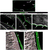
(A) Model of the lens capsule and its relationship to other compartments of the eye. The anterior capsule (red) associates with the aqueous humor and faces the cornea; the equatorial capsule (green) is directly linked to the ciliary zonules (CZ) which extend from the ciliary body (CB); and the posterior capsule (yellow) associates with the vitreous humor and faces the retina. The diagram represents the E15 eye, at which point the lens has matured and is the latest stage of development examined in these studies. All four regions of fiber cell differentiation depicted here are present by E9. Whole embryonic chick eyes E5 (B-D), E9 (E-G), and E15 (H-J) were sectioned and stained for the three primary basement membrane proteins, laminin111 (B, E, and H, red), perlecan (C, F, and I, green), and collagen IV (D, G, and J, purple). Overview images of the entire lens were acquired at each embryonic stage examined, using the same settings to demonstrate the changes in expression/localization during development. Laminin111 and perlecan were present in all regions of the lens capsule at each developmental stage examined (B-I), their intensity lowest in the thinner anterior capsule region. While perlecan intensity and distribution appear to increase in the equatorial zone by E15, laminin localization is diminished in all regions of the capsule by this developmental time. Collagen IV developmental expression and capsule distribution is distinct from laminin and perlecan, its expression limited to the posterior capsule at E5 and greatly increasing throughout all regions of the lens capsule by E15. Mag bar 50μm (B-D), 100μm (E-G) and 200μm (H-J).
Figure 2.
Figure 2.. Dynamic basement protein remodeling in the equatorial lens capsule during development.
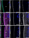
Cryosections of embryonic chick eyes were immunolabeled for three primary basement membrane proteins, laminin111 (A, D, and G, red), perlecan (B, E, and H, green), and collagen IV (C, F, and I, purple) at E5 (A-C), E9 (D-F), and E15 (G-I). Each section was co-labeled with DAPI to detect lens equatorial epithelial cell nuclei (blue). Confocal z-stacks were acquired in the region of the equatorial lens capsule from which the images presented are a single optical plane (0.5 μm). While at E5 and E9 both laminin111 (A, D, open arrowheads) and perlecan (B,E, arrows) are distributed throughout the lens equatorial capsule, by E15 these basement membrane proteins are present in two distinct lamellae (G and H), one at the inner, lens cell-facing side of the capsule, the other at the superficial surface of the capsule where the perlecan lamella is wider than the laminin lamella. In contrast, at early stages of lens development collagen IV is primarily present only in a thin lamella along the cells of the lens equatorial epithelium (C, F, solid arrowheads) and has expanded throughout the equatorial capsule by E15 (I). Mag bar 20μm (A-I).
Figure 3.
Figure 3.. Bilaminar laminin/perlecan domains in the adult mouse equatorial zone.
Cryosection of adult mouse eye immunolabeled for laminin111 (A,C, red) and perlecan (B,C, green), and co-labeled with DAPI to detect lens equatorial epithelial cell nuclei (blue) and imaged by confocal microscopy. The image was acquired as a single optical plane. Both laminin111 (A, arrows) and perlecan (B, arrowheads) are present as two distinct lamellae in the adult mouse lens equatorial capsule, one at the inner, lens cell-facing side of the capsule, the other at the superficial surface of the capsule. Mag bar 20μm.
Figure 4.
Figure 4.. The relationship between basement membrane proteins that comprise the anterior lens capsule.
Cryosections of E15 chick embryo eyes were co-immunolabeled with antibodies to perlecan (green), laminin111 (teal), and collagen IV (purple), in the following combinations: (A) perlecan/laminin111, (D) collagen IV/perlecan, and (G) collagen IV/laminin111. Sections were co-labeled for F-actin (A,D,G; white) and nuclei (A-I; blue). Z-stacks were acquired of the anterior lens capsule by confocal microscopy imaging from which a single optical plane (0.5μm) is shown as an overlay of the co-immunolabeled basement membrane proteins (A,D,G), alongside of which is presented each fluorescent channel in greyscale (B,E-perlecan; C,H-laminin111; F,I-collagen IV). Collagen IV (F,I) extends throughout the anterior capsule and is detected in the anterior epithelium. Laminin (C,H) also spans this region of the lens capsule and is highly coincident with collagen IV (G). In this region of the capsule there is a perlecan gradient, its expression greatest along the inner region of the anterior capsule (E), where it is most highly coincident with collagen IV (D) and laminin (A). An additional perlecan-rich layer occasionally detected at the superficial surface of the anterior lens capsule (B). Mag bar 20μm.
Figure 5.
Figure 5.. Structural imaging of the relationship between basement membrane proteins in the anterior lens capsule.
3D surface structure renderings were created with Imaris software from confocal z-stacks in the region of the anterior lens capsule (denoted by the area within the red rectangle in the model in A) of cryosections of the E15 chick embryo eye immunolabeled for basement membrane proteins laminin (B,C,J,K), perlecan (F,G,L,M) and collagen IV (D,H). The sections were co-immunolabeled for (B-D) laminin (teal)/collagen IV (purple), (F-H) perlecan (green)/collagen IV (purple), or (J-M) laminin (teal)/perlecan (green). Each was co-labeled for F-actin (white) and nuclei (dark blue). (C,G,K,M) are zoomed in structural images of (B,F,J,L), respectively. E and I are presented to demonstrate the surface structure of the basal surface of the anterior epithelial cells shown in C and G, respectively. These structural renderings reveal that laminin has a typical sheet-like structure that is contacted by the basal surfaces of the lens epithelium while perlecan and collagen IV have a basolateral distribution, with expression of collagen IV by the lens epithelium also observed (modeled in N). Z-stack sizes – (B-E) 39 slices; (F-I) 36 slices; (J-M) 36 slices. LC – lens capsule. Mag bar 0.5μm (C,E,G,I,K,M), 1μm (B,D,F,H), and 3μm (J,L).
Figure 6.
Figure 6.. The relationship between basement membrane proteins that comprise the equatorial lens capsule.
Cryosections of E15 chick embryo eyes were co-immunolabeled with antibodies to perlecan (green), laminin111 (teal), and collagen IV (purple), in the following combinations: (A) perlecan/laminin111, (D) collagen IV/perlecan, and (G) collagen IV/laminin111. Sections were co-labeled for F-actin (D,G; white) and nuclei (A-I; blue). Z-stacks were acquired of the equatorial lens capsule by confocal microscopy imaging from which a single optical plane (0.5μm) is shown as an overlay of the co-immunolabeled basement membrane proteins (A,D,G), alongside of which is presented each fluorescent channel in greyscale (B,E-perlecan; C,H-laminin111; F,I-collagen IV). In the equatorial region of the lens capsule there are two distinct laminin and perlecan lamella that are located at the inner and outer surfaces of the capsule and are highly coincident (A), with perlecan (B,E) extending farther toward the center of the capsule than laminin (C,H). Collagen IV is expressed throughout the equatorial capsule (F,I), co-distributing with the perlecan/laminin lamella at the inner and outer capsule surfaces that sandwich the collagen IV-rich region between them (D,G). Collagen IV immunolabeling was most intense in a thin basement membrane zone along the capsule’s innermost surface (F,I), and at times, distinct layers of collagen IV were resolved across this capsule zone (F). Mag bar 20μm.
Figure 7.
Figure 7.. Super-resolution Images of the laminin/perlecan bilateral lamella in the equatorial lens capsule.
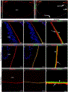
Super-resolution microscopy of cryosections of E15 chick embryo eyes co-immunolabeled for (A,C,D,F) perlecan (green) and (B,C,E,F) laminin111 (red), or immunolabeled for (G,H) nidogen (teal) imaged at the lens equatorial capsule. D,E,F,H are zoomed in regions of A,B,C,G, respectively. Images are a 0.21 μm optical slice from a confocal z-stack. There is significant co-localization of the laminin and perlecan lamella at the inner and outer surfaces of the lens (C,F). In contrast, nidogen spans the equatorial capsule region (G,H). Mag bar: (A,B,C,G) 20μm; (D,E,F,H) 10μm.
Figure 8.
Figure 8.. LN111 producing cells associated the superficial surface of the outer laminin lamella in the equatorial lens capsule.
(A,B) Single optical slice (0.33 μm) of a confocal z-stack of an intact E15 chick embryo lens immunolabeled for laminin111 (red) and co-labeled for cell nuclei (blue). This image, approximately 143μm deep into the lens equatorial zone from the outside edge of the lens equator, demonstrates a LN111-expressing cell localized along and integrated with the superficial surface of the outer LN111 lamella of the lens capsule. B is zoomed in region of A. Mag bar: (A) 20μm; (B) 10μm.
Figure 9.
Figure 9.. Structural imaging of the relationship between basement membrane proteins in the equatorial lens capsule.
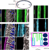
3D surface structure renderings were created with Imaris software from confocal z-stacks in the region of the equatorial lens capsule (denoted by the area within the red rectangle in the model in A) of cryosections of the E15 chick embryo eye immunolabeled for basement membrane proteins perlecan (B,C,H, green), laminin111 (D,E,I, teal) and collagen IV (F, purple). The sections were immunolabeled for (B,C) perlecan (green), or represent sections co-immunolabeled for (D-G) laminin (teal)/collagen IV (purple), or (H,I) perlecan (green)/laminin (teal). (B-G) were co-labeled for F-actin (white) and nuclei (dark blue), with G presented to show the structure of the basal surface of the equatorial epithelial cells depicted in E. (C,E) are zoomed in structural images of (B,D), respectively. The structural images of laminin and perlecan provide the first look of the organization of the bilateral localization of the inner and outer laminin/perlecan basement membrane lamellae at the inner and outer surfaces of the lens equatorial capsule (modeled in J). While collagen IV is present throughout the entire width of the equatorial lens capsule (F), the structural organization of collagen IV at the inner and outer domains of this capsular zone parallel that of laminin (D,E). Z-stack sizes – (D-G) 44 slices; (B,C) 58 slices; (H,I) 54 slices. LC- lens capsule. Mag bar 0.7μm (E, G-I), 0.8μm (C), and 5μm (B, D, F).
Figure 10.
Figure 10.. The relationship between basement membrane proteins that comprise the posterior lens capsule.
Cryosections of E15 chick embryo eyes were co-immunolabeled with antibodies to perlecan (green), laminin111 (teal), and collagen IV (purple), in the following combinations: (A) perlecan/laminin111, (D) collagen IV/perlecan, and (G) collagen IV/laminin111. Sections were co-labeled for F-actin (D,G; white). Z-stacks were acquired of the posterior lens capsule by confocal microscopy imaging from which a single optical plane (0.5μm) is shown as an overlay of the co-immunolabeled basement membrane proteins (A,D,G), alongside of which is presented each fluorescent channel in greyscale (B,E-perlecan; C,H-laminin111; F,I-collagen IV). Perlecan is present as two distinct lamellae in the posterior capsule (B,E), overlapping with both laminin (A) and collagen IV (D), both basement membrane proteins that span this capsule zone (G-I). Mag bar 20μm.
Figure 11.
Figure 11.. Structural imaging of the relationship between basement membrane proteins in the posterior lens capsule.
3D surface structure renderings were created with Imaris software from confocal z-stacks in the region of the posterior lens capsule (denoted by the area within the red rectangle in the model in A) of cryosections of the E15 chick embryo eye immunolabeled for basement membrane proteins laminin (B,C,H,I), perlecan (D,E,J,K), and collagen IV (F). The sections were immunolabeled for (B,C) laminin, or co-immunolabeled for (D-G) perlecan (green)/collagen IV (purple), or (H-K) laminin (teal)/perlecan (green). (B-G) were co-labeled for F-actin (white), with G presented to show the structure of the basal surface of the lens fiber cells depicted in E. (C,E,I,K) are zoomed in structural images of (B,D,H,J), respectively. These structural renderings show that the bilateral organization of perlecan continues into the posterior region of the lens capsule, a wide outer lamella and a thin inner lamella (D,E,J,K), both of which are located within the laminin (H,I) and collagen IV (F) domains that span the posterior capsule (modeled in L). Z-stack sizes – (B,C) 39 slices; (D-G) 44 slices; (H-K) 47 slices. LC – lens capsule. Mag bar 1μm (C, E, G, I, and K) and 3μm (B, D, F, H, and J).
Figure 12.
Figure 12.. Localization of the matrix protein tenascin-C to both the lens capsule and a closely linked extracapsular zone.
Cryosections of E15 chick embryo eyes were labeled with tenascin-C (TN-C) alone (A) or co-labeled for tenascin-C and laminin (B-L). Nuclei were labeled with DAPI (B,F, blue). (A) Low magnification image of tenascin-C distribution across the lens capsule. Confocal imaging of tenascin-C/laminin colocalization in the upper regions of the equatorial zone where it borders with the lens anterior region (B-E) and along the lens equator (F-L). (B,F) single optical planes (0.5 μm), (C,D,G,H,J,K) 3D images from confocal z-stacks with D,H,K showing then TN-C channel alone that appears in co-labeling with laminin in C,G,J, respectively, and (E,I,L) 3D surface structure with tenascin-C rendered transparent (arrowheads). C-E and G-L are zoomed in regions of the boxed regions in B and F, respectively. Z-stack sizes – (C) 30 slices; (G,H,J,K) 41 slices. TN-C – tenascin-C; EQ – equatorial zone. Mag bar 3μm (G-L), 4μm (C-E), 20μm (B,F), and 200μm (A).
Figure 13.
Figure 13.. Fibrillin-2 defines an extracapsular matrix region linked to the lens capsule.
Cryosections of E15 chick embryo eyes were labeled for fibrillin-2 alone (A) or co-labeled for fibrillin-2 (green) and F-actin (white), which defines the cytoarchitecture of the lens cells (B-F). An overview of fibrillin-2 labeling (A) shows it is associated with the posterior and equatorial regions of the lens capsule, ending near the border between the equatorial and anterior regions of the lens. Confocal z-stacks were acquired (B,E) in the upper regions of the equatorial zone where it borders with the anterior of the lens, (C,F) along the lens equator, and (D) in the posterior zone of the lens. Images represent either a single optical plane (0.5 μm) of the z-stack (B-D) or 3D surface structure renderings of the z-stacks (E,F). In the equatorial and posterior regions of the lens fibrillin-2 (B-F, arrowheads) is identified just outside a gap that represents a region of the lens capsule (B-F, LC, arrows), separating the fibrillin matrix from the lens cells. The 3D surface renderings along the lens equator highlight both the structure of the fibrillin extracapsular matrix, its position relative to the lens equatorial epithelium and the termination of the extracapsular fibrillin matrix as it extends towards the lens anterior. Z-stack sizes – (E) 33 slices; (F) 36 slices. LC-lens capsule; CB-ciliary body; EQ – equatorial zone. Mag bar 3μm (E,F), 20μm (B-D), and 200μm (A).
Figure 14.
Figure 14.. Tenascin-C integrates the lens capsule and the fibrillin-2-rich extracapsular matrix zone.
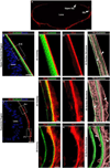
Low magnification of cryosection of E15 chick eye co-immunolabeled for tenascin- C and fibrillin-2 (A). Confocal images of cryosections of E15 chick embryo eyes acquired (B-E) in the upper regions of the equatorial zone where it borders with the anterior of the lens, (F-I) along the lens equator, and (J-M) in the posterior zone of the lens as z-stacks following co- labeling for tenascin-C, fibrillin-2, F-actin, and nuclei. A single optical plane (0.5μm) is shown as an overlay of the co-immunolabeled matrix proteins tenascin-C (red), fibrillin-2 (green), with F-actin (white) and nuclei (blue) (B,F,J) alongside of which is presented each fluorescent channel in greyscale (C,G,K-tenascin-C; D,H,L-fibrillin-2) with nuclei (blue). (E,I,M) 3D reconstructions of the confocal z-stacks created with Imaris 3D View showing tenascin-C (red), fibrillin-2 (green), and nuclei (blue). Arrows in E,I indicate tenascin-C in the capsule and in a closely associated extracapsular matrix region. Arrowhead in M denotes the coincidence of tenascin-C and fibrillin-2 in the posterior extracapsular zone, arrow in M shows tenascin-C extending beyond the extracapsular matrix at the interface with the vitreous. Z-stack sizes – (E) 43 slices; (I) 19 slices; (M) 38 slices. EQ – equatorial zone. Mag bar 5μm (M), 20μm (B-M) and 200μm (A).
Figure 15.
Figure 15.. Coincidence of fibronectin and fibrillin-2 in the extracapsular matrix compartment linked to the lens capsule surface.
Low magnification of E15 chick eye cryosection immunolabeled for fibronectin (A) and co-labeled for fibrillin-2 (B). Confocal images of cryosections of E15 chick embryo eyes were acquired (C-E) in the upper regions of the equatorial zone where it borders with the anterior of the lens, (F-H) along the lens equator, and (I-K) in the posterior zone of the lens as z-stacks following co-labeling for fibronectin, fibrillin-2, and nuclei. A single optical plane (0.5μm) is shown of immunolabeling for fibronectin (red) with nuclei (blue) (C,F,I) and with fibrillin-2 (green) overlay (blue) (D,G,J). (E,H,K) 3D reconstructions of the confocal z-stacks created with Imaris 3D View showing fibronectin (red), fibrillin-2 (green), and nuclei (blue). The arrow in E indicates the presence of fibronectin in the lens capsule. The arrowheads in E,H,K indicate the colocalization of fibronectin with fibrillin-2 in the extracapsular zone. The arrows in H and K indicate the extension of fibronectin beyond the fibrillin-2 extracapsular zone in the equatorial and posterior aspects of the lens, respectively. Z-stack sizes – (E) 29 slices; (H) 9 slices; (K) 26 slices. Mag bars 20μm (C-K) and 200μm (A and B).
Figure 16.
Figure 16.. Fibrillin-2 extracapsular matrix is closely associated with the lens basement membrane capsule.
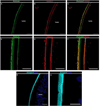
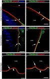
Cryosections of E15 chick embryo eyes were co-labeled for (A,C,E) fibrillin-2 (green), laminin (red) and nuclei (blue), or (B,D,F) fibrillin-2 (green) and perlecan (red). Images are a single optical plane (0.5 μm) of confocal z-stacks acquired in (A,B) the upper equatorial zone at its border with the lens anterior, (C,D) at the lens equator (EQ), and (E,F) in the lens posterior region. The extracapsular fibrillin-2 matrix zone is closely associated with laminin and perlecan along the superficial layer of the lens basement membrane capsule (arrowheads). Arrows point to regions labeling for the basement membrane proteins alone, with the arrow on the right in C indicating the very close apposition of fibrillin-2 with the outer laminin basement membrane lamella. CB – ciliary body, CZ – ciliary zonules. Mag bar 20μm.
Figure 17.
Figure 17.. Super-resolution images of the laminin/fibrillin-2 and perlecan/fibrillin-2 in the equatorial lens capsule.
Super-resolution microscopy of cryosections of E15 chick embryo eyes co-immunolabeled for (A-D) fibrillin-2 (green) and either (A,B) laminin111 (red), or (C,D) perlecan (red) and imaged in the upper region of the lens equatorial zone near its border with the anterior of the lens (A,C) or directly at the lens equator (B,D). Each image is a 0.21 μm optical slice. Fibrillin-2 is closely associated with the superficial surface of the outer laminin/perlecan lamella of the lens capsule. Mag bar 10 μm.
Figure 18.
Figure 18.. 3D surface structure renderings of the integration of fibrillin-2 with the superficial surface of the lens basement membrane capsule.
Cryosections of E15 chick embryo eyes were co-labeled for (A,B) fibrillin-2 (green) and laminin111 (red). Images are 3D surface structure renderings created from the z-stacks of which single optical planes are shown in Figure 16A. A and B are of the same region, with laminin rendered transparent in B. Closed arrowhead (indicating fibrillin-2) and arrow (indicating laminin) are placed at an identical place in A and B. Open arrowheads in B reveal fibrillin-2 fibrils that were inserted within the outer laminin basement membrane lamella that are revealed when laminin is rendered transparent. The closed arrowhead indicates a fibrillin-2 fibril in the extracapsular matrix region localized along the superficial surface of LN111. Z-stack sizes – (A,B) 34 slices. Mag bar 5μm.

Similar articles

Cited by

References

    1. Danysh BP, and Duncan MK (2009) The lens capsule. Exp Eye Res 88, 151–164 - PMC - PubMed
    1. Parmigiani C, and McAvoy J. (1984) Localisation of laminin and fibronectin during rat lens morphogenesis. Differentiation 28, 53–61 - PubMed
    1. Cummings CF, and Hudson BG (2014) Lens capsule as a model to study type IV collagen. Connect Tissue Res 55, 8–12 - PMC - PubMed
    1. Wu W, Tholozan FM, Goldberg MW, Bowen L, Wu J, and Quinlan RA (2014) A gradient of matrix-bound FGF-2 and perlecan is available to lens epithelial cells. Exp Eye Res 120, 10–14 - PMC - PubMed
    1. Bystrom B, Virtanen I, Rousselle P, Gullberg D, and Pedrosa-Domellof F. (2006) Distribution of laminins in the developing human eye. Invest Ophthalmol Vis Sci 47, 777–785 - PubMed

Publication types

MeSH terms

LinkOut - more resources