. 2020 Dec 31;18(12):e3000971.
doi: 10.1371/journal.pbio.3000971. eCollection 2020 Dec.

A comprehensive atlas of white matter tracts in the chimpanzee

Affiliations

A comprehensive atlas of white matter tracts in the chimpanzee

Katherine L Bryant et al. PLoS Biol. .

Abstract

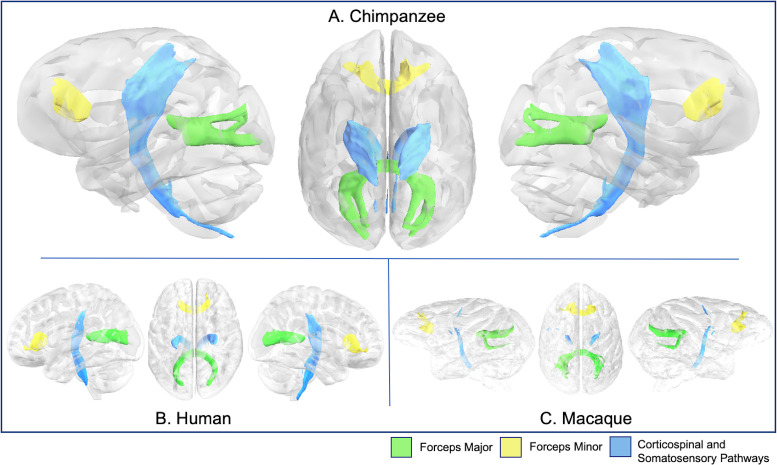
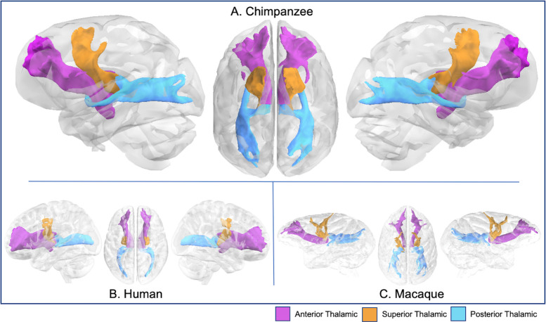
Chimpanzees (Pan troglodytes) are, along with bonobos, humans' closest living relatives. The advent of diffusion MRI tractography in recent years has allowed a resurgence of comparative neuroanatomical studies in humans and other primate species. Here we offer, in comparative perspective, the first chimpanzee white matter atlas, constructed from in vivo chimpanzee diffusion-weighted scans. Comparative white matter atlases provide a useful tool for identifying neuroanatomical differences and similarities between humans and other primate species. Until now, comprehensive fascicular atlases have been created for humans (Homo sapiens), rhesus macaques (Macaca mulatta), and several other nonhuman primate species, but never in a nonhuman ape. Information on chimpanzee neuroanatomy is essential for understanding the anatomical specializations of white matter organization that are unique to the human lineage.


Conflict of interest statement

The authors have declared that no competing interests exist.

Figures

Fig 1
Fig 1. Chimpanzee tractography recipes.
Seed ROIs (yellow), target ROIs (dark blue), exclusion masks (white), stop masks (fuchsia). Left hemisphere protocols are displayed in radiological convention. Protocols are available at https://git.fmrib.ox.ac.uk/rmars/chimpanzee-tractography-protocols/-/tree/master/recipes/chimpanzee. CSP, corticospinal and somatosensory pathway; IFOF, inferior fronto-occipital fascicle; ILF, inferior longitudinal fascicle; MdLF; middle longitudinal fascicle; ROI, region of interest; SLF, superior longitudinal fascicle; VOF, vertical occipital fascicle.
Fig 2
Fig 2. Human tractography recipes.
Seed ROIs (yellow), target ROIs (dark blue), exclusion masks (white), stop masks (fuchsia). Left hemisphere protocols are displayed in radiological convention. Protocols are available at https://git.fmrib.ox.ac.uk/rmars/chimpanzee-tractography-protocols/-/tree/master/recipes/human. CSP, corticospinal and somatosensory pathway; IFOF, inferior fronto-occipital fascicle; ILF, inferior longitudinal fascicle; MdLF; middle longitudinal fascicle; ROI, region of interest; SLF, superior longitudinal fascicle; VOF, vertical occipital fascicle.
Fig 3
Fig 3. Macaque tractography recipes.
Seed ROIs (yellow), target ROIs (dark blue), exclusion masks (white), stop masks (fuchsia). Left hemisphere protocols are displayed in radiological convention. Protocols are available at https://git.fmrib.ox.ac.uk/rmars/chimpanzee-tractography-protocols/-/tree/master/recipes/macaque. IFOF, inferior fronto-occipital fascicle; ILF, inferior longitudinal fascicle; MdLF; middle longitudinal fascicle; ROI, region of interest; SLF, superior longitudinal fascicle; VOF, vertical occipital fascicle.
Fig 4
Fig 4
Arcuate fascicle in chimpanzee (A), human (B), and rhesus macaque (C). Tract volumes are available at https://git.fmrib.ox.ac.uk/rmars/chimpanzee-tractography-protocols/-/tree/master/tract_volumes.
Fig 5
Fig 5
SLFs I, II, and III in chimpanzee (A), human (B), and rhesus macaque (C). Tract volumes are available at https://git.fmrib.ox.ac.uk/rmars/chimpanzee-tractography-protocols/-/tree/master/tract_volumes. SLF, superior longitudinal fascicle.
Fig 6
Fig 6
Major fascicles. that course through the temporal lobe (IFOF, ILF, MdLF, and UF) in chimpanzee (A), human (B), and rhesus macaque (C). Tract volumes are available at https://git.fmrib.ox.ac.uk/rmars/chimpanzee-tractography-protocols/-/tree/master/tract_volumes. IFOF, inferior fronto-occipital fascicle; ILF, inferior longitudinal fascicle; MdLF; middle longitudinal fascicle; UF, uncinate fascicle.
Fig 7
Fig 7
Cingulum and fornix in chimpanzee (A), human (B), and rhesus macaque (C). Tract volumes are available at https://git.fmrib.ox.ac.uk/rmars/chimpanzee-tractography-protocols/-/tree/master/tract_volumes.
Fig 8
Fig 8
Anterior commissure, frontal aslant, MCP, and VOF in chimpanzee (A), human (B), and rhesus macaque (C). Tract volumes are available at https://git.fmrib.ox.ac.uk/rmars/chimpanzee-tractography-protocols/-/tree/master/tract_volumes. MCP, middle cerebellar peduncle; VOF, vertical occipital fascicle.
Fig 9
Fig 9
CSP, forceps major, and minor in chimpanzee (A), human (B), and rhesus macaque (C). Tract volumes are available at https://git.fmrib.ox.ac.uk/rmars/chimpanzee-tractography-protocols/-/tree/master/tract_volumes. CSP, corticospinal and somatosensory pathway.
Fig 10
Fig 10
Anterior, superior, and posterior thalamic radiations in chimpanzee (A), human (B), and rhesus macaque (C). Tract volumes are available at https://git.fmrib.ox.ac.uk/rmars/chimpanzee-tractography-protocols/-/tree/master/tract_volumes.
Fig 11
Fig 11
Optic and acoustic radiations in chimpanzee (A), human (B), and rhesus macaque (C). Tract volumes are available at https://git.fmrib.ox.ac.uk/rmars/chimpanzee-tractography-protocols/-/tree/master/tract_volumes.

References

    1. Catani M, Thiebaut de Schotten M. A diffusion tensor imaging tractography atlas for virtual in vivo dissections. Cortex. 2008;44:1105–32. 10.1016/j.cortex.2008.05.004 - DOI - PubMed
    1. Mori S, Oishi K, Jiang H, Jiang L, Li X, Akhter K, et al. Stereotaxic white matter atlas based on diffusion tensor imaging in an ICBM template. Neuroimage. 2008;40:570–82. 10.1016/j.neuroimage.2007.12.035 - DOI - PMC - PubMed
    1. Schmahmann JD, Pandya DN, Wang R, Dai G, D’Arceuil HE, de Crespigny AJ, et al. Association fibre pathways of the brain: parallel observations from diffusion spectrum imaging and autoradiography. Brain. 2007;130:630–53. 10.1093/brain/awl359 - DOI - PubMed
    1. Gao Y, Parvathaneni P, Schilling KG, Wang F, Stepniewska I, Xu Z, et al. A 3D high resolution ex vivo white matter atlas of the common squirrel monkey (Saimiri sciureus) based on diffusion tensor imaging. Proc SPIE Int Soc Opt Eng. 2016;9784 10.1117/12.2217325 - DOI - PMC - PubMed
    1. Hecht EE, Gutman DA, Bradley BA, Preuss TM, Stout D. Virtual dissection and comparative connectivity of the superior longitudinal fasciculus in chimpanzees and humans. Neuroimage. 2015;108:124–37. 10.1016/j.neuroimage.2014.12.039 - DOI - PMC - PubMed

Publication types

MeSH terms