Review
. 2020 Dec 29;8(1):3.
doi: 10.3390/bioengineering8010003.

Collagen Structure-Function Mapping Informs Applications for Regenerative Medicine

Affiliations
Review

Collagen Structure-Function Mapping Informs Applications for Regenerative Medicine

James D San Antonio et al. Bioengineering (Basel). .

Abstract

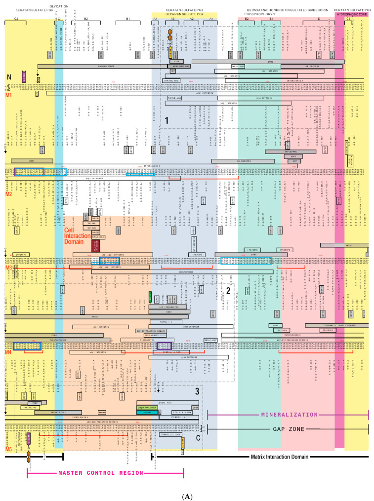
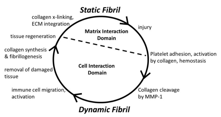
Type I collagen, the predominant protein of vertebrates, assembles into fibrils that orchestrate the form and function of bone, tendon, skin, and other tissues. Collagen plays roles in hemostasis, wound healing, angiogenesis, and biomineralization, and its dysfunction contributes to fibrosis, atherosclerosis, cancer metastasis, and brittle bone disease. To elucidate the type I collagen structure-function relationship, we constructed a type I collagen fibril interactome, including its functional sites and disease-associated mutations. When projected onto an X-ray diffraction model of the native collagen microfibril, data revealed a matrix interaction domain that assumes structural roles including collagen assembly, crosslinking, proteoglycan (PG) binding, and mineralization, and the cell interaction domain supporting dynamic aspects of collagen biology such as hemostasis, tissue remodeling, and cell adhesion. Our type III collagen interactome corroborates this model. We propose that in quiescent tissues, the fibril projects a structural face; however, tissue injury releases blood into the collagenous stroma, triggering exposure of the fibrils' cell and ligand binding sites crucial for tissue remodeling and regeneration. Applications of our research include discovery of anti-fibrotic antibodies and elucidating their interactions with collagen, and using insights from our angiogenesis studies and collagen structure-function model to inform the design of super-angiogenic collagens and collagen mimetics.

Keywords: angiogenesis; connective tissue; extracellular matrix; fibrosis; hemostasis; interactome; ligand binding; microfibril; therapeutic antibodies; type I collagen; type III collagen.


Conflict of interest statement

The authors declare no conflict of interest.

Figures

Figure 1
Figure 1
Type I collagen assembly and structure. (A). Segment of Type I collagen fibril visualized by transmission electron microscopy. One molecular repeat or D-period (D) is indicated. The positive stain microscopy bands, a–e, are as indicated below the image; arrow indicates left border of overlap zone. This fibril preparation was used to localize heparin-binding sites; thus, heparin-gold particles appear as dark circles bound to the fibril. Originally published in San Antonio et al., 1994, J. Cell Biol., 125, 1179–1188. (B). Fibril schematic depicted as negatively-stained TEM preparation where gap regions are dark and overlap regions light. Microfibril schematic shows the Hodge-Petruska scheme [20] of packing where collagen molecules (numbered 1–5 for molecular (M) segments as in M1, M2, etc.) are staggered so that every five M segment does not traverse the entire D-period. Select collagen functional domains (right) are marked along the length of the collagen molecule. (C). A single D period of a single microfibril is shown beneath the microfibril schematic. The C-terminal telopeptide (marked in green on the top of the microfibril) and the rest of monomer 5 is orientated towards the outside of the fibril. The side view is from an observers’ perspective from a neighboring microfibril. Note the molecular segments are relatively straight in the overlap zone but re-organize towards the end of the gap zone especially in the region of the supertwist in the vicinity of the gap zone’s discoidin domain receptor 2 (DDR2) binding site. Figure segments reprinted with permission from [21].
Figure 2
Figure 2
(A) Type I collagen interactome. Human collagen primary sequences were from GenBank, accession #s: a1(I), NP000079; a2(I), NP000080 and aligned as described [23] (Figure 1). Ligand binding sites are indicated by rectangular boxes adjacent to relevant collagen sequences. Gray boxes denote ligand binding to the monomer. Non-shaded boxes denote ligand binding to one α chain. Major ligand binding regions (MLBR) 1, 2, and 3 are designated. Disease-associated mutations are indicated next to affected residues. Broad cross-fibril ligand binding regions are delineated by color-shaded overlays. (B) Legend to type I collagen interactome (A) [22], listing abbreviations of mapped sites; literature citations are in [22]. Human mutation data date to 2013. This figure was modified from research originally published in the J. Biol. Chem. © the American Society for Biochemistry and Molecular Biology [22].
Figure 2
Figure 2
(A) Type I collagen interactome. Human collagen primary sequences were from GenBank, accession #s: a1(I), NP000079; a2(I), NP000080 and aligned as described [23] (Figure 1). Ligand binding sites are indicated by rectangular boxes adjacent to relevant collagen sequences. Gray boxes denote ligand binding to the monomer. Non-shaded boxes denote ligand binding to one α chain. Major ligand binding regions (MLBR) 1, 2, and 3 are designated. Disease-associated mutations are indicated next to affected residues. Broad cross-fibril ligand binding regions are delineated by color-shaded overlays. (B) Legend to type I collagen interactome (A) [22], listing abbreviations of mapped sites; literature citations are in [22]. Human mutation data date to 2013. This figure was modified from research originally published in the J. Biol. Chem. © the American Society for Biochemistry and Molecular Biology [22].
Figure 3
Figure 3
Domain model of collagen fibril function. Type I collagen interactome (Figure 2) and X-ray diffraction fibril model (Figure 4) suggests the conditional domain model of collagen fibril function. Depending on the physiological state of the tissue, the fibril predominantly supports structural duties including intermolecular crosslinking, proteoglycan binding, and biomineralization via the matrix interaction domain; alternatively, dynamic biological processes such as hemostasis, collagen remodeling, and cell adhesion are supported via the cell interaction domain, as schematically shown in Figure 5. This figure was originally published in the J. Biol. Chem. © the American Society for Biochemistry and Molecular Biology [22].
Figure 4
Figure 4
Ranking of ligand accessibilities to crucial binding sites and functional sequences in the native and proteolyzed type I collagen fibril. (A) Key functional domains of collagen were mapped onto a model of the fibril surface viewed from the fibril’s exterior. A molecular accessibility ranking of various ligands to their binding sites was determined for the static fibril, and following MMP-1 cleavage of M4 and “opening up” of the fibril. (B) View of the fibril’s GFOGER and Von Willebrand’s Factor (vWF)-binding sites following MMP-1 cleavage of M4. Reprinted with permission from [50].
Figure 5
Figure 5
Schematic of conditional cell and matrix interaction domain model of type I collagen fibril function. See text and Figure 3 for details.
Figure 6
Figure 6
Analysis of type III collagen interactome suggests a “flexi-rod” model of fibril structure. Top: schematic of type III collagen fibril with sites for cell interactions and remodeling is flanked by intermolecular crosslinks (X) and a hemostasis domain (H). Bottom: Clusters of atypical collagen sequences of lower stability (springs) are interspersed with rigid zones (rods) hosting crucial biologic functions. Reprinted with permission from [74].
Figure 7
Figure 7
X-ray diffraction model of the C-terminal telopeptide region of the rat type I collagen microfibril. The truncated α2(I) chain is shown in cyan (see text). The electron density defining the C-terminal region is seen to terminate at the ends of the C-telopeptides, where the α1(I) chains, in dark blue, fold back on themselves. The anti-fibrotic antibody binding sequence on human collagen—the α2Ct epitope—can be accommodated within the limits of the C-terminus of the rat protein, however the homologous sequence on the rat α2(I) chain is not found in the structural electron density model (gray wireframe, C-terminal electron density indicated; red arrow). To enable molecular modeling of scFv antibody-collagen interactions with rat collagen, the α2(I) chain of the rat protein was extended C-terminally to include the α2Ct epitope of the rat/human sequence in the molecular model shown in Figure 8 (see text and Table 1).
Figure 8
Figure 8
Preliminary molecular simulation of docking between the scFv antibody and the α2Ct epitope of rat collagen. Lower figure: C-terminus of the rat type I collagen model; α1(I) chains, purple and green; α2(I) chain, cyan, α2Ct epitope, dark cyan. Upper figure: scFv antibody with highly water accessible “exposed” regions (mesh surface) color-scaled towards the red end of the spectrum; green is intermediate and blue is not exposed. Antibody subunits show potentially complementary binding interactions at α2Ct epitope (see text).
Figure 9
Figure 9
Apical collagen gel induces angiogenesis of human endothelial cells. Confluent monolayers of cells were overlain with a type I collagen gel at 0 h. At 2–4 h, cell streaming and reorganization occurred. At 6–8 h, cultures were at least about 50% reorganized and at 12–24 h, capillary tube formation was complete. Bar = 50 μm. Reprinted with permission from [106].
Figure 10
Figure 10
Anti-integrin antibody coated beads induce angiogenesis in the absence of collagen. Endothelial cells were exposed to 0.5 × 10⁷ beads/cm2 polystyrene beads (3 μm diameter) coated with bovine serum albumin as a negative control (A,B), anti-α2β1 integrin antibodies (C,D), and collagen (E,F). At 24 h, monolayers were rinsed and photographed. BSA beads showed no activity (A) and interacted with cells poorly (B). Anti-α2β1 integrin antibody-coated beads induced tube formation similar to that of an apical collagen gel ((C), see Figure 9) and interacted extensively with cells (D). Collagen-coated beads induced angiogenesis (E) and also interacted extensively with cells (F). Top row: Bar = 100 μm. Bottom row: 20× objective. Bead diameters = 3 μm. Reprinted with permission from [106].

Similar articles

Cited by

References

    1. Kadler K.E., Baldock C., Bella J., Boot-Handford R.P. Collagens at a glance. J. Cell Sci. 2007;120 (Pt 12):1955–1958. doi: 10.1242/jcs.03453. - DOI - PubMed
    1. Marini J.C., Forlino A., Cabral W.A., Barnes A.M., San Antonio J.D., Milgrom S., Hyland J.C., Korkko J., Prockop D.J., De Paepe A., et al. Consortium for osteogenesis imperfecta mutations in the helical domain of type I collagen: Regions rich in lethal mutations align with collagen binding sites for integrins and proteoglycans. Hum. Mutat. 2007;28:209–221. doi: 10.1002/humu.20429. - DOI - PMC - PubMed
    1. Piez K.A., Reddi A.H. Extracellular Matrix Biochemistry. Elsevier; New York, NY, USA: 1984. p. xvi.473p
    1. Jacenko O., Olsen B.R., LuValle P. Organization and regulation of collagen genes. Crit. Rev. Eukaryot. Gene Expr. 1991;1:327–353. - PubMed
    1. Ricard-Blum S. The collagen family. Cold Spring Harb. Perspect. Biol. 2011;3:a004978. doi: 10.1101/cshperspect.a004978. - DOI - PMC - PubMed

LinkOut - more resources