. 2020 Dec 29;11(1):15.
doi: 10.3390/life11010015.

Fast Diffusion of the Unassembled PetC1-GFP Protein in the Cyanobacterial Thylakoid Membrane

Affiliations

Fast Diffusion of the Unassembled PetC1-GFP Protein in the Cyanobacterial Thylakoid Membrane

Radek Kaňa et al. Life (Basel). .

Abstract

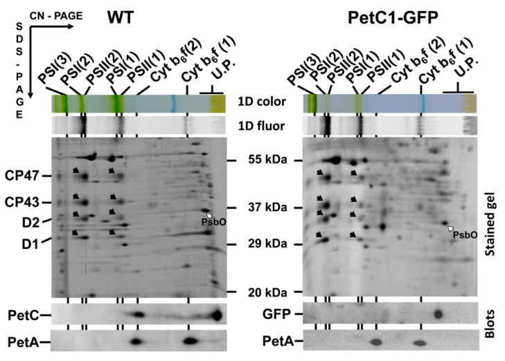
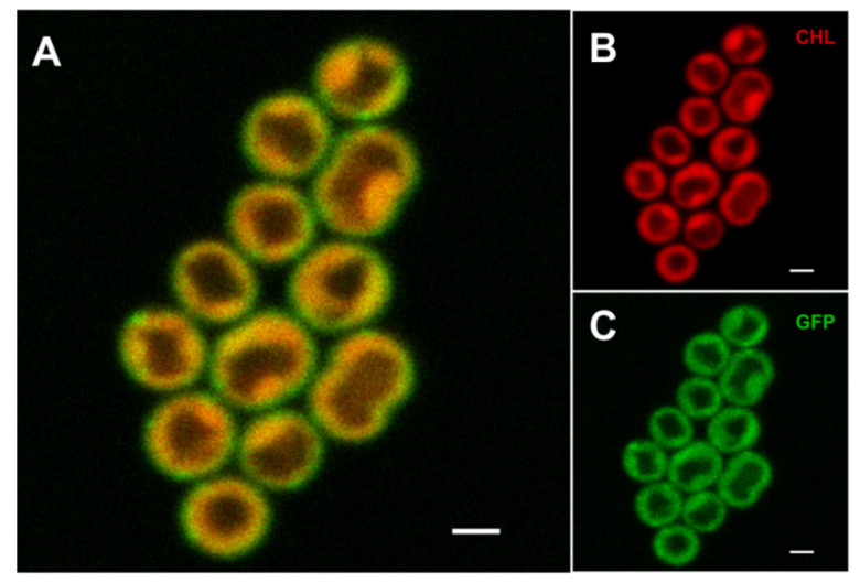
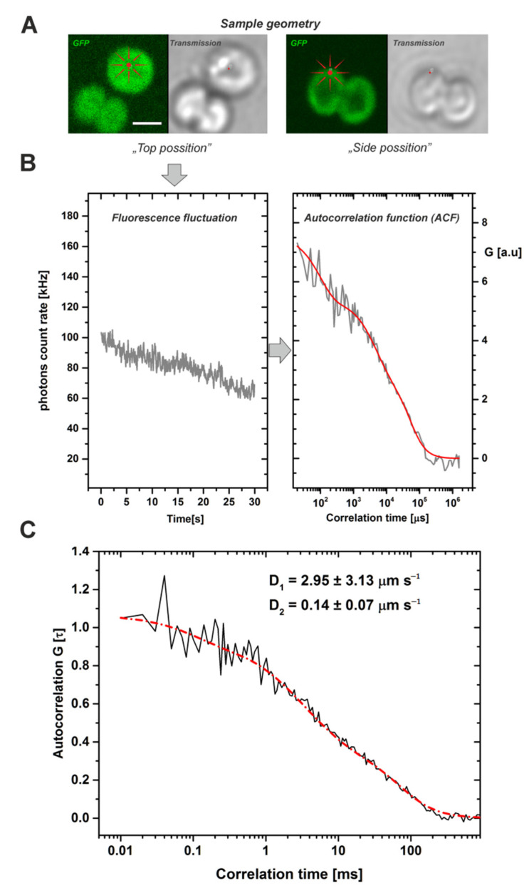
Biological membranes were originally described as a fluid mosaic with uniform distribution of proteins and lipids. Later, heterogeneous membrane areas were found in many membrane systems including cyanobacterial thylakoids. In fact, cyanobacterial pigment-protein complexes (photosystems, phycobilisomes) form a heterogeneous mosaic of thylakoid membrane microdomains (MDs) restricting protein mobility. The trafficking of membrane proteins is one of the key factors for long-term survival under stress conditions, for instance during exposure to photoinhibitory light conditions. However, the mobility of unbound 'free' proteins in thylakoid membrane is poorly characterized. In this work, we assessed the maximal diffusional ability of a small, unbound thylakoid membrane protein by semi-single molecule FCS (fluorescence correlation spectroscopy) method in the cyanobacterium Synechocystis sp. PCC6803. We utilized a GFP-tagged variant of the cytochrome b6f subunit PetC1 (PetC1-GFP), which was not assembled in the b6f complex due to the presence of the tag. Subsequent FCS measurements have identified a very fast diffusion of the PetC1-GFP protein in the thylakoid membrane (D = 0.14 - 2.95 µm2s-1). This means that the mobility of PetC1-GFP was comparable with that of free lipids and was 50-500 times higher in comparison to the mobility of proteins (e.g., IsiA, LHCII-light-harvesting complexes of PSII) naturally associated with larger thylakoid membrane complexes like photosystems. Our results thus demonstrate the ability of free thylakoid-membrane proteins to move very fast, revealing the crucial role of protein-protein interactions in the mobility restrictions for large thylakoid protein complexes.

Keywords: FCS; cyanobacteria; photosynthesis; proteins mobility; thylakoids.


Conflict of interest statement

The authors declare no conflict of interest.

Figures

Figure 1
Figure 1
2D analysis of membrane proteins of WT and GFP-PetC1 expressing strains. Membranes isolated from cells were analyzed by 2D CN/SDS-PAGE in combination with immunoblotting. Dimeric and monomeric Cyt b6-f complexes were identified using anti PetA antibody, PetC in WT was identified using general anti PetC antibody. Designation of complexes: PSI(3), PSI(2), PSI(1), trimeric, dimeric and monomeric PSI complexes, resp.; PSII(2) and PSII(1), dimeric and monomeric PSII core complexes, resp.; Cyt b6-f(2) and Cyt b6-f(1), dimeric and monomeric cytochrome b6-f complexes, resp.; U.P. unassembled proteins. The large PSII proteins CP47, CP43, D2 and D1 within PSII monomers and dimers (black arrows) and the unassembled PsbO (empty arrow) were identified previously by mass spectrometry [40,41] and by immunoblotting [42]. Each loaded sample contained 5 µg of CHL.
Figure 2
Figure 2
Localization of the free PetC1-GFP fusion protein in Synechocystis cells. The combined fluorescence picture (A) is formed from the red chlorophyll autofluorescence (B)—red channel and from the green GFP fluorescence of PetC1-GFP protein (Panel C). Bars represent 1 µm.
Figure 3
Figure 3
Typical work-flow of FCS measurements leading to calculation of diffusion coefficients for the PetC1-GFP protein in Synechocystis thylakoids. The fluorescence fluctuations of the GFP signal have been detected at a single point, in photon counting mode (detection range 500–530 nm) during 30 s of data acquisition. (Panel A): Single cell images of Synechocystis thylakoids (GFP fluorescence, transmission) with two different geometrical orientations: (i) top position mode used for the FSC measurements, it means from the face aligned membrane layer (see image on left); (ii) Side position mode (right). Scale bar 2 µm. (Panel B): Typical fluorescence fluctuations of GFP signal at a single point of a cell in the top position mode (left). These fluctuations were used after the bleaching correction to construct an auto-correlation function ACF (right). Data represent one single experiment (grey) and the result of the fit (red). (Panel C): The averaged cross-correlation functions from measured cells. Black line represents experimental data of calculated autocorrelation functions, red line shows 2D model of diffusion with two diffusion components of PetC1-GFP, D1 = 2.954 µm s−1, D2 = 0.14 µm s−1. Number of cells used for analysis was n = 79. For more details, see Table 1 and Supplementary Figure S1.

Similar articles

Cited by

References

    1. Busch K.B., Deckers-Hebestreit G., Hanke G.T., Mulkidjanian A.Y. Dynamics of bioenergetic microcompartments. Biol. Chem. 2013;394:163–188. doi: 10.1515/hsz-2012-0254. - DOI - PubMed
    1. Liu H., Zhang H., Niedzwiedzki D.M., Prado M., He G., Gross M.L., Blankenship R.E. Phycobilisomes Supply Excitations to Both Photosystems in a Megacomplex in Cyanobacteria. Science. 2013;342:1104–1107. doi: 10.1126/science.1242321. - DOI - PMC - PubMed
    1. Kirchhoff H. Diffusion of molecules and macromolecules in thylakoid membranes. Biochim. Biophys. Acta Bioenerg. 2014;1837:495–502. doi: 10.1016/j.bbabio.2013.11.003. - DOI - PubMed
    1. Steinbeck J., Ross I.L., Rothnagel R., Gabelein P., Schulze S., Giles N., Ali R., Drysdale R., Sierecki E., Gambin Y., et al. Structure of a PSI-LHCI-cyt b6f supercomplex in Chlamydomonas reinhardtii promoting cyclic electron flow under anaerobic conditions. Proc. Natl. Acad. Sci. USA. 2018;115:10517–10522. doi: 10.1073/pnas.1809973115. - DOI - PMC - PubMed
    1. Zhao L.S., Huokko T., Wilson S., Simpson D.M., Wang Q., Ruban A.V., Mullineaux C.W., Zhang Y.Z., Liu L.N. Structural variability, coordination and adaptation of a native photosynthetic machinery. Nat. Plants. 2020;6:869–882. doi: 10.1038/s41477-020-0694-3. - DOI - PubMed