. 2020 Dec 29;10(1):58.
doi: 10.3390/plants10010058.

Shoot Regeneration Is Not a Single Cell Event

Affiliations

Shoot Regeneration Is Not a Single Cell Event

Patharajan Subban et al. Plants (Basel). .

Abstract

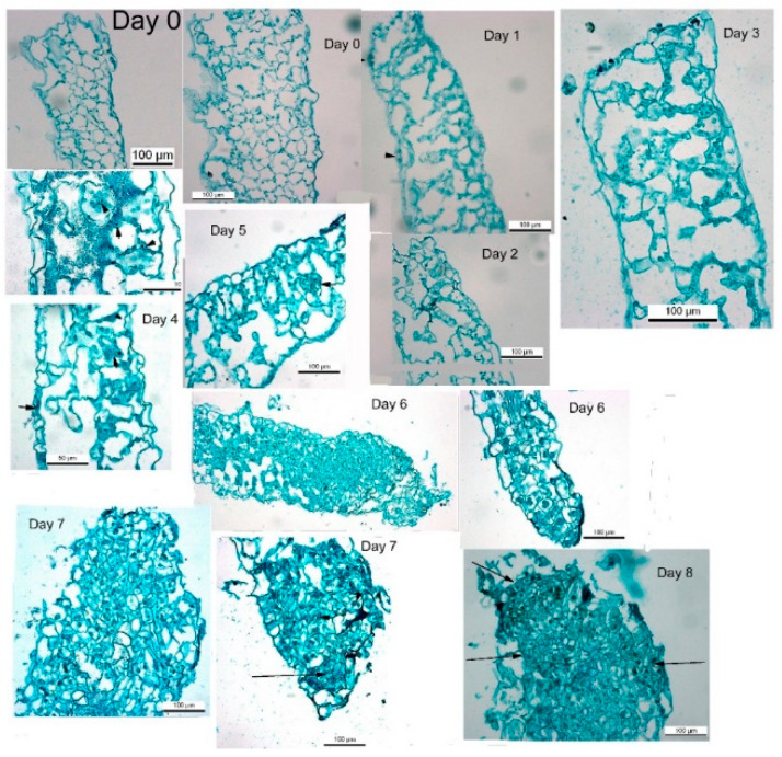
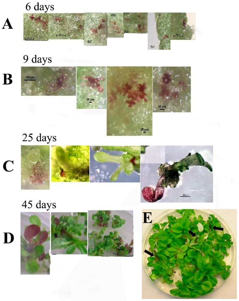
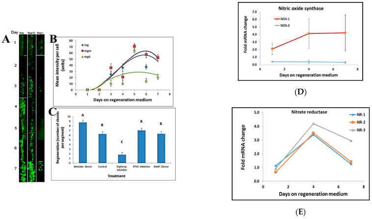
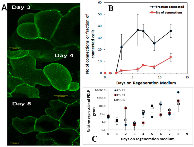
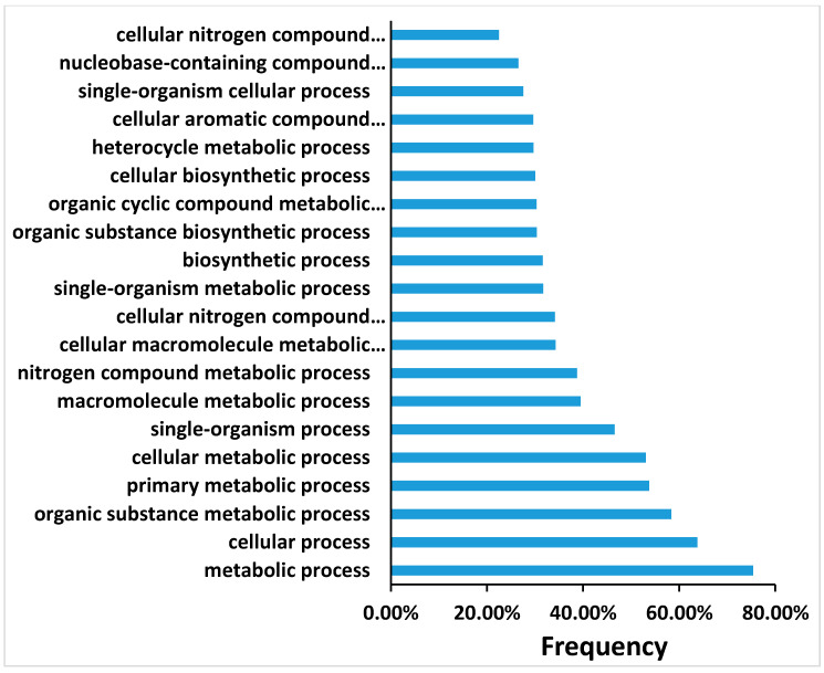
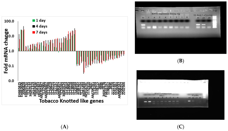
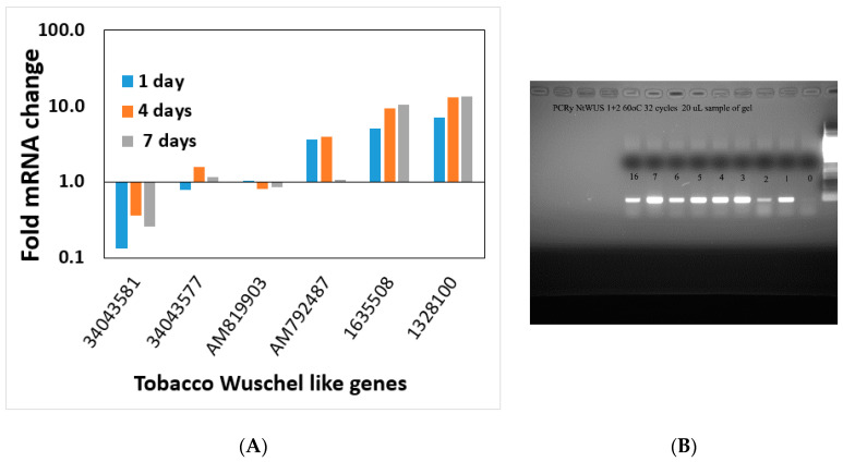
Shoot regeneration is a key tool of modern plant biotechnology. While many researchers use this process empirically, very little is known about the early molecular genetic factors and signaling events that lead to shoot regeneration. Using tobacco as a model system, we found that the inductive events required for shoot regeneration occur in the first 4-5 days following incubation on regeneration medium. Leaf segments placed on regeneration medium did not produce shoots if removed from the medium before four days indicating this time frame is crucial for the induction of shoot regeneration. Leaf segments placed on regeneration medium for longer than five days maintain the capacity to produce shoots when removed from the regeneration medium. Analysis of gene expression during the early days of incubation on regeneration medium revealed many changes occurring with no single expression pattern evident among major gene families previously implicated in developmental processes. For example, expression of Knotted gene family members increased during the induction period, whereas transcription factors from the Wuschel gene family were unaltered during shoot induction. Expression levels of genes involved in cell cycle regulation increased steadily on regeneration medium while expression of NAC genes varied. No obvious possible candidate genes or developmental processes could be identified as a target for the early events (first few days) in the induction of shoot regeneration. On the other hand, observations during the early stages of regeneration pointed out that regeneration does not occur from a single cell but a group of cells. We observed that while cell division starts just as leaf segments are placed on regeneration medium, only a group of cells could become shoot primordia. Still, these primordia are not identifiable during the first days.

Keywords: regeneration induction; shoot regeneration; tobacco.


Conflict of interest statement

The authors declare no conflict of interest.

Figures

Figure 1
Figure 1
Tobacco leaf segments regeneration. (A1) Tobacco leaf segments were placed on MS nutrient medium or Regeneration medium, including auxin and cytokinin or MS medium (see Materials and Methods) containing auxin only (4.44 M IAA). Thirty days after placement on a specific medium, the segments were pictured. (A2) Close-up of leaf segments from the plates in (A1). (A3) Leaf segments from transgenic tobacco expressing a cytokinin reporter gene ARR5::GUS show responsiveness to cytokinin 30 days on the respective media described in (A1). (B) Time on regeneration medium that is required to induce shoots from competent tobacco leaf segments. Leaf segments were placed on regeneration medium for the indicated time and then transferred to MS medium for 30 days. Percent of shoots forming leaf segment was recorded after 30 days on MS medium + SE. Each point represents three plates with 20 segments. (C) Chlorophyll content of leaf segments on MS or regeneration medium. Leaf segments were placed on agar plates with MS only (MS) or with added growth regulators (REG), and chlorophyll was extracted and determined from each segment. Chlorophyll content per gram fresh weight from leaf segment was recorded on ten segments every day + SE. (D) Area of leaf segments explants during the first two weeks on MS or REG medium. Leaf segments were placed on agar plates with MS salts only (MS) or with added growth regulators (REG), and the area of segments was recorded daily to measure cell proliferation. Size of leaf segment was recorded 20 segments every day on the two media + SE.
Figure 2
Figure 2
Cross sections of leaf segments taken during the first 8 days on REG medium. Arrowhead in Day 1 indicates stomata and arrows in Days 4, 5, 7, and 8 indicate cell clusters with cell divisions which will may form shoots.
Figure 3
Figure 3
(AD) Leaves of transformed tobacco show chimerism even on selection medium after transformation with tomato ScANT1 gene construct and selection on Kanamycin. (EI) Pictures of tobacco leaves transformed with the ScANT1 construct. (J) PCR analysis of genomic DNA isolated from Red or Green sections of the leaves in (EI).
Figure 4
Figure 4
Bright light pictures of leaf segments transformed with the 35S::ScANT1 construct, taken at various time points following transformation and explant growth on regeneration medium without Kanamycin selection. (A) leaf segments showing pigmented cells 6 days after transformation. (B) leaf segments showing pigmented cells 9 days after transformation. (C) tissue showing pigmented section 25 days after transformation. (D,E) plants showing pigmented leave 45 days after transformation. Arrows point to the colored leaves in the plate.
Figure 5
Figure 5
NO has a role in shoot regeneration. (A) NO staining at the border of leaf segments placed on regeneration medium in the presence of NO inhibitor (Reg+I), NO donor (Reg+D) and control (Reg). (B) Stain intensity per cell at the borders of leaf segments during the induction period of shoot induction. NO inhibitor (Regd), NO donor (Regm), and control (Reg). (C) Effect of NO donors (S-Nitroso-N-acetyl-DL-penicillamine; Molsidomine), NO scavenger (PTIO), and NO synthesis inhibitor (Diphenyleneiodonium) on shoot regeneration. Different letters (A, B, C) define statistically different results, p(f) was 7.65 × 10⁻³⁵ using Tukey test. (D) Transcript level of Nitric Oxide Synthase1 increases during shoot induction while transcript level of Nitric Oxide Synthase2 is unchanged. The data presented are from the Differential Gene Expression experiment. (E) Transcript level of all Nitrate reductase genes increase during incubation on Reg medium. The data presented are from the Differential Gene Expression experiment. Reg = regeneration medium; regm = regeneration medium with added Molsidomine; regd = regeneration medium with added Diphenyleneiodonium. NR = nitrate reductase; NOS = nitric oxide synthase. Error bars indicated standard error of 50 cells in (B); 20 segment in 3 replicated in (C); and 3 biological replicates in (D,E).
Figure 6
Figure 6
Plasmodesmata during shoot regeneration. (A) Leaf segment from tobacco plants that are homozygous to the transgene 35S::AtPDLP5-GFP were placed on Reg medium and samples were removed every day and the new cells were visualized and analyzed using confocal microscopy. (B) The number of PD connections per cell was counted and fraction of cells with PD from all new cells was counted. (C) Expression pattern of three tobacco PDLP genes during the shoot induction period is depicted. Error bars indicated standard error of 50 cells in B; and 3 biological replicates in (C).
Figure 7
Figure 7
A sample of transcript analyses of top 20 biological processes that upregulate at day 4 of the induction of shoot regeneration. Only some of the processes were fitted on the Y-axis.
Figure 8
Figure 8
(A) Transcript pattern of Knotted gene family members during the induction period of shoot regeneration. The gene numbers are depicted on the x-axis. (B) expression of NtKnotted1 during the induction period. (C) expression of NtKnotted2 during the induction period.
Figure 9
Figure 9
(A) Transcript pattern of Wuschel gene family (WOX) members during the induction period of shoot regeneration. The gene numbers are depicted on the x-axis. (B) Expression of NtWus during the shoot induction period on Reg medium.
Figure 10
Figure 10
The orientation of leaf segment affects shoot regeneration and percent regeneration. (A) Number of shoots regenerated from leaf segments placed with the segment above the agar Reg medium (Up); leaf segments placed with the segment on the agar and the plates turned upside down, thus, the segment are beneath the agar Reg medium (Down) but still exposed to the air on one side or leaf segments placed with the segment above the agar Reg medium, and the plate was rotated at 30 rotations per min (Rotating). Results are the mean ± se of five repetitions representing 802 leaf segments. Different letters (A, B) define statistically different results (B) Percent regeneration from leaf segments placed as in (A), percent shooting was calculated as the number of segments that produced at least one shoot out of a total number of the segment in a plate. Results are the mean ± se of five repetitions for each treatment. Different letters (A, B) define statistically different results (C) Effect of positioning leaf segments containing the auxin reporter transgene IAA/AUX3::GUS above or beneath the agar medium on IAA responsiveness; up and down as in A. Each point is the mean ± se of five measurements of GUS activity done on ten individual leaf segments.

Similar articles

Cited by

References

    1. Vogel G. How does a single somatic cell become a whole plant? Science. 2005;309:86. doi: 10.1126/science.309.5731.86. - DOI - PubMed
    1. Birnbaum K.D., Sánchez Alvarado A. Slicing across kingdoms: Regeneration in plants and animals. Cell. 2008;22:697–710. doi: 10.1016/j.cell.2008.01.040. - DOI - PMC - PubMed
    1. Fehér A. Somatic embryogenesis—Stress-induced remodeling of plant cell fate. Biochim. Biophys. Acta. 2015;1849:385–402. doi: 10.1016/j.bbagrm.2014.07.005. - DOI - PubMed
    1. Steward F.C. Growth and Organized Development of Cultured Cells. III. Interpretations of the Growth from Free Cell to Carrot Plant. Am. J. Bot. 1958;45:709–713. doi: 10.1002/j.1537-2197.1958.tb10600.x. - DOI
    1. Zimmerman J.L. Somatic embryogenesis: A model for early development in higher plants. Plant Cell. 1993;5:1411–1423. doi: 10.2307/3869792. - DOI - PMC - PubMed

LinkOut - more resources