. 2020 Dec 29;13(1):37.
doi: 10.3390/v13010037.

Broadly Active Antiviral Compounds Disturb Zika Virus Progeny Release Rescuing Virus-Induced Toxicity in Brain Organoids

Affiliations

Broadly Active Antiviral Compounds Disturb Zika Virus Progeny Release Rescuing Virus-Induced Toxicity in Brain Organoids

Aleksandra Pettke et al. Viruses. .

Abstract

RNA viruses have gained plenty of attention during recent outbreaks of Severe acute respiratory syndrome coronavirus 2 (SARS-CoV-2), Zika virus (ZIKV), and Ebola virus. ZIKV is a vector borne Flavivirus that is spread by mosquitoes and it mainly infects neuronal progenitor cells. One hallmark of congenital ZIKV disease is a reduced brain size in fetuses, leading to severe neurological defects. The World Health Organization (WHO) is urging the development of new antiviral treatments against ZIKV, as there are no efficient countermeasures against ZIKV disease. Previously, we presented a new class of host-targeting antivirals active against a number of pathogenic RNA viruses, such as SARS-CoV-2. Here, we show the transfer of the image-based phenotypic antiviral assay to ZIKV-infected brain cells, followed by mechanism-of-action studies and a proof-of-concept study in a three-dimensional (3D) organoid model. The novel antiviral compounds showed a therapeutic window against ZIKV in several cell models and rescued ZIKV-induced neurotoxicity in brain organoids. The compound's mechanism-of-action was pinpointed to late steps in the virus life cycle, impairing the formation of new virus particles. Collectively, in this study, we expand the antiviral activity of new small molecule inhibitors to a new virus class of Flaviviruses, but also uncover compounds' mechanism of action, which are important for the further development of antivirals.

Keywords: Zika virus; antivirals; brain organoids; mode-of-action; pathogenic RNA viruses.


Conflict of interest statement

The authors declare no competing interest.

Figures

Figure 1
Figure 1
Image-based screening on Zika virus (ZIKV)-infected cells identifies TH3289 and TH6744 as active small molecule inhibitors against ZIKV. (A) Experimental workflow of phenotypic antiviral testing of small molecule compounds. Nuclei count was used as an indicator of cytotoxicity, viral antibody staining was quantified to assess the percentage of virus infected cells and virus titer was quantified by end-point dilution assay. (B) U87 cells were infected with ZIKV (MOI 10) and treated with structural analogs of TH3289 at 10 µM concentration for 48 h. Infected cells were stained for DAPI and ZIKV NS1 and analyzed by high-throughput imaging. Virus titers from supernatants were determined by end-point dilution assay and inhibition of viral titer was calculated relative to dimethyl sulfoxide (DMSO). Data are presented as a mean of two technical replicates per compound performed in n = 1 biological replicate. Representative images from ZIKV-infected U87 cells treated with DMSO or TH3289 (upper image panels; Scale bar equals 100 µm) and corresponding example of titrated ZIKV from the end-point dilution assay (lower image panels) are shown. DAPI in grey, ZIKV NS-1 in magenta. (C) The inhibition of viral titers of Hazara virus (HAZV) and ZIKV from both assays was correlated. In the HAZV assay, SW13 cells were infected with HAZV (MOI 10) and treated with 10 μM of compounds from the in-house library for 24 h. Virus titer from supernatant was determined by end-point dilution assay. Cells were stained for DAPI (in blue) and HAZV NP (in green) and analyzed by high-throughput imaging. Data are presented as a mean of two technical replicates per compound performed in n = 1 biological replicate. (D) Chemical structures of top hits from ZIKV and HAZV antiviral screenings and their respective inhibition on viral titers.
Figure 2
Figure 2
Series of antiviral compounds have a therapeutic window and rescue ZIKV-induced CPE in cellular models. (AE) U87 cells were infected with ZIKV (MOI 1) and treated with increasing doses of (A) TH3289, (B) TH5264, (C) TH6744, or (D) TH5487 for 48 h. Cell viability was determined by nuclei count (in blue), infected cells by ZIKV NS1 staining (in orange) and virus titer by an end-point dilution assay (in black). Data are presented as mean ± SD from n = 3 independent replicates. (E) Curve fitting was performed to calculate EC50 values for living cells, infected cells and viral titer. (F) Vero cells were infected with ZIKV (MOI 10) and treated with 12.5 μM TH6744 or DMSO for 48 h. Cells were stained for ZIKV NS1 (in green), ZIKV Capsid (in red) and DAPI (in blue). Images of n = 1 biological replicate. In the overview image, scale bar equals 100 μm and in the close-up 25 μm. (G) U87 cells were infected with ZIKV (MOI 1) and treated with 10 μM TH6744 or DMSO for 24, 48, or 72 h. Cells were stained for Pan-Flavi (in green), Golgi marker GM130 (in red), ER marker KDEL (in yellow), and DAPI (in blue). Images of n = 1 biological replicate. Scale bar equals 50 μm. (H) Vero cells were infected with ZIKV (MOI 0.1) and treated with increasing doses of TH6744 for 24 h. Viral titer was measured from the supernatants by end-point dilution assay on Vero cells and the development of CPE were monitored by visual assessment. Data presented as mean ± SD from n = 2 biological replicates.
Figure 3
Figure 3
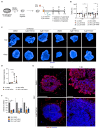
TH6744 and TH5487 treatment reverses ZIKV-induced neurotoxicity and limits ZIKV propagation in three-dimensional (3D) cerebral organoids. (A) Experimental workflow of ZIKV infection on brain organoid model. (BF) Organoids were infected with ZIKV (2 × 10⁴ particles/organoid) followed by treatment with 5 μM TH6744, 5 μM TH5487 or DMSO. (B) Organoids were dissociated to single cells at 7 or 10 dpi and cell viability was measured by Resazurin assay. Viability is shown relative to uninfected DMSO-treated control at respective timepoints. One datapoint represents one organoid. Data are presented as a mean ± SD of n = 2 biological replicates. Statistical significance was determined using two-way ANOVA with Dunnett’s multiple comparison analysis. * p < 0.05, ** p < 0.01, (C) ZIKV-infected and uninfected whole organoids treated with TH6744 or DMSO for 7 or 10 dpi, stained by Hoechst and imaged by high-content confocal microscopy. Representative images of n = 2 biological replicates per condition. The scale bar equals 500 μm. Hoechst in blue. (D) ZIKV-infected organoids that were treated with TH6744 or DMSO were fixed at 7 dpi, cryosectioned and stained with Hoechst and ZIKV NS1 protein antibody. ZIKV NS1 intensity was quantified using Cellprofiler software. Data are presented as single values and mean ± SD from n = 2 biological replicates. Statistical significance was determined using one-way ANOVA with Dunnett’s multiple comparison analysis. * p < 0.05, ** p < 0.01. (E) ZIKV-infected organoids treated with TH6744 or DMSO were fixed at 7 dpi, cryosectioned, stained with Hoechst and ZIKV NS1 protein antibody and imaged by confocal microscopy. Representative images of n = 2 biological replicates per condition. Scale bar equals 500 μm in overview image and 100 μm in close-up. ZIKV NS1 protein in red, Hoechst in blue. (F) Viral titer from ZIKV-infected and compound treated organoids at indicated time points was determined by an end-point dilution assay. Viral titer is presented as relative to ZIKV-infected DMSO-treated control at respective timepoints. Data are presented as a mean ± SD from n = 2 biological replicates. Statistical significance was determined using two-way ANOVA with Sidak’s multiple comparison analysis. * p < 0.05, ** p < 0.01.
Figure 4
Figure 4
TH6744 disturbs late replication cycle steps in ZIKV life cycle. (AD) U87 cells were infected with ZIKV (MOI 1) and treated with indicated compounds for 24, 48 or 72 h. (A) ZIKV RNA levels inside the cells were quantified by qPCR. (B) Number of ZIKV-infected cells was quantified by virus infectivity assay. (C) ZIKV RNA levels in the supernatant were quantified by one-step qRT-PCR. (D) ZIKV titers were quantified by end-point dilution assay. (E) Schematic overview of time-of-addition experimental setup in U87 cells. (F) U87 cells were infected with ZIKV (MOI 10) for a total of 24 h and treated with DMSO control, 10 μM TH6744 or 100 μM Ribavirin for indicated periods during early (entry), late (budding) or throughout replication cycle (post-inoculation). ZIKV titers were quantified by end-point dilution assay. (G) Schematic overview of budding experimental setup. (H) U87 cells were infected with ZIKV (MOI 10) for 22 h and treated with indicated TH6744 doses for 2 h. Intracellular ZIKV particles were obtained by mechanical cell lysis and extracellular ZIKV particles from the supernatants and both were quantified by end-point dilution assay. (I) Average fractions of intracellular and extracellular infectious ZIKV particles of the total particles quantified in (H). (AD,F,H) Data are expressed as a mean ± SD from at least n = 3 biological replicates. Statistical significance was determined by using one-way ANOVA with Dunnett’s multiple comparison analysis. * p < 0.05, ** p < 0.01, *** p < 0.001.

Similar articles

Cited by

References

    1. WHO Prioritizing Diseases for Research and Development in Emergency Contexts. [(accessed on 26 November 2020)]; Available online: https://www.who.int/activities/prioritizing-diseases-for-research-and-de....
    1. Gulland A. Zika virus is a global public health emergency, declares WHO. BMJ. 2016;352:i657. doi: 10.1136/bmj.i657. - DOI - PubMed
    1. Hoen B., Schaub B., Funk A.L., Ardillon V., Boullard M., Cabié A., Callier C., Carles G., Cassadou S., Césaire R., et al. Pregnancy Outcomes after ZIKV Infection in French Territories in the Americas. N. Engl. J. Med. 2018 doi: 10.1056/NEJMoa1709481. - DOI - PubMed
    1. Souza B.S.F., Sampaio G.L.A., Pereira C.S., Campos G.S., Sardi S.I., Freitas L.A.R., Figueira C.P., Paredes B.D., Nonaka C.K.V., Azevedo C.M., et al. Zika virus infection induces mitosis abnormalities and apoptotic cell death of human neural progenitor cells. Sci. Rep. 2016 doi: 10.1038/srep39775. - DOI - PMC - PubMed
    1. Cugola F.R., Fernandes I.R., Russo F.B., Freitas B.C., Dias J.L.M., Guimarães K.P., Benazzato C., Almeida N., Pignatari G.C., Romero S., et al. The Brazilian Zika virus strain causes birth defects in experimental models. Nature. 2016 doi: 10.1038/nature18296. - DOI - PMC - PubMed

Publication types