Review
. 2020 Dec 29;13(1):88.
doi: 10.3390/nu13010088.

Effects of Coffee and Its Components on the Gastrointestinal Tract and the Brain-Gut Axis

Affiliations
Review

Effects of Coffee and Its Components on the Gastrointestinal Tract and the Brain-Gut Axis

Amaia Iriondo-DeHond et al. Nutrients. .

Abstract

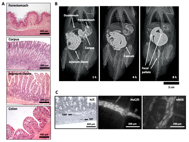
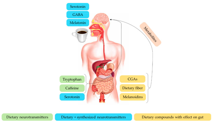
Coffee is one of the most popular beverages consumed worldwide. Roasted coffee is a complex mixture of thousands of bioactive compounds, and some of them have numerous potential health-promoting properties that have been extensively studied in the cardiovascular and central nervous systems, with relatively much less attention given to other body systems, such as the gastrointestinal tract and its particular connection with the brain, known as the brain-gut axis. This narrative review provides an overview of the effect of coffee brew; its by-products; and its components on the gastrointestinal mucosa (mainly involved in permeability, secretion, and proliferation), the neural and non-neural components of the gut wall responsible for its motor function, and the brain-gut axis. Despite in vitro, in vivo, and epidemiological studies having shown that coffee may exert multiple effects on the digestive tract, including antioxidant, anti-inflammatory, and antiproliferative effects on the mucosa, and pro-motility effects on the external muscle layers, much is still surprisingly unknown. Further studies are needed to understand the mechanisms of action of certain health-promoting properties of coffee on the gastrointestinal tract and to transfer this knowledge to the industry to develop functional foods to improve the gastrointestinal and brain-gut axis health.

Keywords: brain–gut axis; caffeine; coffee; coffee by-products; dietary fiber; enteric; gastrointestinal; melanoidins; mucosa; myenteric.


Conflict of interest statement

The authors declare no conflict of interest.

Figures

Figure 1
Figure 1
(A) Histological appearance of the wall of forestomach, corpus, jejunum-ileum (the longest part of the small intestine), and colon. (B) Organs of the rat gastrointestinal tract visualized by radiographic methods at different time points after intragastric barium administration in a conscious rat. Since rats do not vomit, barium can only progress towards the anus: 1 h after contrast, the two parts of the rat stomach (forestomach and corpus) can be distinguished, as well as the duodenum and the jejunum-ileum; 4 h after contrast, the stomach and small intestine can still be partially seen but now the cecum is filled with contrast; 8 h after contrast, the stomach and small intestine are barely seen, but the cecum is well filled with contrast and some fecal pellets are present within the colon. (C) Microscopic images showing the appearance of the enteric nervous system: left, location of the submucous (SMP) and the myenteric plexuses (MP) within the rat ileal wall in histological sections, stained with hematoxylin/eosin (H/E); middle and right, whole-mount or “sheet-like” preparations (from guinea pig ileum), obtained after dissecting away mucosa, submucosa, and circular muscle, leaving behind only the longitudinal muscle layer with the myenteric plexus attached; whole-mount preparations were processed immunohistochemically to show all the neurons with the pan-neuronal marker HuC/D (middle), or the specific subpopulation of neurons immunoreactive to neuronal nitric oxide synthase (nNOS), for which both somata and nerve fibers, but not nuclei, can be distinguished (right).
Figure 2
Figure 2
Intracellular recording from myenteric neurons. A fixed whole-mount preparation, processed immunohistochemically to show calretinin positive neurons, is used to illustrate how electrical activity of myenteric neurons would be recorded using current clamp electrophysiological modality. Calretinin immunoreactivity in guinea pig ileum whole-mount preparations allows one to distinguish the different components of the myenteric plexus: the primary component, which includes the myenteric ganglia and the internodal strands; the secondary branches that run circumferentially; and the tertiary plexus, a web of fine nerves that correspond to axons derived from excitatory longitudinal muscle motor neurons [165]. The intracellular recording electrode is represented in green (right)—this electrode allows for the recording of neuronal electrical activity, as well as direct intracellular stimulation of the cell with depolarizing or hyperpolarizing continuous or pulsed current, and marker injection to allow the impaled neuron to be visualized after immunohistochemical processing. The electrode for focal, extracellular stimulation is represented in red (left); this is placed on top of a circumferential internodal nerve strand. If the strand carries an axon (represented as a dotted line) that synapses on the impaled neuron, then focal stimuli (represented as a red blast symbol) will cause neurotransmitter release from the axon terminal and a postsynaptic potential on the impaled neuron (see Figure 3 for morphological and electrophysiological classifications of myenteric neurons).
Figure 3
Figure 3
Morphological and electrophysiologic features of myenteric neurons and effect of caffeine on AH/II neurons. By use of intracellular recording methods illustrated in Figure 2, two main classes of myenteric neurons can be distinguished. According to morphology (left), neurons are classified as Dogiel type I (top) or Dogiel type II (bottom). These neurons broadly correspond to electrophysiological types S and AH, respectively. S neurons display short sodium-dependent APs, whereas APs of AH neurons are wider and depend on the entry of both Na+ and Ca2+, displaying a “hump” during the falling phase of the AP, due to Ca2+ entry. S neurons respond to single focal electrical stimuli with fast excitatory postsynaptic potentials (f-EPSPs), which are not seen in AH neurons, although both classes may respond to trains of focal stimulation with slow excitatory postsynaptic potentials (s-EPSPs). Finally, AH neurons display a s-AHP, due to K+ efflux dependent on the increase in intracellular free Ca2+ ([Ca2+]i) released from ryanodine-dependent stores. This s-AHP is increased and prolonged by caffeine, making these neurons, which are intrinsic peripheral nerve afferents, less excitable. Abbreviations: AP, action potential; f-EPSP, fast excitatory postsynaptic potential; RP, resting potential; s-AHP, slow after hyperpolarization; s-EPSP, slow excitatory postsynaptic potentials. Light grey blocks with dotted border, artifact of the stimulus; red blast symbol, focal stimulus.
Figure 4
Figure 4
Effect of coffee compounds on the brain–gut axis. Abbreviations: CGAs, chlorogenic acids; GABA, γ-amino butyric acid.

References

    1. Loomis D., Guyton K.Z., Grosse Y., Lauby-Secretan B., El Ghissassi F., Bouvard V., Benbrahim-Tallaa L., Guha N., Mattock H., Straif K. Carcinogenicity of Drinking Coffee, Mate, and Very Hot Beverages. Lancet Oncol. 2016;17:877–878. doi: 10.1016/S1470-2045(16)30239-X. - DOI - PubMed
    1. Grosso G., Godos J., Galvano F., Giovannucci E.L. Coffee, Caffeine, and Health Outcomes: An Umbrella Review. Annu. Rev. Nutr. 2017;37:131–156. doi: 10.1146/annurev-nutr-071816-064941. - DOI - PubMed
    1. Grosso G., Stepaniak U., Polak M., Micek A., Topor R., Stefler D., Szafraniec K., Pajak A. Europe PMC Funders Group Coffee Consumption and Risk of Hypertension in the Polish Arm of the HAPIEE Cohort Study. Eur. J. Clin. Nutr. 2016;70:109–115. doi: 10.1038/ejcn.2015.119. - DOI - PMC - PubMed
    1. Liebeskind D.S., Sanossian N., Fu K.A., Wang H.-J., Arab L. The Coffee Paradox in Stroke: Increased Consumption Linked with Fewer Strokes. Nutr. Neurosci. 2016;19:406–413. doi: 10.1179/1476830515Y.0000000035. - DOI - PubMed
    1. Poole R., Kennedy O.J., Roderick P., Fallowfield J.A., Hayes P.C., Parkes J. Coffee Consumption and Health: Umbrella Review of Meta-Analyses of Multiple Health Outcomes. BMJ. 2017;359:j5024. doi: 10.1136/bmj.j5024. - DOI - PMC - PubMed