. 2021 Jan 12;118(2):e2020241118.
doi: 10.1073/pnas.2020241118.

Engineering exosome polymer hybrids by atom transfer radical polymerization

Affiliations

Engineering exosome polymer hybrids by atom transfer radical polymerization

Sushil Lathwal et al. Proc Natl Acad Sci U S A. .

Abstract

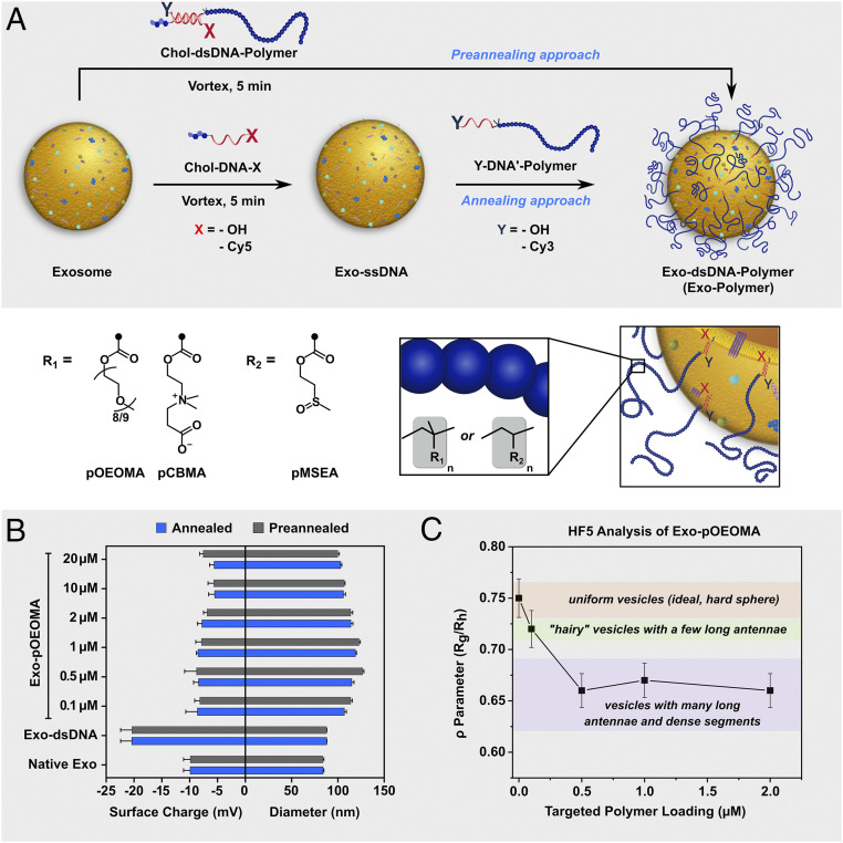
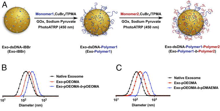
Exosomes are emerging as ideal drug delivery vehicles due to their biological origin and ability to transfer cargo between cells. However, rapid clearance of exogenous exosomes from the circulation as well as aggregation of exosomes and shedding of surface proteins during storage limit their clinical translation. Here, we demonstrate highly controlled and reversible functionalization of exosome surfaces with well-defined polymers that modulate the exosome's physiochemical and pharmacokinetic properties. Using cholesterol-modified DNA tethers and complementary DNA block copolymers, exosome surfaces were engineered with different biocompatible polymers. Additionally, polymers were directly grafted from the exosome surface using biocompatible photo-mediated atom transfer radical polymerization (ATRP). These exosome polymer hybrids (EPHs) exhibited enhanced stability under various storage conditions and in the presence of proteolytic enzymes. Tuning of the polymer length and surface loading allowed precise control over exosome surface interactions, cellular uptake, and preserved bioactivity. EPHs show fourfold higher blood circulation time without altering tissue distribution profiles. Our results highlight the potential of precise nanoengineering of exosomes toward developing advanced drug and therapeutic delivery systems using modern ATRP methods.

Keywords: ATRP; exosome; polymer; polymer biohybrid.


Conflict of interest statement

The authors declare no competing interest.

Figures

Fig. 1.
Fig. 1.
Preparation of EPHs using DNA tethers. Chol-DNA embeds into the exosome membrane to form Exo-ssDNA (single-stranded DNA) species with DNA strands orienting outward. Hybridization of complementary DNA block copolymer (DNA′-Polymer) to the DNA tethers on Ex-ssDNA species generates Exo-dsDNA-Polymer (Exo-Polymer) by the “grafting-to” strategy. Alternatively, for the “grafting-from” strategy, a complementary DNA initiator (DNA′-Initiator) functionalized with the α-bromoisobutyrate group is hybridized with the DNA tethers, followed by surface-initiated ATRP to prepare Exo-Polymer species.
Fig. 2.
Fig. 2.
”Grafting-to” strategy to prepare EPHs. (A) Schematic showing polymer functionalization of the exosome membrane by the annealing and preannealing approach. Chol-DNA-X embeds into the exosome membrane (Exo-ssDNA), and complementary Y-DNA′-polymer can be hybridized to Exo-ssDNA to prepare EPHs by the annealing approach. Alternatively, for the preannealing approach, Chol-DNA-X and Y-DNA′-Polymer can be hybridized before tethering to exosomes. (B) Plot showing size and surface charge of EPHs prepared by both the annealing and preannealing approach with varying surface loading of DNA′-pOEOMA30K (0 µM to 20 µM). Bars indicate mean ± SEM (n = 3 independent experiments). (C) Plot showing the ρ parameter (Rg/Rh) of Exo-pOEOMA species with varying surface polymer loading. Rg and Rh values were determined by the HF5 method (SI Appendix, Fig. S4). Bars indicate mean ± SD (n = 4 independent measurements).
Fig. 3.
Fig. 3.
”Grafting-from” strategy to prepare EPHs. (A) Schematic showing the grafting of polymers directly from the exosome surface by oxygen-tolerant blue light-mediated PhotoATRP. The ATRP initiator tethered to the exosome lipid membrane (Exo-dsDNA-iBBr) initiates polymer chains to prepare homopolymers (Exo-dsDNA-Polymer1), which can be subsequently chain extended to prepare block copolymers (Exo-dsDNA-Polymer1-b-Polymer2). (B) DLS plot showing size distribution of native exosomes and EPHs after grafting block copolymer with two pOEOMA blocks from the exosome surface. (C) Plot showing size distribution of native exosomes and EPHs after grafting pOEOMA from the exosome surface and further extending the polymer chains with a cationic pDMAEMA block.
Fig. 4.
Fig. 4.
Assessment of surface accessibility of EPHs. (A) Schematic showing the exosome surface protein CD63-mediated binding of Cy5-labeled Exo-pOEOMA (Exo-pOEOMA-Cy5) onto anti-CD63 beads. The binding of Exo-pOEOMA-Cy5 species was evaluated with varying MWs (Mn = 10,000, 20,000, 30,000 g/mol) and surface loadings (0 to 5 µM) of pOEOMA. (Inset) Polymer surface loadings by varying DNA′-pOEOMA concentration can influence the accessibility of CD63 protein on the Exo-pOEOMA-Cy5 surface. (BD) Graphs showing mean fluorescence intensity (MFI) of anti-CD63 beads-bound Exo-pOEOMA-Cy5 with different lengths of pOEOMA—10,000 (B), 20,000 (C), 30,000 (D)—and varying surface loadings of polymers. Accessibility of DNA tethers to nucleases is assessed by treating beads-bound Exo-pOEOMA with DNase-I for 1 h at 37 °C. The drop in the MFI postnuclease treatment highlights the degradation of DNA tethers. Bars indicate MFI ± SEM (n = 3 independent experiments). A.U. = arbitrary unit. (E) Schematic showing the binding assay of AS1411 aptamer-functionalized Exo-pOEOMA on the nucleolin protein-functionalized surface using QCM. (F) Plot showing frequency changes (ΔF) for the surface binding of Exo-pOEOMA-AS1411 species with constant loading of pOEOMA30K (1 µM) but varying AS1411 loading −1 µM (low) and 10 µM (high). Bars indicate the mean ± SEM (n = 3 independent experiments), ns, no significant difference; **P = 0.003; *P < 0.05.
Fig. 5.
Fig. 5.
Effect of polymer functionalization on the stability of exosomes. (A) EPHs can be reversibly functionalized with polymers using a photocleavable DNA tether. The p-nitrophenyl group in between the cholesterol and DNA sequence induces reversibility to EPHs. (B) Plot showing percent total column eluant radioactivity (count/minute) of different size exclusion chromatography fractions of 125I-labeled native exosomes and EPHs after incubation with trypsin at 37 °C for 1 h. Shown is the stability of exosomal surface proteins against trypsin by size exclusion chromatography. EPHs prepared using photocleavable DNA tethers (Exo-pc-pOEOMA30K and Exo-pc-pCBMA) showed no degradation of surface proteins. After irradiation of EPHs with UV light (365 nm) for 2 min, removal of polymer from exosome surface showed protein degradation post 60-min incubation at 37 °C. (C) Plot showing the change in the average diameter of native exosomes and Exo-pOEOMA30K (1 µM polymer loading) after incubation at 4 °C and 37 °C in 1x PBS buffer for 30 d. The study was repeated four times, each with samples in triplicate. ns, no significant difference; ****P < 0.0001.
Fig. 6.
Fig. 6.
Assessment of bioactivity of EPHs in vitro. (A) Schematic showing the in vitro assessment of bioactivity of EPHs and bioactive cargo-loaded EPHs. (B) Plot comparing the cell internalization efficiency of native exosomes and EPHs with different lengths of pOEOMA polymer (1 µM loading) in HEK293 cells after 6 h. (C) Plot showing the internalization efficiency of EPHs with different polymers—pOEOMA, pCBMA, pMSEA—in HEK293 cells after 6 h. To inhibit two major pathways of exosome internalization, cells were treated with heparin and methyl-β-cyclodextrin. A drop in the cellular uptake of both native exosomes and EPHs highlights a similar internalization mechanism. (D) Images showing the proangiogenesis property of MSC-derived exosomes and corresponding Exo-pOEOMA30K at 1 µM polymer loading. Similar increase in the branch points and tube length was observed in two cells lines—LECs and HUVECs. (E) Images showing the osteogenic property of BMP2-loaded exosomes and BMP2-loaded Exo-pOEOMA30K at 1 µM polymer loading. Both species promoted the expression of the ALP marker in 72 h. (F) Quantification of increase in the branch points and tube length in LECs and HUVECs by MSC-derived exosomes and Exo-pOEOMA30K species. (G) Plot showing the quantification of ALP expression by BMP2-loaded exosomes and Exo-pOEOMA30K species. (H) Plot showing the antiinflammatory effects of curcumin-loaded exosomes and curcumin-loaded Exo-pOEOMA30K at 1 µM polymer loading. Similar activity was observed for native exosomes and EPHs. All bars indicate mean ± SEM, n = 3 independent experiments. ***P < 0.001; ns, no significant difference.
Fig. 7.
Fig. 7.
In vivo pharmacokinetics and biodistribution of EPHs. (A) Plot showing the percent fluorescent intensity of ExoGlow-labeled exosomes and EPHs (Exo-pOEOMA, Exo-pCBMA, Exo-pMSEA) in mice blood at different time intervals following intravenous injection through the tail vein. The results are expressed as mean ± SEM of eight mice. (B) Plot showing the percentage accumulation of exosome and EPHs in different organs of mice after 24 h. The results are expressed as mean ± SEM of six mice. GI, gastrointestinal.

Similar articles

Cited by

References

    1. Yáñez-Mó M., et al. , Biological properties of extracellular vesicles and their physiological functions. J. Extracell. Vesicles 4, 27066 (2015). - PMC - PubMed
    1. Maas S. L. N., Breakefield X. O., Weaver A. M., Extracellular vesicles: Unique intercellular delivery vehicles. Trends Cell Biol. 27, 172–188 (2017). - PMC - PubMed
    1. Kourembanas S., Exosomes: Vehicles of intercellular signaling, biomarkers, and vectors of cell therapy. Annu. Rev. Physiol. 77, 13–27 (2015). - PubMed
    1. Hong C.-S., et al. , Circulating exosomes carrying an immunosuppressive cargo interfere with cellular immunotherapy in acute myeloid leukemia. Sci. Rep. 7, 14684 (2017). - PMC - PubMed
    1. Colombo M., Raposo G., Théry C., Biogenesis, secretion, and intercellular interactions of exosomes and other extracellular vesicles. Annu. Rev. Cell Dev. Biol. 30, 255–289 (2014). - PubMed

Publication types

LinkOut - more resources