. 2021 Jan-Jun:296:100250.
doi: 10.1074/jbc.RA120.015307. Epub 2021 Jan 9.

Cross-seeding between the functional amyloidogenic CRES and CRES3 family members and their regulation of Aβ assembly

Affiliations

Cross-seeding between the functional amyloidogenic CRES and CRES3 family members and their regulation of Aβ assembly

Hoa Quynh Do et al. J Biol Chem. 2021 Jan-Jun.

Abstract

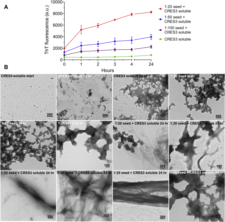
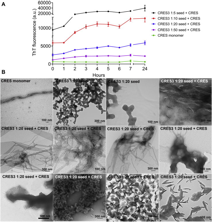
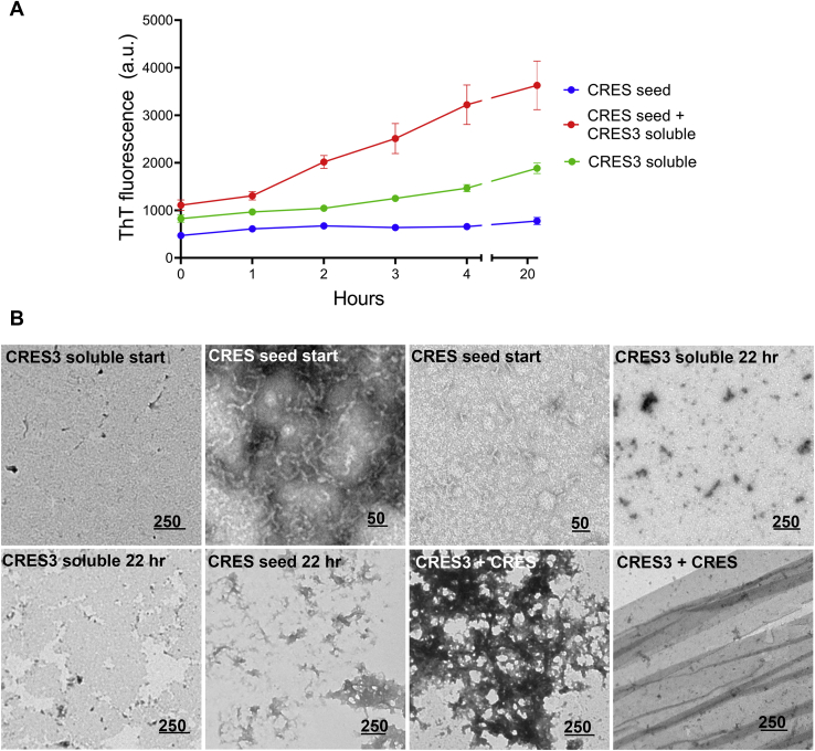
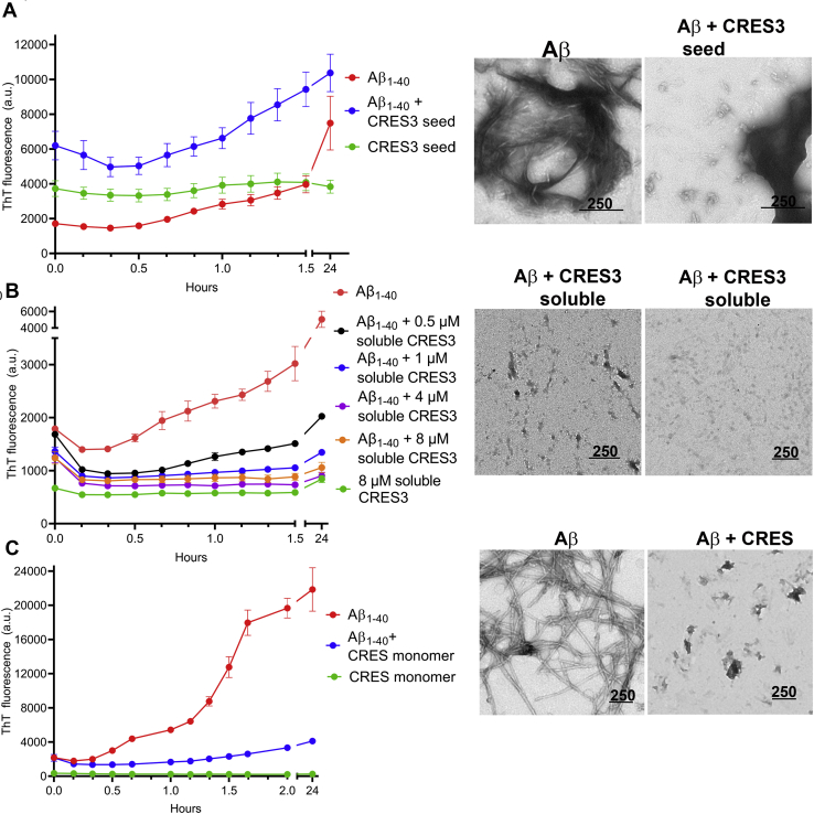
Accumulating evidence shows that amyloids perform biological roles. We previously showed that an amyloid matrix composed of four members of the CRES subgroup of reproductive family 2 cystatins is a normal component of the mouse epididymal lumen. The cellular mechanisms that control the assembly of these and other functional amyloid structures, however, remain unclear. We speculated that cross-seeding between CRES members could be a mechanism to control the assembly of the endogenous functional amyloid. Herein we used thioflavin T assays and negative stain transmission electron microscopy to explore this possibility. We show that CRES3 rapidly formed large networks of beaded chains that possessed the characteristic cross-β reflections of amyloid when examined by X-ray diffraction. The beaded amyloids accelerated the amyloidogenesis of CRES, a less amyloidogenic family member, in seeding assays during which beads transitioned into films and fibrils. Similarly, CRES seeds expedited CRES3 amyloidogenesis, although less efficiently than the CRES3 seeding of CRES. These studies suggest that CRES and CRES3 hetero-oligomerize and that CRES3 beaded amyloids may function as stable preassembled seeds. The CRES3 beaded amyloids also facilitated assembly of the unrelated amyloidogenic precursor Aβ by providing a surface for polymerization though, intriguingly, CRES3 (and CRES) monomer/early oligomer profoundly inhibited Aβ assembly. The cross-seeding between the CRES subgroup members is similar to that which occurs between bacterial curli proteins suggesting that it may be an evolutionarily conserved mechanism to control the assembly of some functional amyloids. Further, interactions between unrelated amyloidogenic precursors may also be a means to regulate functional amyloid assembly.

Keywords: Aβ; CRES subgroup; amyloid; cross-seeding; cystatin; epididymis; mouse.


Conflict of interest statement

Conflict of interest The authors declare that they have no conflicts of interest with the contents of this article.

Figures

Figure 1
Figure 1
CRES3 forms SDS-sensitive and SDS-resistant aggregates.A, Coomassie-stained SDS-PAGE of CRES3 supernatant (S) and pellet (P) (left panel) and western blot analysis of CRES3 pellet using an anti-CRES3 antibody (right panel). Following dilution into aqueous buffer and adjustment to neutral pH, CRES3 formed a white precipitate that was further separated into a soluble (supernatant) and insoluble (pellet) fraction by centrifugation. Arrows indicate CRES3 monomer and putative dimer, tetramer, and oligomers. B, dynamic light scattering of CRES3 soluble fraction showed particles >1000 nm diameter suggestive of oligomers. Data represent the mean ±SD diameter of particles from three independent CRES3 preparations. C, circular dichroism of CRES3 soluble fraction predicted a protein with mixed secondary structure containing antiparallel β-sheets. CD spectral curve shows experimental (red dotted line) and fitted data (blue solid line) from a representative CRES3 preparation. Table shows the mean ± SEM secondary structure as predicted from the spectral data by the BeStSel algorithm from n = 3 independent CRES3 preparations. D, negative stain TEM revealed a mixture of early amyloid assemblies in the CRES3 soluble/supernatant fraction while the insoluble/pellet contained only branched beaded chains. Data are representative of three independent CRES3 protein preparations. Similar branched beaded chains were detected by TEM in CD-1 mouse epididymal luminal fluid following binding and elution from the PAD ligand (epididymis). The beaded chain amyloids were not present in PAD pull-down of buffer only (data not shown). Scale bar, nm. E, X-ray diffraction of CRES3 seeds showed 4.6 and 10.6 Å reflections characteristic of amyloid.
Figure 2
Figure 2
Stability and structural changes in the CRES3 aggregates after exposure to different denaturants. CRES3 pellet fractions containing beaded chains were exposed to 0.5% SDS for 20 min, 70% formic acid (FA) for 30 min, and 90% DMSO for 45 min, and structures examined by negative stain TEM. Control, CRES3 pellet incubated in 50 mM HEPES, 100 mM NaCl, pH 7.4 for 30 min. 1.5, 2.5 years, CRES3 beaded chains after storage in 50 mM HEPES, 100 mM NaCl, pH 7.4 at 4 °C for 1.5 and 2.5 years. Scale bar, nm.
Figure 3
Figure 3
Two-dimensional solid-state NMR 13C-13C spectrum of CRES3 analyzed with the aid of chemical shifts predicted from two homology models.A, structural model for the CRES3 monomer based on the X-ray crystal structure of CRES (pdbcode:6UIO) (26). A sequence alignment of mouse CRES, CRES2, CRES3, cystatin E2, and cystatin C was used to compute a homology model with PROMALS3D (28) and Modeller (29). Disulfide bonds are indicated in orange. B, chemical shifts (indicated by x) for the homology model were predicted with SHIFTX-2, converted into 13Cα-13C side-chain peak lists using FANDAS, and overlaid onto a 2D DARR (12 ms) 13C–13C correlation spectrum of U-15N,13C-CRES3 (blue). The predicted chemical shifts for β-sheet and turn residues in the protein agree well with the DARR spectrum. However, chemical shifts predicted from the α-helix (red in [A] and red boxes in [B]) agree poorly with the experimental spectrum. C, structural model for CRES3 with only β-sheet and turn geometries allowed. 13C labeled sites for Thr and Val shown in cyan and magenta, respectively. D, predicted cross-peaks from the structure in (C) agree well with the experimental spectrum, indicating β-sheet, and not α-helical structure is largely present. This includes the canonical β-sheet chemical shift correlations for Thr 13Cα-13Cγ2 and Thr 13Cβ-13C γ2 (cyan coloring of predicted peaks) and Val 13Cα-13Cγ1,2 canonical β-sheet chemical shift correlations (magenta coloring of predicted peaks).
Figure 4
Figure 4
Self-seeding of CRES3. A, different dilutions of CRES3 seeds (1:20, 1:50, 1:100) were added to 10 μM CRES3 soluble fraction and assembly of amyloid monitored over time using a ThT plate assay. After 4 h the plates were sealed to prevent evaporation and then read again after 24 h total incubation. Data are presented as mean ± SEM of three independent experiments using three different CRES3 protein preparations. The ThT values for the individual seeding reactions are shown in Figure S3. The error bars for the CRES3 soluble fraction are too small to be detected. B, negative stain TEM of the CRES3 1:20 ThT seeding reactions after 24 h. Start, samples of the CRES3 soluble fraction and seed before incubation with ThT. Scale bar, nm.
Figure 5
Figure 5
CRES3 beaded chains template amyloid assembly in CRES C48A monomer.A, different amounts of CRES3 seeds (1:5, 1:10, 1:20, 1:50) were added to 10 μM CRES C48A monomer and assembly of amyloid monitored over time using a ThT plate assay. After 7 h the plates were sealed to prevent evaporation and then read again after 24 h total incubation. The data are presented as the mean ± SEM of three independent experiments using three different CRES3 and CRES C48A preparations. The error bars for the CRES C48A monomer are too small to be detected. The ThT values for the individual seeding reactions are shown in Figure S4. B, negative stain TEM of the ThT CRES3 1:20/CRES C48A seeding reactions after 24 h. Scale bar, nm.
Figure 6
Figure 6
CRES seeds template amyloid assembly in the CRES3 soluble fraction.A, CRES seeds (1:20) were added to 10 μM soluble CRES3 and assembly of amyloid followed with time by ThT fluorescence. After 4 h the plates were sealed to prevent evaporation and read again after 22 h total incubation. Data shown are the mean ± SEM of three independent experiments using 2–3 different CRES and CRES3 preparations. Error bars for CRES seed and CRES3 soluble fraction alone are too small to be detected. B, negative stain TEM of the CRES3 soluble and CRES seed samples alone at time 0 (start) and after 22 h and of the CRES-CRES3 seeded samples after 22 h incubation at RT. Scale bar, nm.
Figure 7
Figure 7
CRES3 and CRES regulate Aβ1-40amyloidogenesis.A, CRES3 seed (1:20) or (B) 0.5–8 μM soluble CRES3 was added to 8 μM Aβ and ThT fluorescence determined over 1.5 h. The plates were sealed and then read again after 24 h. Data shown are the mean ± SEM of 9–12 replicates from three independent experiments. The ThT values for the individual soluble CRES3/Aβ reactions are shown in Figure S5. Aliquots of the ThT reactions were examined by TEM after 24 h. Additional TEM images are shown in Figures S6 and S7. C, 8 μM CRES C48A monomer was added to 8 μM Aβ and ThT fluorescence determined over 2 h. The plates were sealed and then read again after 22 h. Data shown are the mean ± SEM of six replicates from two independent experiments. Additional TEM images are shown in Figure S8. (A–C), Scale bar, nm.

References

    1. Fowler D.M., Koulov A.V., Balch W.E., Kelly J.W. Functional amyloid - from bacteria to humans. Trends Biochem. Sci. 2007;32:217–224. - PubMed
    1. Pham C.L.L., Kwan A.H., Sunde M. Functional amyloid: Widespread in nature, diverse in purpose. Amyloids Health Dis. 2014;56:207–219. - PubMed
    1. Fowler D.M., Koulov A.V., Alory-Jost C., Marks M.S., Balch W.E., Kelly J.W. Functional amyloid formation within mammalian tissue. PLoS Biol. 2006;4:100–107. - PMC - PubMed
    1. Egge N., Muthusubramanian A., Cornwall G.A. Amyloid properties of the mouse egg zona pellucida. PLoS One. 2015;10 - PMC - PubMed
    1. Guyonnet B., Egge N., Cornwall G.A. Functional amyloids in the mouse sperm acrosome. Mol. Cell. Biol. 2014;34:2624–2634. - PMC - PubMed

Publication types

LinkOut - more resources