Review
. 2020 Dec 15:13:586169.
doi: 10.3389/fnmol.2020.586169. eCollection 2020.

Molecular Imaging of Tau Protein: New Insights and Future Directions

Affiliations
Review

Molecular Imaging of Tau Protein: New Insights and Future Directions

Rocco Pizzarelli et al. Front Mol Neurosci. .

Abstract

Tau is a microtubule-associated protein (MAPT) that is highly expressed in neurons and implicated in several cellular processes. Tau misfolding and self-aggregation give rise to proteinaceous deposits known as neuro-fibrillary tangles. Tau tangles play a key role in the genesis of a group of diseases commonly referred to as tauopathies; notably, these aggregates start to form decades before any clinical symptoms manifest. Advanced imaging methodologies have clarified important structural and functional aspects of tau and could have a role as diagnostic tools in clinical research. In the present review, recent progresses in tau imaging will be discussed. We will focus mainly on super-resolution imaging methods and the development of near-infrared fluorescent probes.

Keywords: Alzheimer’s disease; BODIPY; near-infrared fluorescent probes; neurodegeneration; super-resolution imaging; tau; tauopathies.


Conflict of interest statement

The authors declare that the research was conducted in the absence of any commercial or financial relationships that could be construed as a potential conflict of interest.

Figures

Figure 1
Figure 1
Stochastic optical reconstruction microscopy (STORM) image of beta-amyloid plaque and neurofibrillary tangles (NFT) in Alzheimer’s disease (AD) patient brain slice. (A) Confocal microscopy image of beta-amyloid aggregates (in red) and neuro-fibrillary tangles (A1) from an AD patient brain specimen. Super-imposed images are shown in panel (A2). Same senile plaque as in panel (A), imaged by combining fluorescence microscopy for beta-amyloid aggregate (B) with STORM microscopy for NFT (B1). Super-imposed images are shown in panel (B2). Note the difference in resolution between image in panels (A2) and (B2). Modified from Codron et al. (2020) under the Creative Commons CC-BY-NC-ND.
Figure 2
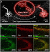
Figure 2
4,4-difluoro-4-bora3a,4a-diaza-s-indacene (BODIPY)-based probe binds to neurofibrillary tangles in a mouse model of AD. (A) Near-infrared probes (NIR) Tau 1&2 bind to NFT structures. (B) Immunostaining showing the colocalization between pTau (green signal) and Tau1 (red signal) in the CA1 (upper panels) and CA3 (lower panels) hippocampal region of a mouse treated with okadaic acid. Colocalization of the two signals is shown. Adapted with permission from Verwilst et al. (2017). Copyright 2017 American Chemical Society.

Similar articles

Cited by

References

    1. Arendt T., Stieler J. T., Holzer M. (2016). Tau and tauopathies. Brain Res. Bull. 126, 238–292. 10.1016/j.brainresbull.2016.08.018 - DOI - PubMed
    1. Barbier P., Zejneli O., Martinho M., Lasorsa A., Belle V., Smet-Nocca C., et al. . (2019). Role of tau as a microtubule-associated protein: structural and functional aspects. Front. Aging Neurosci. 11:204. 10.3389/fnagi.2019.00204 - DOI - PMC - PubMed
    1. Benda A., Aitken A., Davies D. S., Whan R., Goldsbury C. (2016). STED imaging of tau filaments in Alzheimer’s disease cortical gray matter. J. Struct. Biol. 195, 345–352. 10.1016/j.jsb.2016.07.003 - DOI - PubMed
    1. Betzig E., Patterson G. H., Sougrat R., Lindwasser O. W., Olenych S., Bonifacino J. S., et al. . (2006). Imaging intracellular fluorescent proteins at nanometer resolution. Science 313, 1642–1645. 10.1126/science.1127344 - DOI - PubMed
    1. Biswas S., Kalil K. (2018). The microtubule-associated protein tau mediates the organization of microtubules and their dynamic exploration of actin-rich lamellipodia and filopodia of cortical growth cones. J. Neurosci. 38, 291–307. 10.1523/JNEUROSCI.2281-17.2017 - DOI - PMC - PubMed

LinkOut - more resources