Review
. 2020 Dec 15:11:595635.
doi: 10.3389/fphar.2020.595635. eCollection 2020.

Cannabidiol as a Therapeutic Target: Evidence of its Neuroprotective and Neuromodulatory Function in Parkinson's Disease

Affiliations
Review

Cannabidiol as a Therapeutic Target: Evidence of its Neuroprotective and Neuromodulatory Function in Parkinson's Disease

Felipe Patricio et al. Front Pharmacol. .

Abstract

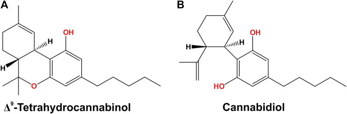
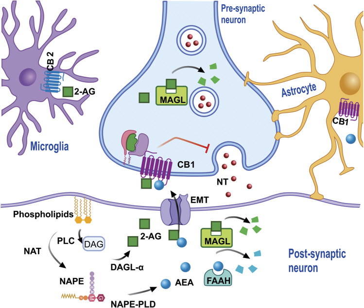
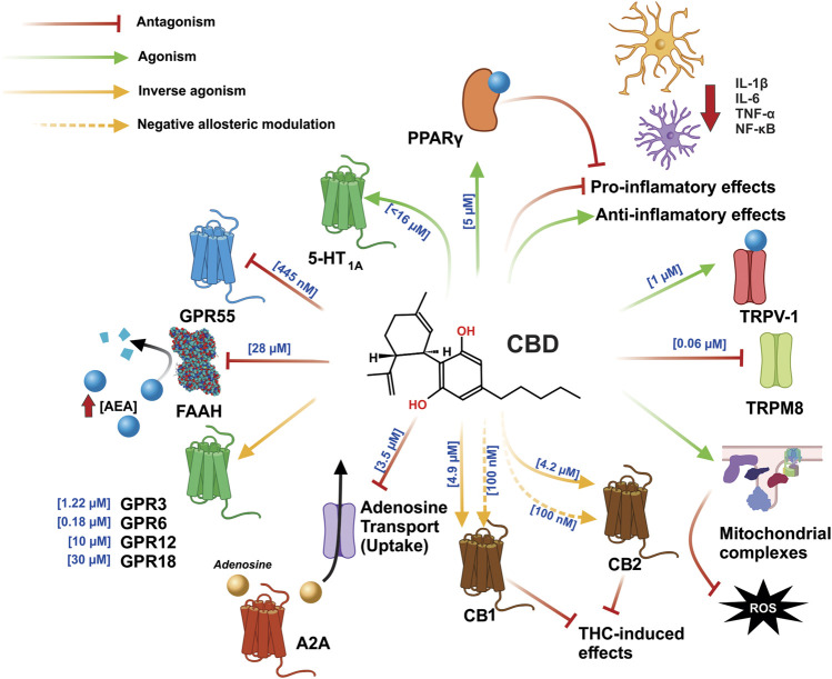
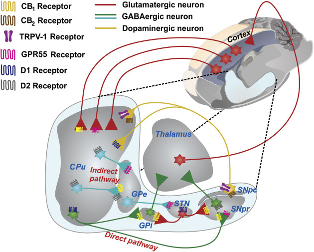
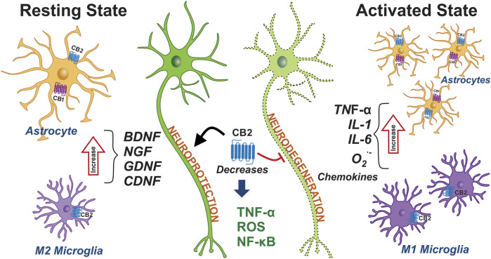
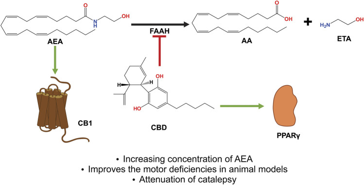
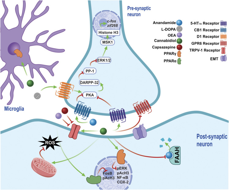
The phytocannabinoids of Cannabis sativa L. have, since ancient times, been proposed as a pharmacological alternative for treating various central nervous system (CNS) disorders. Interestingly, cannabinoid receptors (CBRs) are highly expressed in the basal ganglia (BG) circuit of both animals and humans. The BG are subcortical structures that regulate the initiation, execution, and orientation of movement. CBRs regulate dopaminergic transmission in the nigro-striatal pathway and, thus, the BG circuit also. The functioning of the BG is affected in pathologies related to movement disorders, especially those occurring in Parkinson's disease (PD), which produces motor and non-motor symptoms that involving GABAergic, glutamatergic, and dopaminergic neural networks. To date, the most effective medication for PD is levodopa (l-DOPA); however, long-term levodopa treatment causes a type of long-term dyskinesias, l-DOPA-induced dyskinesias (LIDs). With neuromodulation offering a novel treatment strategy for PD patients, research has focused on the endocannabinoid system (ECS), as it participates in the physiological neuromodulation of the BG in order to control movement. CBRs have been shown to inhibit neurotransmitter release, while endocannabinoids (eCBs) play a key role in the synaptic regulation of the BG. In the past decade, cannabidiol (CBD), a non-psychotropic phytocannabinoid, has been shown to have compensatory effects both on the ECS and as a neuromodulator and neuroprotector in models such as 6-hydroxydopamine (6-OHDA), 1-methyl-4-phenyl-1,2,3,6-tetrahydropyridine (MPTP), and reserpine, as well as other PD models. Although the CBD-induced neuroprotection observed in animal models of PD has been attributed to the activation of the CB1 receptor, recent research conducted at a molecular level has proposed that CBD is capable of activating other receptors, such as CB2 and the TRPV-1 receptor, both of which are expressed in the dopaminergic neurons of the nigro-striatal pathway. These findings open new lines of scientific inquiry into the effects of CBD at the level of neural communication. Cannabidiol activates the PPARγ, GPR55, GPR3, GPR6, GPR12, and GPR18 receptors, causing a variety of biochemical, molecular, and behavioral effects due to the broad range of receptors it activates in the CNS. Given the low number of pharmacological treatment alternatives for PD currently available, the search for molecules with the therapeutic potential to improve neuronal communication is crucial. Therefore, the investigation of CBD and the mechanisms involved in its function is required in order to ascertain whether receptor activation could be a treatment alternative for both PD and LID.

Keywords: cannabidiol (CBD); l-DOPA-induced dyskinesia; neuromodulatory; neuroprotective; parkinson’s diasese.


Conflict of interest statement

The authors declare that the research was conducted in the absence of any commercial or financial relationships that could be construed as a potential conflict of interest.

Figures

FIGURE 1
FIGURE 1
Chemical structures, (A) THC and (B) CBD, the main phytocannabinoids extracted from the Cannabis plant THC, tetrahydrocannabinol; CBD, cannabidiol.
FIGURE 2
FIGURE 2
eCB is synthesized from membrane phospholipids. NAT synthesizes the precursor NAPE, which subsequently, through the action of PLD, produces AEA in the cytoplasm of the post-synaptic neuron (or neuron spine). AEA leaves the cytoplasm and enters the synaptic space via diffusion and/or the action of EMT in order that, once it has onside, AEA activates the cannabinoid receptors which inhibit the release of NT. The degradation of AEA in EMT is regulated by FAAH, which produces metabolites such as AA and ETA. 2-AG requires the formation of the DAG precursor by PLC, which then through the action of diacylglycerol lipase α, and together with arachidonic acid generates 2-AG, which then leaves the synaptic space to activate cannabinoid receptors, which are also present in the microglia and/or astrocytes, and can be degraded by MAGL both in the pre and post-synapse, generating AA and Gro as metabolites. Abbreviations: eCB, endocannabinoids; NAT, N-acyl transferase; NAPE, N-acyl-phosphatidylethanolamine; PLD, phospholipase D; AEA, anandamide; NT, neurotransmitters; EMT, endocannabinoid membrane transporter; FAAH, fatty acid amide hydrolase; AA, arachidonic acid; ETA, ethanolamine; 2-AG, 2- Arachidonoylglycerol; DAG, diacylglycerol; PLC, phospholipase C; MAGL, monoacylglycerol lipase.
FIGURE 3
FIGURE 3
CBD exerts an agonist-like effect on the PPARγ, TRPV1, CB1, and CB2 receptors, by inhibiting the enzyme that degrades AEA and FAAH, leading to increased AEA concentration and greater interaction with said receptors. In addition, CBD inhibits GPR55 and TRPM8 and exerts an effect as an inverse antagonist on the GPR3, GPR6, GPR12, CB1, and CB2 receptors; moreover, in CB1 and CB2, it can function as a negative allosteric modulator, which is involved in blocking the effects of THC. The anti-inflammatory effects of CBD function by directly decreasing the synthesis of pro-inflammatory cytokines and increasing the synthesis of anti-inflammatory cytokines. CBD also reduces inflammation by stimulation PPARγ. Part of its antioxidant effects are achieved via the increased activity of mitochondrial complexes I, II, II-III, and IV. Abbreviations: CBD, cannabidiol; PPARγ, peroxisome proliferator activated receptor γ; TRPV1, transient potential receptor V1; CB1, cannabinoid receptor type 1; CB2, cannabinoid receptor type 2; AEA, anandamide; FAAH, fatty acid amide hydrolase; GPR55, G protein coupled receptor 55; TRPM8, transient potential receptor M8; GPR3, G protein coupled receptor 3; GPR6, G protein coupled receptor 6; GPR12, G protein coupled receptor 12; THC, tetrahydrocannabinol.
FIGURE 4
FIGURE 4
The basal ganglia network and its wide expression of receptors in presynaptic neurons. A schematic representing the GABAergic and glutamatergic connections in a sagittal section of the rat brain is shown. The CPu, the main input nucleus of the circuit, receives cortical projections of glutamatergic neurons. The expression of D1 receptors in the striatum forms the direct pathway of the BG circuit, which is projected toward the GPi and SNpr. The expression of the D2 receptors forms the indirect pathway, which is projected toward the GPe and subsequently toward the STN, which then sends projections to the GPi and SNpr. The projections that emerge from the exit nuclei direct the thalamus and return the processed information to the cerebral cortex. The expression of CB1 receptors mostly occurs in both GABAergic and glutamatergic neurons. GPR55 is present in the GPe, CPu, STN, and SNpc. CB2 and TRPV-1 are the sole in terminals of the SNpc. Abbreviations: CPu, Striatum or caudate-putamen; GPi, Internal globus pallidus; SNpr, Substantia nigra pars reticulata; SNpc, Substantia nigra pars compacta; GPe, External globus pallidus; STN, subthalamic nucleus.
FIGURE 5
FIGURE 5
Expression of cannabinoid receptors in glial cells and their role in PD. The phenotypes of microglia and astrocytes are schematically represented under non-inflammatory conditions, namely in a resting state. The microglia and astrocytes are shown in pro-inflammatory conditions and in an active state. The activation of microglia and astrocytes promotes and triggers the neuroinflammation that contributes to PD. The activation of the CB1 and CB2 receptors may decrease the inflammation seen in PD. Abbreviations: CB1, cannabinoid receptor type 1; CB2, cannabinoid receptor type 2; BDNF, brain-derived neurotrophic factor; NGF, nerve growth factor; GDNF, glial cell line-derived neurotrophic factor; CDNF, cerebral dopamine neurotrophic factor; ROS, reactive oxygen species; NF-κB, nuclear factor kappa B; IL-1, interleukin-1; IL-6, interleukin-1; O2-, Superoxide anion; TNF-α, tumor necrosis factor alpha.
FIGURE 6
FIGURE 6
CBD acts as an indirect agonist of the CB1 receptor by inhibiting the enzyme that degrades AEA and FAAH, thus increasing the concentration of said eCB, which has a greater affinity to activating CB1, thus exerting neuroprotective and antiparkinsonian effects. In addition, CBD activates the PPARγ receptors, which have been shown to be involved in neuroinflammatory processes. Abbreviations: CBD, cannabidiol; CB1, cannabinoid receptor type 1; AEA, anandamide; FAAH, fatty acid amide hydrolase; eCB, endocannabinoids; PPARγ, peroxisome proliferator activated receptor γ.
FIGURE 7
FIGURE 7
The chronic administration of l-DOPA leads to a sensitization of D1 receptors, which maintain the over-activation of PKA in LIDs. PKA regulates the pathway that activates DARPP-32, which inhibits the modification by PP-1, of ERK1/2 signaling, which acts on nuclear targets, such as MSK1, and, along with histone H3, regulates the expression of early genes such as c-fos and zif268. CBD exerts antidyskinetic effects by increasing AEA concentration by inhibiting of FAAH, thus stimulating the CB1 receptors, which decrease PKA activity. The CB1 requiere the co-administration of a TRPV1 inhibitor (CPZ), because they stimulate TRPV1 via AEA and CBD, both of which generate opposite effects to the activation of CB1. Furthermore, increased OEA is generated via the inhibition of FAAH, an endocannabinoid able to block TRPV1 and stimulate PPARσ receptors, reducing biochemical markers such as FosB and pAcH3. In addition, CBD activate the 5-HT1A receptor, a receptor that had previously only been implicated in the anticataleptic effect of CBD. By activating PPARγ receptors, CBD reduces the levels of molecular markers involved in LIDs, such as pERK, pAcH3, NF-Kβ and COX-2, while it also generates an anti-inflammatory effect by stimulating said receptors, which are present in the glia. Furthermore, CBD is able to reduce oxidative damage, decreasing the production of ROS by increasing the activity of mitochondrial complexes. The inverse agonism that CBD exerts on GPR6 could form part of its antidyskinetic mechanisms. Abbreviations: l-DOPA, L-3,4-Dihydroxyphenylalanine; D1, Dopamine receptor 1; PKA, cAMP-dependent protein kinase; LID, l-DOPA-induced dyskinesias; DARPP-32–32 KDa Phosphoprotein regulated by cAMP and dopamine; PP-1, phosphoprotein 1; ERK, extracellular signal-regulated kinase; MSK-1, mitogen and stress regulated protein kinase; CBD, cannabidiol; AEA, anandamide; FAAH, fatty acid amide hydrolase; TRPV1, transient potential receptor V1; CPZ, cpazazepine; OEA, oleoylethanolamide; PPARα, peroxisome proliferator activated receptor α; pAcH3, Histone 3 phosphoacetylation; 5HT1A, Serotonin receptor 1A; PPARγ, peroxisome proliferator activated receptor γ; NF-Kβ, nuclear factor Kβ; COX-2, cyclooxygenase 2.

References

    1. Albin R. L., Young A. B., Penney J. B. (1989). The functional anatomy of basal ganglia disorders. Trends Neurosci. 12, 366–375. 10.1016/0166-2236(89)90074-x - DOI - PubMed
    1. Alexander G. E., Crutcher M. D. (1990). Functional architecture of basal ganglia circuits: neural substrates of parallel processing. Trends Neurosci. 13 (7), 266–271. 10.1016/0166-2236(90)90107-l - DOI - PubMed
    1. Alexi T., Borlongan C. V., Faull R. L., Williams C. E., Clark R. G., Gluckman P. D., et al. (2000). Neuroprotective strategies for basal ganglia degeneration: Parkinson's and Huntington's diseases. Prog. Neurobiol. 60 (5), 409–470. 10.1016/s0301-0082(99)00032-5 - DOI - PubMed
    1. Alhouayek M., Masquelier J., Muccioli G. G. (2018). Lysophosphatidylinositols, from cell membrane constituents to GPR55 ligands. Trends Pharmacol. Sci. 39 (6), 586–604. 10.1016/j.tips.2018.02.011 - DOI - PubMed
    1. Almási R., Szoke E., Bölcskei K., Varga A., Riedl Z., Sándor Z., et al. (2008). Actions of 3-methyl-N-oleoyldopamine, 4-methyl-N-oleoyldopamine and N-oleoylethanolamide on the rat TRPV1 receptor in vitro and in vivo . Life Sci. 82 (11-12), 644–651. 10.1016/j.lfs.2007.12.022 - DOI - PubMed