

Prevalence and morphology of multiple roots, root canals and C-shaped canals in mandibular premolars from cone-beam computed tomography images in a Thai population
- PMID: 33384798
- PMCID: PMC7770317
- DOI: 10.1016/j.jds.2020.06.010
Prevalence and morphology of multiple roots, root canals and C-shaped canals in mandibular premolars from cone-beam computed tomography images in a Thai population
Abstract
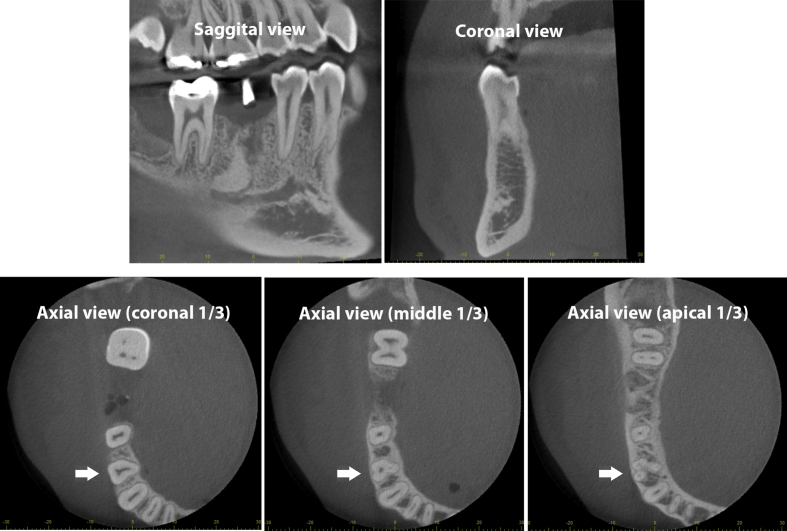
Background/purpose: Variations in root and root canal morphology of mandibular premolars of Thais has not been reported, and understanding these variations enhances endodontic success. The purpose was to investigate prevalence and morphology of multiple roots, root canals and C-shaped canals in mandibular premolars in a Thai population from cone-bean computed tomography (CBCT) images.
Materials and methods: A total of 349 first mandibular premolars and 416 second premolars from CBCT images with 0.125-mm voxel size and 60 × 60 mm field of view were evaluated. Number of roots, root canals, and C-shaped canals were recorded and statistically analyzed using chi-square test. Root canal configurations were defined according to the Vertucci's classification. Levels and distances of separated multiple canals were reported.
Results: Multiple roots in mandibular first premolars were found at 5.73% while none of second premolars had. Multiple root canals were found in the first premolars at 19.48% and the second premolars at 3.85%. C-shaped canals (C1/C2) were found in the first premolars at 3.72% and the second premolars at 0.48%. All parameters in the first premolars were significantly higher than in the second premolars (p < 0.01). The majority of multiple root canals were defined as Vertucci's type V (1-2 canals). Multiple root canals were frequently separated at the middle level of roots about 6.5-7.0 mm from the cementoenamel junction.
Conclusion: Prevalence of multiple roots/root canals and C-shaped canals in mandibular first premolars were significantly higher than in mandibular second premolars. Level of separation in multiple root canals was frequently at the mid-root level.
Keywords: C-shaped canal; Cone-beam computed tomography; Mandibular premolars; Root canal morphology; Vertucci's classification.
© 2020 Association for Dental Sciences of the Republic of China. Publishing services by Elsevier B.V.
Conflict of interest statement
The authors deny any conflicts of interest related to this study.
Figures

References
-
- Siqueira J.F. Aetiology of root canal treatment failure: why well-treated teeth can fail. Int Endod J. 2001;34:1–10. - PubMed
-
- Gorni F.G., Gagliani M.M. The outcome of endodontic retreatment: a 2-yr follow-up. J Endod. 2004;30:1–4. - PubMed
-
- Vertucci F.J. Root canal morphology and its relationship to endodontic procedures. Endod Topics. 2005;10:3–29.
-
- Cleghorn B.M., Christie W.H., Dong C.C. The root and root canal morphology of the human mandibular second premolar: a literature review. J Endod. 2007;33:1031–1037. - PubMed
LinkOut - more resources
Full Text Sources
Other Literature Sources
Miscellaneous
