

Multicolor Scanning Laser Ophthalmoscopy Strengthens Surgeons' Preoperative Decision-Making and Intraoperative Performance on Epiretinal Membrane
- PMID: 33384890
- PMCID: PMC7757626
- DOI: 10.1167/tvst.9.13.36
Multicolor Scanning Laser Ophthalmoscopy Strengthens Surgeons' Preoperative Decision-Making and Intraoperative Performance on Epiretinal Membrane
Abstract
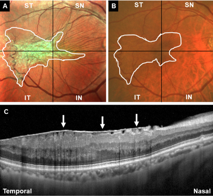
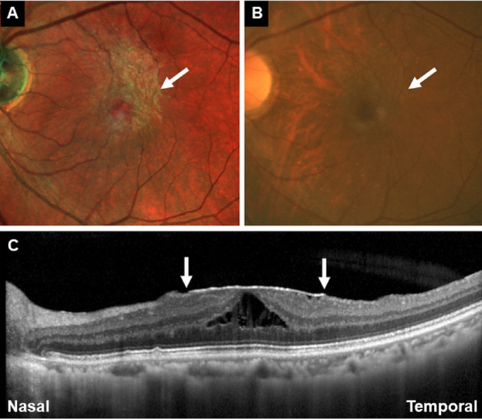
Purpose: To determine whether multicolor scanning laser ophthalmoscopy (MC-SLO) was better than color fundus photography (CFP) to enhance residents and specialists' preoperative decision-making and intraoperative performance on the epiretinal membrane (ERM).
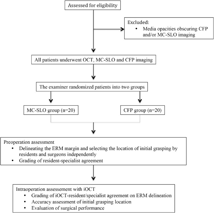
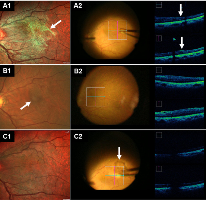
Methods: Consecutive patients with idiopathic ERM were recruited prospectively. All the patients underwent MC-SLO and CFP imagings and were randomized into MC-SLO (n = 20) and CFP (n = 20) groups. Preoperatively, residents and specialists were required to have ERM delineation and select an optimal location for initial ERM peeling independently, based on the MC-SLO (MC-SLO group) or CFP (CFP group) images. Intraoperative optical coherence tomography (iOCT) was introduced to evaluate the accuracy.
Results: Preoperatively, residents and specialists acted more effectively in ERM delineation and selection of initial grasping location in the MC-SLO group (both P < 0.001). In the MC-SLO group, higher resident-specialist agreements were achieved in ERM delineation (P = 0.002) and selection of initial grasping location (P = 0.035). The iOCT revealed greater interobserver (iOCT-resident and iOCT-specialist) agreements of ERM delineation in MC-SLO group (P < 0.001 and = 0.027, respectively). Surgeons acted more effectively on completely peeling the ERM in the MC-SLO group (P < 0.001).
Conclusions: MC-SLO provided a better visual reference for residents and specialists in ERM delineation and the selection of an initial grasping location for the surgery, compared with CFP.
Translational relevance: MC-SLO is able to help surgeons achieve better intraoperative performance and shorten the learning process for residents.
Keywords: color fundus photography; epiretinal membrane; intraoperative optical coherence tomography; multicolor scanning laser ophthalmoscopy; pars plana vitrectomy.
Copyright 2020 The Authors.
Conflict of interest statement
Disclosure: Z. Zhang, None; M. Li, None; Y. Sun, None; Y. Wei, None; S. Zhang, None
Figures
Similar articles
-
MORE EFFECTIVE SCREENING FOR EPIRETINAL MEMBRANES WITH MULTICOLOR SCANNING LASER OPHTHALMOSCOPE THAN WITH COLOR FUNDUS PHOTOGRAPHS.Retina. 2020 Jul;40(7):1412-1418. doi: 10.1097/IAE.0000000000002595. Retina. 2020. PMID: 31180985
-
Comparison of MultiColor fundus imaging and colour fundus photography in the evaluation of epiretinal membrane.Acta Ophthalmol. 2019 Jun;97(4):e533-e539. doi: 10.1111/aos.13978. Epub 2018 Nov 22. Acta Ophthalmol. 2019. PMID: 30565886
-
High-resolution imaging of the photoreceptor layer in epiretinal membrane using adaptive optics scanning laser ophthalmoscopy.Ophthalmology. 2011 May;118(5):873-81. doi: 10.1016/j.ophtha.2010.08.032. Epub 2010 Nov 12. Ophthalmology. 2011. PMID: 21074858
-
THE EFFECT OF INTERNAL LIMITING MEMBRANE PEELING ON IDIOPATHIC EPIRETINAL MEMBRANE SURGERY, WITH A REVIEW OF THE LITERATURE.Retina. 2017 May;37(5):873-880. doi: 10.1097/IAE.0000000000001263. Retina. 2017. PMID: 27617536 Review.
-
Vitrectomy with or without internal limiting membrane peeling for idiopathic epiretinal membrane: A meta-analysis.PLoS One. 2017 Jun 16;12(6):e0179105. doi: 10.1371/journal.pone.0179105. eCollection 2017. PLoS One. 2017. PMID: 28622372 Free PMC article. Review.
Cited by
-
Progress of Imaging in Diabetic Retinopathy-From the Past to the Present.Diagnostics (Basel). 2022 Jul 11;12(7):1684. doi: 10.3390/diagnostics12071684. Diagnostics (Basel). 2022. PMID: 35885588 Free PMC article. Review.
-
Commentary: Utility of multicolour imaging in identifying tractional membranes over the retina.Indian J Ophthalmol. 2022 Feb;70(2):470-471. doi: 10.4103/ijo.IJO_2485_21. Indian J Ophthalmol. 2022. PMID: 35086218 Free PMC article. No abstract available.
-
Enhancing Precision and Clarity with New Digital Color Assistant in 3D Heads-Up Vitreoretinal Surgery.Ophthalmol Ther. 2025 Apr;14(4):805-814. doi: 10.1007/s40123-025-01106-1. Epub 2025 Feb 17. Ophthalmol Ther. 2025. PMID: 39962031 Free PMC article.
-
SLOctolyzer: Fully Automatic Analysis Toolkit for Segmentation and Feature Extracting in Scanning Laser Ophthalmoscopy Images.Transl Vis Sci Technol. 2024 Nov 4;13(11):7. doi: 10.1167/tvst.13.11.7. Transl Vis Sci Technol. 2024. PMID: 39514218 Free PMC article.
-
Evaluation of macular vascular density and foveal avascular zone changes by optical coherence tomography angiography (OCT-A) after intravitreal dexamethasone implant in diabetic macular edema resistant to Anti-VEGF treatment.Int Ophthalmol. 2022 Nov;42(11):3579-3588. doi: 10.1007/s10792-022-02374-7. Epub 2022 Jun 23. Int Ophthalmol. 2022. PMID: 35737210
References
-
- Wise GN. Clinical features of idiopathic preretinal macular fibrosis. Schoenberg Lecture. Am J Ophthalmol. 1975; 79: 349–347. - PubMed
-
- Wickham L, Konstantinidis L, Wolfensberger TJ. Epiretinal membranes, vitreoretinal traction, and cystoid macular edema. In: Schachat AP, (Eds.) Ryan's Retina, 6th ed. Amsterdam: Elsevier; 2018: 2194–2212.
-
- Sandali O, El Sanharawi M, Basli E, et al. .. Epiretinal membrane recurrence: incidence, characteristics, evolution, and preventive and risk factors. Retina. 2013; 33: 2032–2038. - PubMed
Publication types
MeSH terms
LinkOut - more resources
Full Text Sources
Miscellaneous
