. 2020 Dec 18:8:e10595.
doi: 10.7717/peerj.10595. eCollection 2020.

Sesamoids in Caudata and Gymnophiona (Lissamphibia): absences and evidence

Affiliations

Sesamoids in Caudata and Gymnophiona (Lissamphibia): absences and evidence

María Laura Ponssa et al. PeerJ. .

Abstract

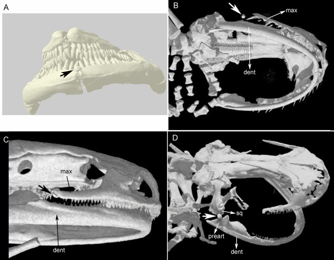
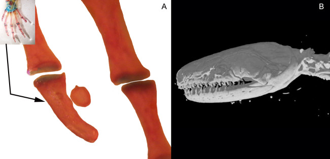
An integrative definition of sesamoid bones has been recently proposed, highlighting their relationship with tendons and ligaments, their genetic origin, the influence of epigenetic stimuli on their development, and their variable tissue composition. Sesamoid bones occur mainly associated with a large number of mobile joints in vertebrates, most commonly in the postcranium. Here, we present a survey of the distribution pattern of sesamoids in 256 taxa of Caudata and Gymnophiona and 24 taxa of temnospondyls and lepospondyls, based on dissections, high-resolution X-ray computed tomography from digital databases and literature data. These groups have a pivotal role in the interpretation of the evolution of sesamoids in Lissamphibia and tetrapods in general. Our main goals were: (1) to contribute to the knowledge of the comparative anatomy of sesamoids in Lissamphibia; (2) to assess the evolutionary history of selected sesamoids. We formally studied the evolution of the observed sesamoids by optimizing them in the most accepted phylogeny of the group. We identified only three bony or cartilaginous sesamoids in Caudata: the mandibular sesamoid, which is adjacent to the jaw articulation; one located on the mandibular symphysis; and one located in the posterior end of the maxilla. We did not observe any cartilaginous or osseous sesamoid in Gymnophiona. Mapping analyses of the sesamoid dataset of urodeles onto the phylogeny revealed that the very conspicuous sesamoid in the mandibular symphysis of Necturus beyeri and Amphiuma tridactylum is an independent acquisition of these taxa. On the contrary, the sesamoid located between the maxilla and the lower jaw is a new synapomorphy that supports the node of Hydromantes platycephalus and Karsenia coreana. The absence of a mandibular sesamoid is plesiomorphic to Caudata, whereas it is convergent in seven different families. The absence of postcranial sesamoids in salamanders might reveal a paedomorphic pattern that would be visible in their limb joints.

Keywords: Amphibians; Heterotopic elements; Homology; Mandibular sesamoid; Optimization.


Conflict of interest statement

Virginia Abdala is an Academic Editor for PeerJ.

Figures

Figure 1
Figure 1. Cranial sesamoids in salamanders.
Sesamoid in the mandibular symphysis of Necturus beyeri (UF177187), view of stl surface model of a cranial CT scan from MorphoSource, dataset DOI 10.17602/M2/M39589 (A). Skulls of: Hydromantes platycephalus (uncatalogued), image credit: Digimorph.org (B), and Karsenia koreana (DRV5033) with a sesamoid between the posterior end of the maxilla and the lower jaw joint, image credit: Digimorph.org (C); Peradactylodon persicus (MVZ241494) with a sesamoid in the quadrate-prearticular joint, image credit: Digimorph.org (D). The largest arrows show the sesamoids. dent, dentary; max, maxilla; preart, preartricular; sq, squamosal.
Figure 2
Figure 2. Cranial sesamoids in salamanders.
Mandibular sesamoid in the quadrate-prearticular joint of the skull of Siren intermedia (FMNH84082) (A); Neurergus crocatus (FMNH19629) (B); Ambystoma mexicanum (FMNH22888) under white (C) and fluorescent lighting (photo credit: Jennifer Y. Lamb) (D); Ambystoma jeffersonianum (FMNH196112) (E); Necturus maculosus (FMNH21523) (F). Scale bar = 1 mm. White arrow = indicates the ligament surrounding the sesamoid in detail of the Ambystoma mexicanum mandibular joint.
Figure 3
Figure 3. Cranial sesamoids in salamanders.
Mandibular sesamoid in the quadrate-prearticular joint of the skull of Notophthalmus viridiscens (FMNH93537) (A); Notophthalmus meridionalis (FMNH93536) under white (B) and fluorescent light (photo credit: Jennifer Y. Lamb) (C and D). Scale bar = 1 mm.
Figure 4
Figure 4. Accessory structure lateral to the terminal and subterminal phalanx of hands in Ambystoma mexicanum (FMNH22888) (A); and surrounding the skull of Ichthyophis bannanicus (MVZ236728) image credit: Digimorph.org (B).
Figure 5
Figure 5. Salamander phylogeny used in the optimization analysis of mandibular sesamoid characters follows relationships proposed by Bonnet & Blair (2017), and Pyron & Wiens (2011) for species not included in the most recent phylogeny.
Mapping of sesamoid of the mandibular symphysis is indicated in green; mapping of mandibular sesamoid, located between the quadrate and prearticular bone is indicated in red; mapping of sesamoid between the skull and the lower jaw, located at the posterior end of the maxilla is indicated in blue.
Figure 6
Figure 6. Examples of joints in salamanders and frogs showing the difference in the complexity of the joint surfaces.
(A) Manus of the facultative paedomorphic species Pleurodeles waltl (FML30803) (B) Manus of the paedomorphic species Siren intermedia (FMNH84082) (C) Manus of the anuran species Leptodactylus latinasus (FML30832), showing the glide sesamoids in the inter-phalangeal joints. (D) Hindlimb of Pleurodeles waltl (FML30803), the simple articular surfaces of the knee and ankle joints are evident. (E) Ankle and (F) Knee joints of Leptodactylus bufonius (FML30833) where the related sesamoids are visible. Scale bar = 1 mm.
Figure 7
Figure 7. Three discussed hypotheses of interrelationships of the three modern amphibian groups and their relationships to possible Paleozoic records.
(A) Polyphyly hypothesis simplified from Ruta & Coates (2007); (B) temnospondyl hypothesis (Trueb & Cloutier, 1991; Milner, 1993; Ruta, Coates & Quicke, 2003); (C) lepospondyl hypothesis (Laurin & Reisz, 1997; Laurin, 1998; Vallin & Laurin, 2004). Red bars indicate the presence of postcranial sesamoids (PS) as has been known until present.

Similar articles

Cited by

References

    1. Abdala V, Vera MC, Amador LI, Fontanarrosa G, Fratani J, Ponssa ML. Sesamoids in tetrapods: the origin of new skeletal morphologies. Biological Reviews. 2019;94(6):2011–2032. doi: 10.1111/brv.12546. - DOI - PubMed
    1. Abdala V, Vera MC, Ponssa ML. On the presence of the patella in frogs. Anatomical Record. 2017;300(10):1747–1755. doi: 10.1002/ar.23629. - DOI - PubMed
    1. Adriaens D, Verraes W. Ontogeny of the osteocranium in the African catfish, Clarias gariepinus Burchell, 1822 (Siluriformes: Clariidae): ossification sequence as a response to functional demands. Journal of Morphology. 1998;235:183–237. doi: 10.1002/(SICI)1097-4687(199803)235:3<183::AID-JMOR2>3.0.CO;2-8. - DOI - PubMed
    1. Alberch P, Alberch J. Heterochronic mechanisms of morphological diversification and evolutionary change in the neotropical salamander, Bolitoglossa occidentalis (Amphibia: Plethodontidae) Journal of Morphology. 1981;167(2):249–264. doi: 10.1002/jmor.1051670208. - DOI - PubMed
    1. Agnarsson I, Coddington JA. Quantitative tests of primary homology. Cladistics. 2008;24:51–61. doi: 10.1111/j.1096-0031.2007.00168.x. - DOI

LinkOut - more resources