

Insight Into Nanoliposomes as Smart Nanocarriers for Greening the Twenty-First Century Biomedical Settings
- PMID: 33384988
- PMCID: PMC7770187
- DOI: 10.3389/fbioe.2020.579536
Insight Into Nanoliposomes as Smart Nanocarriers for Greening the Twenty-First Century Biomedical Settings
Abstract
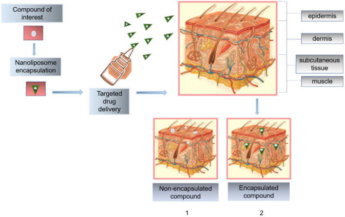
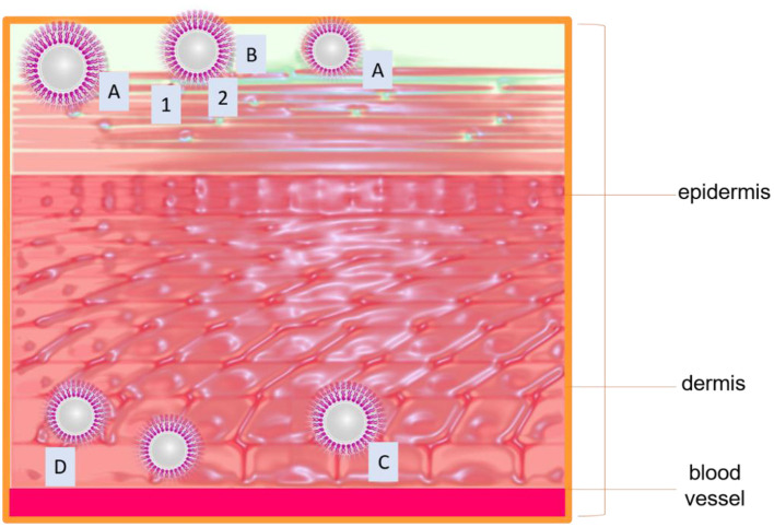
The necessity to develop more efficient, biocompatible, patient compliance, and safer treatments in biomedical settings is receiving special attention using nanotechnology as a potential platform to design new drug delivery systems (DDS). Despite the broad range of nanocarrier systems in drug delivery, lack of biocompatibility, poor penetration, low entrapment efficiency, and toxicity are significant challenges that remain to address. Such practices are even more demanding when bioactive agents are intended to be loaded on a nanocarrier system, especially for topical treatment purposes. For the aforesaid reasons, the search for more efficient nano-vesicular systems, such as nanoliposomes, with a high biocompatibility index and controlled releases has increased considerably in the past few decades. Owing to the stratum corneum layer barrier of the skin, the in-practice conventional/conformist drug delivery methods are inefficient, and the effect of the administered therapeutic cues is limited. The current advancement at the nanoscale has transformed the drug delivery sector. Nanoliposomes, as robust nanocarriers, are becoming popular for biomedical applications because of safety, patient compliance, and quick action. Herein, we reviewed state-of-the-art nanoliposomes as a smart and sophisticated drug delivery approach. Following a brief introduction, the drug delivery mechanism of nanoliposomes is discussed with suitable examples for the treatment of numerous diseases with a brief emphasis on fungal infections. The latter half of the work is focused on the applied perspective and clinical translation of nanoliposomes. Furthermore, a detailed overview of clinical applications and future perspectives has been included in this review.
Keywords: antifungal; biomedical applications; drug-loaded constructs; fabrication strategies; influencing factors; nanocarriers; nanoliposomes; targeted drug delivery.
Copyright © 2020 Aguilar-Pérez, Avilés-Castrillo, Medina, Parra-Saldivar and Iqbal.
Conflict of interest statement
The authors declare that the research was conducted in the absence of any commercial or financial relationships that could be construed as a potential conflict of interest.
Figures
References
-
- Arshad M., Pradhan R. A., Zubair M., Ullah A. (2020). Lipid-derived renewable amphiphilic nanocarriers for drug delivery, biopolymer-based formulations: biomedical and food applications,” in Biopolymer-Based Formulations (Cambridge, MA: Elsevier; ), 283–310. 10.1016/B978-0-12-816897-4.00013-8 - DOI
Publication types
LinkOut - more resources
Full Text Sources
Miscellaneous