. 2021 Feb 10;29(2):210-221.e6.
doi: 10.1016/j.chom.2020.12.002. Epub 2020 Dec 31.

Murine model of colonization with fungal pathogen Candida auris to explore skin tropism, host risk factors and therapeutic strategies

Affiliations

Murine model of colonization with fungal pathogen Candida auris to explore skin tropism, host risk factors and therapeutic strategies

Xin Huang et al. Cell Host Microbe. .

Abstract

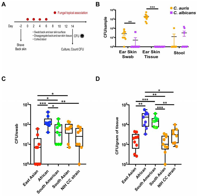
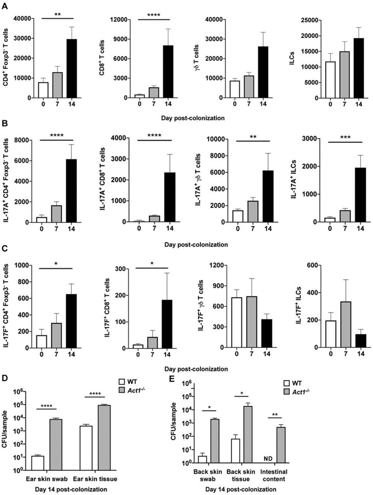
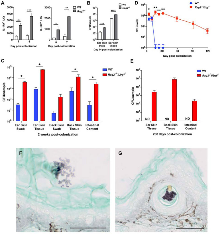
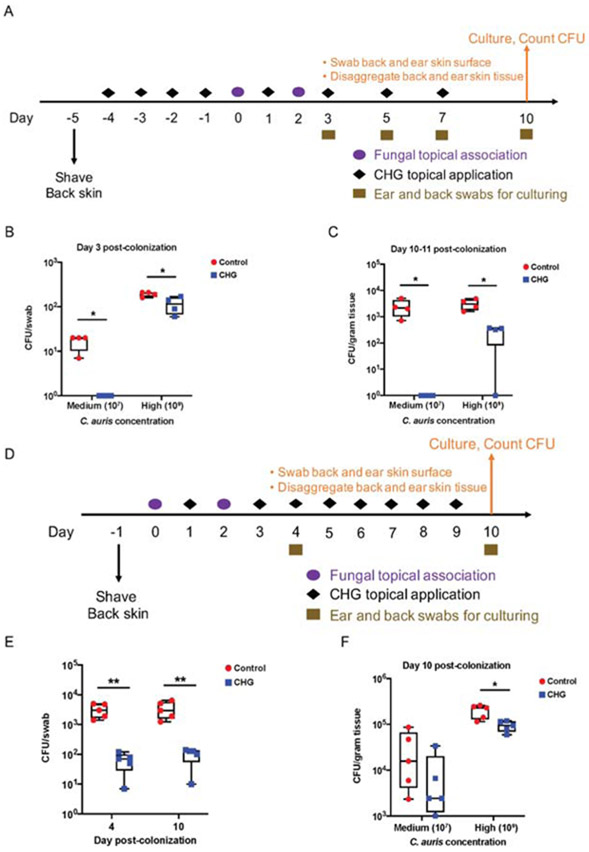
Candida auris is an emerging multi-drug-resistant human fungal pathogen. C. auris skin colonization results in environmental shedding, which underlies hospital transmissions, and predisposes patients to subsequent infections. We developed a murine skin topical exposure model for C. auris to dissect risk factors for colonization and to test interventions that might protect patients. We demonstrate that C. auris establishes long-term residence within the skin tissue compartment, which would elude clinical surveillance. The four clades of C. auris, with geographically distinct origins, differ in their abilities to colonize murine skin, mirroring epidemiologic findings. The IL-17 receptor signaling and specific arms of immunity protect mice from long-term C. auris skin colonization. We further determine that commonly used chlorhexidine antiseptic serves as a protective and decolonizing agent against C. auris. This translational model facilitates an integrated approach to develop strategies to combat the unfolding global outbreaks of C. auris and other skin-associated microbial pathogens.

Keywords: Candida auris; adaptive and innate immunity; epidemiology; fungal pathogen; skin colonization; translational mouse model.


Conflict of interest statement

Declaration of interests The authors declare no competing interests.

Figures

Figure 1.
Figure 1.. Candida auris colonizes skin surface and tissue compartment with clade specificity.
(A) Schematic for experimental design. Fungal cells were topically associated on ear pinna and dorsal back skin every other day four times. Skin swabs, skin tissue and stool were obtained on day 14 and processed for C. auris culturing. (B) C. auris colonizes skin surface and tissue but not the gut compartment. Colony forming units (CFUs) per swab, gram of tissue or gram of stool from wild-type (WT) mice colonized with C. auris (orange) and C. albicans (purple) are plotted. Shown are mean values +/− SEM. Statistics were calculated using the Mann-Whitney U test. N=9 mice for each group, two independent experiments. See also Figure S1A and Table S1. (C and D) C. auris colonization of WT mice with each of the four clades or the NIH Clinical Center strain (South Asian clade) was measured by culturing skin swabs (C) and disaggregated skin tissue (D). Statistics were calculated using one-way ANOVA with Tukey’s post-hoc tests or Games-Howell tests, as appropriate. N=8-9 mice for each group, two independent experiments. See also Figure S2. *: p<0.05, **: p<0.01, ***: p<0.001.
Figure 2.
Figure 2.. C. auris elicits a protective IL-17A/IL-17F response derived from innate and adaptive lymphoid cells.
(A) Accumulation of CD4+ T cells, CD8+ T cells, γδ T cells, and innate lymphoid cells (ILCs) in the skin of WT mice 7 and 14 days after C. auris topical association as compared with naïve WT mice. (B-C) Differences in IL-17A-producing (A) and IL-17F-producing (B) CD4+ T cells, CD8+ T cells, γδ T cells, and ILCs are shown for naïve WT mice, and WT mice 7 and 14 days after topical association with C. auris. (A-C) Total number of cells are shown for each cell type. Statistics were calculated using one-way ANOVA with Tukey’s HSD post-hoc test or Kruskal-Wallis test with Dunn’s multiple comparison test, as appropriate. N=6-9 mice for each group; two independent experiments. Data are shown with mean ± SEM. See also Figures S3, S4A-C and S5. (D) Mice deficient in Act1, an essential adaptor of IL-17R family signaling, are defective in clearing C. auris from the ear skin surface (CFU/swab) and ear skin tissue (CFU/gram of tissue) relative to WT mice, measured 14 days after C. auris topical association. Statistics were calculated using the Mann-Whitney U test. N=10 mice for each group; two independent experiments. Data are shown with mean ± SEM. (E) Compared with WT, mice deficient in Act1 have increased C. auris on the back skin surface (CFU/swab), back skin tissue (CFU/gram of tissue) and intestinal compartments (CFU/gram of intestinal content) on day 14 after topical C. auris association. Statistics were calculated using the Mann-Whitney U test. N=4-5 mice for each group. Data are shown with mean ± SEM. *: p<0.05, **: p<0.01, ***: p<0.001, ****: p<0.0001, ND: not detected.
Figure 3.
Figure 3.. Immunodeficient Rag2−/− and Rag2−/−Il2rg−/− mice exhibit greater propensity for C. auris skin colonization.
(A) IL-17A and IL-17F production by ILCs is increased in the skin of Rag2−/− mice. Shown are numbers of IL-17A-producing (left panel) and IL-17F-producing (right panel) ILCs in the skin of WT and Rag2-deficient mice at steady state and 7 days after C. auris colonization. The statistics were calculated using the t-test or Mann-Whitney U test, as appropriate. N=6-9 mice for each group; two independent experiments. Data are mean ± SEM. (B) Rag2−/− mice are defective in controlling C. auris colonization on the ear skin surface (CFU/swab) and within the tissue compartment (CFU/gram of tissue). The statistics were calculated using the t-test or Mann-Whitney U test, as appropriate. N=9-10 mice for each group; two independent experiments. (C) Immunodeficient Rag2−/−Il2rg−/− mice are defective in clearing C. auris from the ear and back skin surface (CFU/swab) and tissue (CFU/gram of tissue); and intestinal compartments (CFU/gram of intestinal content) as determined by culturing from WT and Rag2−/−Il2rg−/− mice two weeks after topical colonization. The statistics were calculated using the Mann-Whitney U test. N=5 mice for each group, representative of two independent experiments. Data are shown with mean ± SEM. See also Figure S4G-H. (D) Longitudinally, WT and Rag2−/−Il2rg−/− mouse skin swabs were cultured for C. auris on days 4, 10, 17, 24, 32, and extended through days 59, 91 and 119 for Rag2−/−Il2rg−/− mice. The Mann-Whitney U test was used to compare between WT and Rag2−/−Il2rg−/− mice at each time point up to day 32. N=6 mice for each group, representative of three independent experiments. Data are shown with mean ± SEM. (E) Immunodeficient Rag2−/−Il2rg−/− mice are defective in clearing C. auris from the ear and back skin tissue (CFU/gram of tissue); and intestinal compartments (CFU/gram of intestinal content) as measured by culturing from WT and Rag2−/−Il2rg−/− mice 208 days after topical association. No C. auris colonies were cultured from WT mice on day 208. N=6 mice for each group, representative of three independent experiments. Data are shown with mean ± SEM. ND: not detected. (F and G) Grocott-Gomori's methenamine silver stain for fungi from ear pinna sections of a Rag2−/−Il2rg−/− mouse colonized by C. auris 7 days after topical association. Clusters of C. auris cells (purple) can be seen on the skin surface (stratum corneum, F) and located inside a hair follicle (G). 50 μm scale bar is displayed at the bottom right. The brown stain is endogenous mouse skin pigment also observed on control uncolonized mice. See also Figure S1A. *: p<0.05, **: p<0.01, ***: p<0.001, ****: p<0.0001.
Figure 4.
Figure 4.. Skin antiseptic chlorhexidine protects WT mice from C. auris colonization and promotes C. auris decolonization of Rag2−/−Il2rg−/− mice.
(A) Schematic for experimental design to model chlorhexidine gluconate (CHG) skin bathing. Wipes impregnated with CHG were used daily for four days to ‘bathe’ the mouse skin before topical association. Cotton gauze saturated with sterile PBS was used as procedural control. C. auris cells were topically associated onto mouse ear pinna and dorsal back skin at two concentrations: 107 CFU as medium concentration, and 109 CFU as high concentration. After topical association, CHG was applied every other day for the duration of the experiment. Skin swabs were taken during the experiment and skin tissues were disaggregated at the end of the experiment and processed for C. auris culturing. (B) CHG bathing protects WT mouse skin from C. auris topical exposure (medium and high concentrations) as measured by culturing skin swabs. N=4 mice for each group, representative of two independent experiments. (C) CHG bathing protects WT mouse skin tissue from C. auris residence after topical exposure (medium and high concentrations). N=4 mice for each group, representative of two independent experiments. (D) Schematic for experimental design to model CHG skin bathing to promote C. auris decolonization. After C. auris (medium and high concentrations) topical association, daily bathing with CHG was deployed for one week. Skin swabs were taken during the experiment and skin tissues were disaggregated at the end of the experiment for culturing. (E) CHG bathing promotes C. auris decolonization on Rag2−/−Il2rg−/− ear skin swabs 4 and 10 days after high concentration topical exposure. The statistics were calculated using the t-test. N=5 mice for each group, representative of two independent experiments. (F) CHG bathing promotes C. auris decolonization in Rag2−/−Il2rg−/− resident skin tissue after medium and high concentration topical exposure. N=5 mice for each group, representative of two independent experiments. See also Table S3. (B,C,E,F) The statistics were calculated using the t-test. *: p<0.05, **: p<0.01.

Comment in

  • Exploring Candida auris in its habitat.
    Zhai B, Rolling T, Hohl TM. Zhai B, et al. Cell Host Microbe. 2021 Feb 10;29(2):150-151. doi: 10.1016/j.chom.2021.01.010. Cell Host Microbe. 2021. PMID: 33571440 Free PMC article.

References

    1. Adams E, Quinn M, Tsay S, Poirot E, Chaturvedi S, Southwick K, Greenko J, Fernandez R, Kallen A, Vallabhaneni S, et al. (2018). Candida auris in Healthcare Facilities, New York, USA, 2013-2017. Emerg Infect Dis. 24(10), 1816–1824. - PMC - PubMed
    1. Ben-Ami R, Berman J, Novikov A, Bash E, Shachor-Meyouhas Y, Zakin S, Maor Y, Tarabia J, Schechner V, Adler A, et al. (2017). Multidrug-Resistant Candida haemulonii and C. auris, Tel Aviv, Israel. Emerg Infect Dis. 23(1). - PMC - PubMed
    1. Bishu S, Hernández-Santos N, Simpson-Abelson MR, Huppler AR, Conti HR, Ghilardi N, Mamo AJ and Gaffen SL (2014). The adaptor CARD9 is required for adaptive but not innate immunity to oral mucosal Candida albicans infections. Infect. Immun. 82(3), 1173–1180. - PMC - PubMed
    1. Boisson B, Wang C, Pedergnana V, Wu L, Cypowyj S, Rybojad M, Belkadi A, Picard C, Abel L, Fieschi C, et al. (2013). An ACT1 mutation selectively abolishes interleukin-17 responses in humans with chronic mucocutaneous candidiasis. Immunity. 39(4), 676–686. - PMC - PubMed
    1. Cassir N, Papazian L, Fournier PE, Raoult D, and La Scola B (2015). Insights into bacterial colonization of intensive care patients' skin: the effect of chlorhexidine daily bathing. Eur J Clin Microbiol. 34(5), 999–1004. - PubMed

Publication types

MeSH terms