. 2021 Feb 10:455:165-176.
doi: 10.1016/j.neuroscience.2020.12.014. Epub 2020 Dec 29.

Chronic Ethanol Exposure Potentiates Cholinergic Neurotransmission in the Basolateral Amygdala

Affiliations

Chronic Ethanol Exposure Potentiates Cholinergic Neurotransmission in the Basolateral Amygdala

Sarah E Sizer et al. Neuroscience. .

Abstract

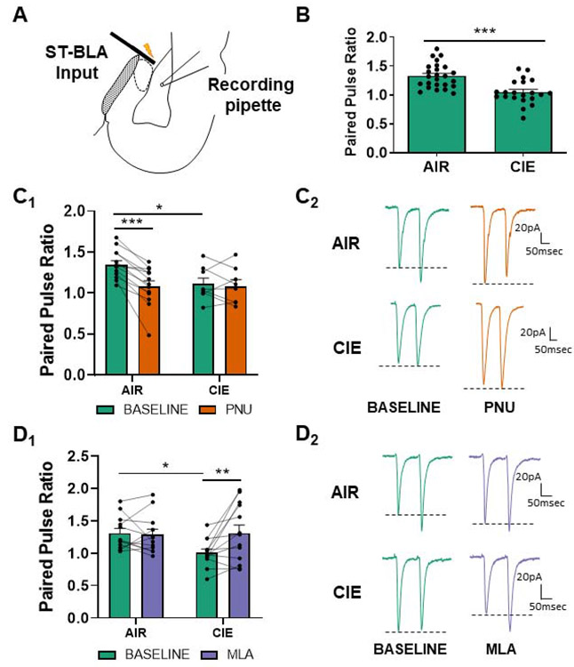
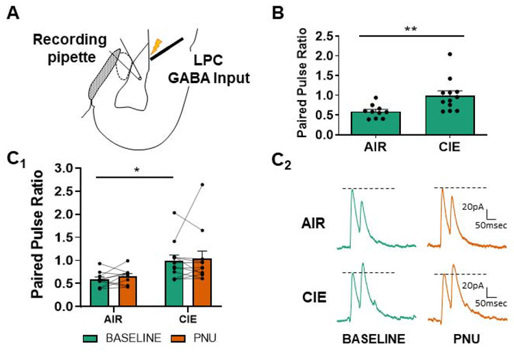
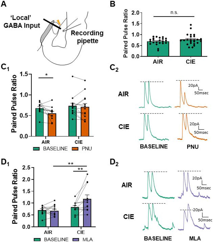
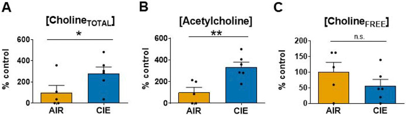
Chronic intermittent ethanol (CIE) exposure dysregulates glutamatergic and GABAergic neurotransmission, facilitating basolateral amygdala (BLA) pyramidal neuron hyperexcitability and the expression of anxiety during withdrawal. It is unknown whether ethanol-induced alterations in nucleus basalis magnocellularis (NBM) cholinergic projections to the BLA mediate anxiety-related behaviors through direct modulation of GABA and glutamate afferents. Following 10 days of CIE exposure and 24 h of withdrawal, we recorded GABAergic and glutamatergic synaptic responses in BLA pyramidal neurons with electrophysiology, assessed total protein expression of cholinergic markers, and quantified acetylcholine and choline concentrations using a colorimetric assay. We measured α7 nicotinic acetylcholine receptor (nAChR) dependent modulation of presynaptic function at distinct inputs in AIR- and CIE-exposed BLA coronal slices as a functional read-out of cholinergic neurotransmission. CIE/withdrawal upregulates the endogenous activity of α7 nAChRs, facilitating release at both GABAergic' local' interneuron and glutamatergic synaptic responses to stria terminalis (ST) stimulation, with no effect at GABAergic lateral paracapsular cells (LPCs). CIE caused a three-fold increase in BLA acetylcholine concentration, with no changes in α7 nAChR or cholinergic marker expression. These data illustrate that α7 nAChR-dependent changes in presynaptic function serve as a proxy for CIE-dependent alterations in synaptic acetylcholine levels. Thus, cholinergic projections appear to mediate CIE-induced alterations at GABA/glutamate inputs.

Keywords: acetylcholine; basolateral amygdala; patch-clamp electrophysiology; presynaptic; withdrawal.


Conflict of interest statement

Conflict of Interest: Authors report no conflict of interest

Figures

Figure 1.
Figure 1.
α7 nAChR activity is upregulated and facilitates the upregulation of glutamate release at stria terminalis (ST) inputs during withdrawal. (A) Schematic showing the typical placement of the stimulating electrode at ST inputs and patch electrode in the basolateral nucleus for electrophysiology recordings. (B) 10 days of CIE and 24 hours of withdrawal (N=21) decreases the paired pulse ratio (see Methods) at ST glutamatergic inputs in comparison to AIR-exposed controls (N=25), as previously reported (t=4.482, p<0.0001; see (Morales et al., 2015)). (C) PNU-282987 (designated PNU in graphs) modulates PPR in AIR-exposed (N=13), but not CIE-exposed BLA neurons (N=8). Bars (C1) represent mean ± SEM of PPRs during baseline and bath application of PNU-282987 (0.5μM). Two-way repeated-measures ANOVA; significant interaction and the main effect of PNU-282987, p<0.05. Bonferroni post-tests confirmed a significant difference between AIR and CIE release probabilities at baseline (t=2.507, * - p<0.05) and following PNU-282987 application in AIR animals (t=6.023, *** - p<0.001). Representative traces (C2) of glutamatergic paired-pulse responses from AIR or CIEneurons at baseline and following drug application. Dashed lines illustrate the drug effect on the second peak amplitude (P2) relative to the first peak amplitude (P1). (D) The α7 nAChR competitive antagonist MLA (10nM) reverses withdrawal-induced decreases in PPR. Two-way repeated-measures ANOVA reveals a main effect of MLA and a significant interaction between exposure and drug (p<0.05). MLA significantly increased PPR in CIE neurons (N=13, D1; Bonferroni post-test, t=3.453, p<0.01) but not in AIR neurons (N=12, p>0.05). Representative traces (D2) of paired-pulse responses from AIR or CIE neurons at baseline and following MLA perfusion.
Figure 2.
Figure 2.
α7 nAChR activity does not modulate LPC GABAergic release probability. (A) Schematic shows the placement of the stimulating electrode at LPC GABA inputs and patch electrode in the basolateral nucleus for electrophysiology recordings. (B) CIE/withdrawal (N=12) increased the paired pulse ratio (decreased release probability; see Methods) at LPC interneurons relative to AIR-exposed controls (N=10) as previously reported (t-test, t=3.098, ** - p<0.01 see (Diaz et al., 2011)). (C) PNU-282987 (labeled PNU in graphs; 0.5μM) does not alter the release probability in either AIR-exposed or CIE neurons. Bars (C1) represent mean ± SEM of PPRs during baseline and following bath application of PNU-282987 (two-way ANOVA with Bonferroni post-test, p>0.05 for all). Representative traces (C2) of LPC GABAergic paired-pulse responses from AIR or CIE neurons at baseline and following PNU-282987. Dashed lines as in Figure 1.
Figure 3.
Figure 3.
α7 nAChRs are tonically active at ‘local’ GABAergic synapses during withdrawal. (A) Schematic showing the typical placement of the stimulating electrode at ‘local’ GABAergic interneurons and patch electrode in the basolateral nucleus for electrophysiology recordings. (B) 10 days of CIE and 24 hours of withdrawal (N=24) does not alter the baseline release probability at local interneurons in comparison to AIR-exposed controls, “n.s.” = not significant (N=22; t-test, t=1.328 , p>0.05) as previously reported (Diaz et al., 2011). (C) PNU-282987 (designated PNU in graphs) decreases the PPR (increases release probability) at ‘local’ GABAergic synapses recorded from AIR-exposed control BLA neurons (N=11) with no effect in CIE neurons (N=13; two-way repeated-measures ANOVA, p<0.05). Bars (C1) represent mean ± SEM of PPRs during baseline and following bath application of PNU-282987 (0.5μM). Representative traces (C2) of ‘local’ GABAergic paired-pulse responses from AIR or CIE neurons at baseline and following drug application. (D) The α7 nAChR antagonist MLA (10nM) enhances PPR (decreases release) at ‘local’ GABA synapses following CIE but does not affect AIR-treated control neurons. Average ± SEM (D1) PPRs during baseline and in the presence of α7 nAChR competitive antagonist MLA. Two-way repeated-measures ANOVA reveals a significant interaction between exposure and drug (p<0.05). PPR significantly increased in CIE neurons (N=11) following application of MLA relative to both CIE baseline (**- p<0.01, Bonferroni post-test) and MLA-treated AIR controls (N=11, ** - p<0.01). There was no effect of MLA treatment in AIR-exposed neurons (p>0.05). Representative traces (D2) of paired-pulse responses from AIR or CIE neurons at baseline and following MLA perfusion.
Figure 4.
Figure 4.
10 day CIE/24 hr WD does not change the protein expression of cholinergic 'markers' within the BLA. Western blot analysis reveals no significant differences in protein expression of (A) α7 nAChR (~56kD; AIR - 100.0±5.5%, N=7, CIE - 92.9±5.7%, N=7; t-test, t=0.9009, p>0.05), (B) vesicular acetylcholine transporter (vAChT, ~56kD; AIR - 100.0±14.2%, N=8, CIE – 108.8±22.9%, N=7; t=0.3446, p>0.05), (C) choline acetyltransferase (ChAT, ~72kD; AIR - 100.0±10.2%, N=8, CIE - 94.3±6.0%, N=8; t=0.4623, p>0.05), and (D) acetylcholinesterase (AChE, ~68kD; AIR - 100.0±7.8%, N=8, CIE - 96.9±4.1%, N=8; t=0.5561, p>0.05). "n.s." = not significant. Band volumes were normalized to total lane protein and expressed as percent of the group mean from the AIR-exposed controls (see Methods). Each analysis includes a representative blot. Approximate position of molecular weight markers on each gel is shown to the left of the blots. * - indicates band analyzed for the statistical comparisons.
Figure 5.
Figure 5.
10 day CIE/24 hr WD significantly increases cholineTOTAL and acetylcholine concentration in the BLA. (A) CholineTOTAL levels (acetylcholine + cholineFREE; see Methods) were significantly higher in CIE homogenates (AIR - 100.0±67.2%, N=5, CIE - 279.8±60.9%, N=6; unpaired t-test, t=1.983, * - p<0.05). (B) Acetylcholine levels were also significantly higher in CIE BLA total homogenates (AIR - 100.0±46.2%, N=5; CIE - 334.6±45.6%, N=6; t-test, t=3.578, ** - p<0.01). (C) CholineFREE levels did not significantly differ between AIR and CIE BLA total homogenates, "n.s." = not significant (AIR - 100.0±31.3%, N=5; CIE - 56.3±20.4%, N=6; t-test, t=1.209, p>0.05). Substrate concentrations were standardized to homogenate total protein, followed by Min.–Max. normalization of raw absorbance values. Values are represent a percentage of the group mean from AIR-exposed controls.

References

    1. Aitta-Aho T, Hay YA, Phillips BU, Saksida LM, Bussey TJ, Paulsen O, Apergis-Schoute J (2018). Basal forebrain and brainstem cholinergic neurons differentially impact amygdala circuits and learning-related behavior. Curr Biol, 28, 2557–2569.e4. - PubMed
    1. Alkondon M, Pereira EF, Wonnacott S, Albuquerque EX (1992). Blockade of nicotinic currents in hippocampal neurons defines methyllycaconitine as a potent and specific receptor antagonist. Mol Pharmacol, 41, 802–8. - PubMed
    1. Andreasen M, Hablitz JJ (1994). Paired-pulse facilitation in the dentate gyrus: A patch-clamp study in rat hippocampus in vitro. J Neurophysiol, 72, 326–36. - PubMed
    1. Arendt T, Allen Y, Marchbanks RM, Schugens MM, Sinden J, Lantos PL, Gray JA (1989). Cholinergic system and memory in the rat: Effects of chronic ethanol, embryonic basal forebrain brain transplants and excitotoxic lesions of cholinergic basal forebrain projection system. Neuroscience, 33, 435–62. - PubMed
    1. Arvidsson U, Riedl M, Elde R, Meister B (1997). Vesicular acetylcholine transporter (vacht) protein: A novel and unique marker for cholinergic neurons in the central and peripheral nervous systems. J Comp Neurol, 378, 454–67. - PubMed

Publication types

LinkOut - more resources