Review
. 2021 Apr:254:110879.
doi: 10.1016/j.cbpa.2020.110879. Epub 2020 Dec 29.

Phylogeny and ontogeny of the renin-angiotensin system: Current view and perspectives

Affiliations
Review

Phylogeny and ontogeny of the renin-angiotensin system: Current view and perspectives

Hiroko Nishimura et al. Comp Biochem Physiol A Mol Integr Physiol. 2021 Apr.

Abstract

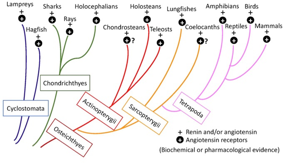
The renin-angiotensin system (RAS) evolved early among vertebrates and remains functioning throughout the vertebrate phylogeny and has adapted to various environments. The RAS is crucial for the regulation of blood pressure, fluid-electrolyte balance and tissue homeostasis. The RAS is also expressed during early ontogeny in renal and extra-renal tissues, and exerts unique vascular growth and differentiation functions. In this brief review, we describe advances from molecular-genetic and whole animal approaches and discuss similarities and unique aspects of the RAS in the context of embryonic development and vertebrates' phylogeny.


Conflict of interest statement

Conflict of Interest

The authors declare that no conflict of interest exists.

Figures

Fig. 1.
Fig. 1.
Diagrammatic presentation of vertebrate phylogenetic tree and presence of biochemical activities of renin and/or angiotensin (+) and molecular, biochemical, or pharmacological evidence for the presence of angiotensin receptors (+ in closed circle). A question mark (?) indicates that the presence of an angiotensin receptor is presumed but not confirmed. (Modified from Nishimura 2017)
Fig. 2:
Fig. 2:
A. Diagrams of nephrons and renin expressing juxtaglomerular granules (JGG) of various non-mammalian vertebrates. JGG are scattered along the afferent arterioles and the small arteries. Distal tubules attach to their parent glomeruli in some species, but no macula densa is recognized except in birds in which a part of characteristics of macula densa cells are seen. (Modified from Nishimura 2017) B. JG apparatus from fetal (late stage) and adult rodents. Adult JG apparatus (JGA) consists of glomerular tuft, intraglomerular and extraglomerular mesangium (navy), afferent and efferent arterioles, and JG cells (green). A part of distal tubule cells at the JGA become taller and narrower, forming a macula densa. Sympathetic nerve fibers (green) are densely innervated on the JG cells of the afferent arteriole. Renin containing- and renin gene expressing cells are located in the afferent arteriole adjacent to the JGA in the adult, but in the fetal kidney, renin-expressing cells extend along and beyond the afferent arterioles (Gomez et al., 1989). C. The table summarizes the components of the JGA structures found in various vertebrate species. Data are from Sokabe et al. 1969; Nishimura et a., 1973; Harris and Gomez 1997.
Fig. 3.
Fig. 3.
A. Renin mRNA measured by quantitative polymerase chain reaction (qPCR) in the kidney (solid column), liver (gray column), and adrenal (open column) of embryonic day 13 (E13) embryo (n = 3) and newborn day 4 (D4) chicks (n = 3). Expression was standardized to the value of D30 kidney (Hoy et al., 2020). Note that the scale of the ordinate varies. Data are expressed as means ± SD. *P < 0.05, **P < 0.01. B. Schematic illustration of renin expressing cells distribution in the mouse during development. In early embryos, renin cells are seen in the large fetal zone of the adrenal gland, as well as in the developing arteries and few tubular cells in the kidney. In the late stage of embryo and new born mouse, renin cells are distributed throughout the arterial trees, intra- and extraglomerular mesangium, and interstitial pericytes. In adult, renin cells are localized at the juxtaglomerular area of the afferent arteriole (Gomez and Sequeira-Lopez, 2018).
Fig. 4:
Fig. 4:
A. Plasma renin activity (PRA) (dotted columns) and blood pressure (BP) (closed circles) responses to an intra-arterial injection of a converting enzyme inhibitor (SQ 14,225) in the conscious toadfish, Opsanus tau (mean ± SEM, n = 8). Time control PRA and BP were measured before the experiments in the same fish. Reproduced from Nishimura and Bailey (1982). B. Development of arterial trees were compared between wild type (left) and angiotensin converting enzyme (ACE) knockout (right) mice. Vascular development was seriously inhibited in ACE-knockout mice. Reproduced from Hilgers et al., 1997.
Fig. 5:
Fig. 5:
A. Suppression of renin secretion from toadfish kidney slices depolarized by 50 mM K+ in the superfusate, simulating the effect of Ca2+ entry via the voltage-gated Ca2+ channel induced by depolarization of presumptive stretch receptor. This renin-suppressing effect was restored by nifedipine, voltage gated Ca2+ channel blocker, but nifedipine effect was inhibited by simultaneous application of a Ca2+ channel agonist (BayK 8644) (Nishimura and Madey, 1989). Flow rate was measured in the effluent. Data are mean ± SEM (n = 10). B. Diagram for cellular mechanism of renin secretion based on the data from the toadfish kidney. Ca2+ movement across cell membrane of renin secretory cells is likely a primary mechanism to regulate renin release, whereas the angiotensin and beta-adrenoceptor-cAMP system shows no effect in this model.

Similar articles

Cited by

References

    1. Barajas L, Latta H, 1967. Structure of the juxtaglomerular apparatus. Circ Res 21(Suppl 2):15–28 - PubMed
    1. Davis JO, Freeman RH,1976. Mechanisms regulating renin release. Physiol. Rev, 56:1–56 - PubMed
    1. Elett F, Lieschke GJ, 2010. Zebafish as a model for vertebrate hematopoiesis. Current Opin. Pharmacol. 10, 563–570. 10.1016/jcoph.2010.05.004 - DOI - PubMed
    1. Gomez RA, 1998. Role of angiotensin in renal vascular development. Kidney Int. 54 (Suppl 67), S12–S16. Doi:10.1046/j.1523-1755.1998.06703.x. - DOI - PubMed
    1. Gomez RA, Sequeira-Lopez ML, 2016. Novel functions of renin precursors in homeostasis and disease. Physiology 31, 25–33. 10.1152/physiol.00039. - DOI - PMC - PubMed

Publication types

LinkOut - more resources