Review
. 2021 Mar;155(3):323-340.
doi: 10.1007/s00418-020-01954-x. Epub 2021 Jan 2.

Diverse functions of myosin VI in spermiogenesis

Affiliations
Review

Diverse functions of myosin VI in spermiogenesis

Przemysław Zakrzewski et al. Histochem Cell Biol. 2021 Mar.

Abstract

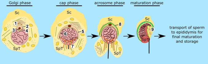
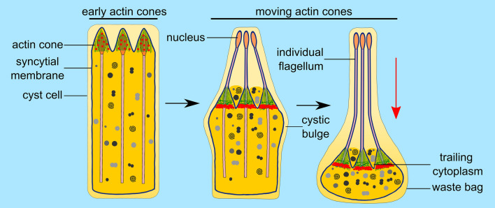
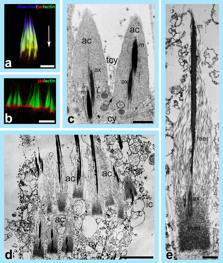
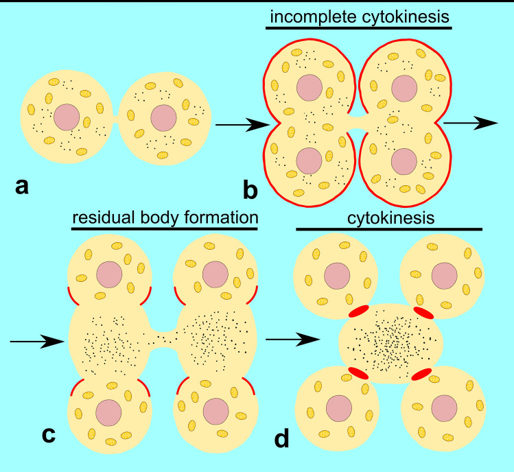
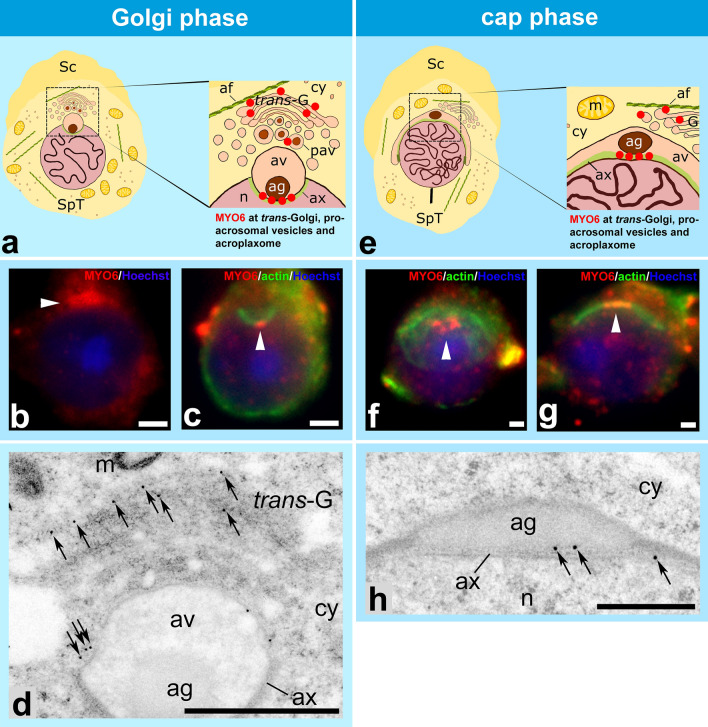
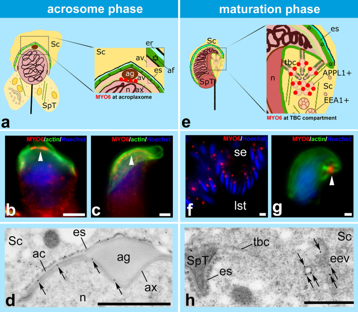
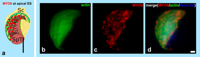
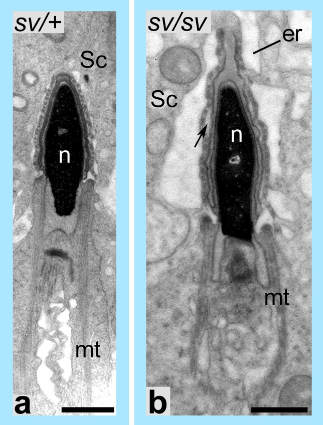
Spermiogenesis is the final stage of spermatogenesis, a differentiation process during which unpolarized spermatids undergo excessive remodeling that results in the formation of sperm. The actin cytoskeleton and associated actin-binding proteins play crucial roles during this process regulating organelle or vesicle delivery/segregation and forming unique testicular structures involved in spermatid remodeling. In addition, several myosin motor proteins including MYO6 generate force and movement during sperm differentiation. MYO6 is highly unusual as it moves towards the minus end of actin filaments in the opposite direction to other myosin motors. This specialized feature of MYO6 may explain the many proposed functions of this myosin in a wide array of cellular processes in animal cells, including endocytosis, secretion, stabilization of the Golgi complex, and regulation of actin dynamics. These diverse roles of MYO6 are mediated by a range of specialized cargo-adaptor proteins that link this myosin to distinct cellular compartments and processes. During sperm development in a number of different organisms, MYO6 carries out pivotal functions. In Drosophila, the MYO6 ortholog regulates actin reorganization during spermatid individualization and male KO flies are sterile. In C. elegans, the MYO6 ortholog mediates asymmetric segregation of cytosolic material and spermatid budding through cytokinesis, whereas in mice, this myosin regulates assembly of highly specialized actin-rich structures and formation of membrane compartments to allow the formation of fully differentiated sperm. In this review, we will present an overview and compare the diverse function of MYO6 in the specialized adaptations of spermiogenesis in flies, worms, and mammals.

Keywords: Actin; C. elegans; Drosophila; Mouse; Myosin VI; Spermiogenesis.


Conflict of interest statement

The authors declare that they have no conflict of interest.

Figures

Fig. 1
Fig. 1
Schematic diagram highlighting the sequential steps of mouse spermiogenesis. During the Golgi phase, proacrosomal vesicles (1) fuse to form the acrosomal vesicle (2), which contains the acrosomal granule (3). The acrosomal vesicle adheres to the nuclear envelope through the acroplaxome (4). During the cap phase, the acrosomal vesicle flattens and spreads over the nucleus to form a cap (5). Acrosomal reshaping and spermatid elongation is mediated by the acroplaxome (6). During the acrosomal phase, the manchette (7) participates in the formation of the sperm flagellum. At this stage, spermatids are attached to Sertoli cells through the apical ES (8) which also supports their movement across the seminiferous tubule. Finally, during the maturation phase, the elongation of the spermatid is completed and most of the cytoplasm and organelles are removed and phagocytosed by Sertoli cells. The TBCs form at the spermatid-Sertoli cell interface (9) and internalize cell–cell junctions supporting the sperm release. Sc Sertoli cell, SpT spermatid
Fig. 2
Fig. 2
Spermatid individualization in Drosophila. In each testicular cyst a syncytial membrane is reorganized into individual membranes encasing 64 spermatids during individualization. This process is driven by actin cones, which assemble around nuclei of spermatids and move synchronously down the length of the axonemes. Early actin cones are made of parallel actin bundles and contain jaguar (ortholog of MYO6; red dots). As they move, the actin cones separate into two distinct domains—at the rear end parallel actin filament bundles predominate, whereas at the front actin filaments form a dense meshwork. At this stage, jaguar concentrates at the front of the actin cones. While the cytoplasm and organelles are extruded from spermatids, the cystic bulge forms. Remnants of the trailing cytoplasm can be observed between the moving actin cones. When the actin cones reach the end of the cyst, excess membrane and cytoplasm are pinched off in the form of the waste bag and the spermatids are left completely encased in individual membranes and with fully formed flagella. Red arrow shows the direction of movement of the actin cones
Fig. 3
Fig. 3
Localization of jaguar (MYO6 ortholog) and ultrastructure of Drosophila actin cones. a Localization of jaguar (red) by immunofluorescence in actin cones (green) at the beginning of spermatid individualization. Jaguar is present throughout the actin cones (yellow indicates overlap between jaguar and actin) that form around the nuclei of spermatids (blue). b Immunofluorescence localization of jaguar (red) in actin cones (visualized in green) at a later stage of spermatid individualization, when jaguar forms a dense band at the front of the moving actin cones. c Ultrastructural analysis of actin cones in a cyst isolated from Drosophila testis. Arrow indicates the syncytial membrane, which progresses downwards to separate the spermatids during individualization. d Ultrastructural visualization of actin cones in the cystic bulge decorated by myosin-II subfragment 1, which highlights the two distinct domains of the actin cones, a dense actin meshwork at the front and parallel actin filaments at the rear. The actin polymerization in the rear region drives cone movement and the actin meshwork at the front ensures exclusion of the cytoplasm and reorganization of the syncytial membrane. e High resolution electron microscope image of a single actin cone decorated by myosin-II subfragment 1. ac actin cone, ax axoneme, cy cytoplasm, ic individualization complex, m mitochondrion, tcy trailing cytoplasm. White arrow in a shows the direction of actin cones movement, which is the same for be. Bars 5 µm (ab, d), 1 µm (c, e)
Fig. 4
Fig. 4
Model of spe-15 (MYO6 ortholog) function during spermatid differentiation in C. elegans. After meiosis, two haploid spermatids remain connected and differentiate by shedding residual cytoplasm in the form of residual body. Spermatid budding is mediated by spe-15 (red). Mitochondria are transported to spermatids, whereas all ribosomes and remaining organelles are packed to the residual body. Spermatids detach from the residual body following the cytokinesis mediated by spe-15 and next mature into spermatozoa (based on Hu et al. 2019a, b)
Fig. 5
Fig. 5
Localization of MYO6 in mouse developing spermatids during the Golgi and cap phases. a During the Golgi phase, MYO6 (red dots) localizes to the trans-Golgi network, proacrosomal vesicles and acroplaxome below the acrosomal granule. b, c Immunofluorescence localization of MYO6 (red) at the Golgi complex (b) and at the acroplaxome (green, c). Arrowhead indicates the Golgi complex in (b) and the area below the acrosomal granule in (c). d Ultrastructural localization of MYO6 using immunogold labeling at the trans-Golgi network and on the surface of the acrosomal vesicles (arrows). e During the cap phase, MYO6 (red dots) localizes to the trans-Golgi network, proacrosomal vesicles and acroplaxome below the acrosomal granule. f and g Immunofluorescence localization of MYO6 (red) at the acroplaxome (green). Arrowheads indicate area below the acrosomal granule. h Ultrastructural localization of MYO6 using immunogold labeling at the acroplaxome below the acrosomal granule (arrows). Panel d is modified from Zakrzewski et al. (2017) (published under CC BY 4.0). af actin filament, ag acrosomal granule, av acrosomal vesicle, ax acroplaxome, cy cytoplasm, m mitochondrion, n nucleus, pav proacrosomal vesicles, trans-G trans-Golgi network, Sc Sertoli cell, SpT spermatid. Bars 1 µm
Fig. 6
Fig. 6
Localization of MYO6 in mouse developing spermatids during the acrosome and maturation phases. a During the acrosome phase, MYO6 (red dots) localizes to the acroplaxome below the acrosomal granule. b, c Immunofluorescence localization of MYO6 (red) at the acroplaxome (actin visualized in green) in elongating spermatids during the acrosome phase. Arrowheads indicate area below the acrosomal granule. d Ultrastructural localization of MYO6 using immunogold labeling at the acroplaxome below the acrosomal granule and the acrosome (arrows). Panel d is modified from Zakrzewski et al. (2017) (published under CC BY 4.0). e During the maturation phase, MYO6 (red dots) is concentrated at the bulbs of the TBCs and APPL1-positive early endosomes. f Immunofluorescence localization of MYO6 (red) at the spermatid-Sertoli cell interface in the seminiferous epithelium on a semi-thin paraffin section. During this stage, maturing spermatids are close to the lumen of the seminifereous tubules. g Immunofluorescence localization of MYO6 (red) in the endocytic compartment of the TBCs (actin visualized in green). h Ultrastructural localization of MYO6 using immunogold labelling in early endosomes in a spermatid during the maturation phase (arrows). ac acrosome, af actin filament, ag acrosomal granule, APPL1 + APPL1-positive early endocytic vesicle, av acrosomal vesicle, ax acroplaxome, EEA1 + EEA1-positive early endosome, eev early endocytic vesicle, er endoplasmic reticulum, es apical ES, lst lumen of seminiferous tubule, n nucleus, Sc Sertoli cell, se seminiferous epithelium, SpT spermatid, tbc tubulobulbar complex. Bars 5 µm (f), 1 µm (bd, g), 500 nm (h)
Fig. 7
Fig. 7
MYO6 localizes to the apical ES. During the maturation phase, MYO6 (red) is also present at the apical ES, where it localizes to actin bundles (visualized in b and d in green) that enclose the maturing spermatids. Sc Sertoli cell, SpT spermatid. Bars 1 µm
Fig. 8
Fig. 8
Loss of MYO6 causes ultrastructural disruptions of the apical ES in maturing spermatids. Ultrastructural analysis of the apical ES of a MYO6-expressing spermatid (a) and MYO6-deficient spermatid (b). In the absence of MYO6 the apical ES appears to be disrupted and detachment of the spermatid head from the apical ES and swelling of the ER can be observed (arrow) (b). er endoplasmic reticulum, mt manchette, n nucleus, Sc Sertoli cell. Bars 1 µm

Similar articles

Cited by

References

    1. Adams A, Vogl AW. High resolution localization of Rab5, EEA1, and nectin-3 to tubulobulbar complexes in the rat testis. Anat Rec (Hoboken) 2017;300:1160–1170. - PubMed
    1. Adams A, Sriram A, Vogl AW. Internalization of intact intercellular junctions in the testis by clathrin/actin-mediated endocytic structures: tubulobulbar complexes. Anat Rec (Hoboken) 2018;301:2080–2085. - PubMed
    1. Ameen N, Apodaca G. Defective CFTR apical endocytosis and enterocyte brush border in myosin VI-deficient mice. Traffic. 2007;8:998–1006. - PubMed
    1. Anahara R, Toyama Y, Maekawa M, Kai M, Ishino F, Toshimori K, Mori C. Flutamide depresses expression of cortactin in the ectoplasmic specialization between the Sertoli cells and spermatids in the mouse testis. Food Chem Toxic. 2006;44:1050–1056. - PubMed
    1. Aschenbrenner L, Lee T, Hasson T. Myo6 facilitates the translocation of endocytic vesicles from cell peripheries. Mol Biol Cell. 2003;14:2728–2743. - PMC - PubMed

LinkOut - more resources