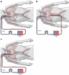
Pediatric and neonatal extracorporeal life support: current state and continuing evolution
- PMID: 33386443
- PMCID: PMC7775668
- DOI: 10.1007/s00383-020-04800-2
Pediatric and neonatal extracorporeal life support: current state and continuing evolution
Abstract
The use of extracorporeal life support (ECLS) for the pediatric and neonatal population continues to grow. At the same time, there have been dramatic improvements in the technology and safety of ECLS that have broadened the scope of its application. This article will review the evolving landscape of ECLS, including its expanding indications and shrinking contraindications. It will also describe traditional and hybrid cannulation strategies as well as changes in circuit components such as servo regulation, non-thrombogenic surfaces, and paracorporeal lung-assist devices. Finally, it will outline the modern approach to managing a patient on ECLS, including anticoagulation, sedation, rehabilitation, nutrition, and staffing.
Keywords: Extracorporeal life support (ECLS); Extracorporeal membrane oxygenation (ECMO); Neonatal; Pediatric; Respiratory failure.
Conflict of interest statement
The authors declare that they have no conflict of interest.
Figures


Similar articles
-
Extracorporeal Life Support Organization (ELSO): 2020 Pediatric Respiratory ELSO Guideline.ASAIO J. 2020 Sep/Oct;66(9):975-979. doi: 10.1097/MAT.0000000000001223. ASAIO J. 2020. PMID: 32701626
-
Extracorporeal life support: updates and controversies.Semin Pediatr Surg. 2015 Feb;24(1):8-11. doi: 10.1053/j.sempedsurg.2014.11.002. Epub 2014 Nov 7. Semin Pediatr Surg. 2015. PMID: 25639803
-
A novel approach to delivery of extracorporeal support using a modified continuous flow ventricular assist device in a mid-volume congenital heart program.Artif Organs. 2021 Jan;45(1):55-62. doi: 10.1111/aor.13840. Artif Organs. 2021. PMID: 33029801
-
Pediatric Extracorporeal Membrane Oxygenation.Crit Care Clin. 2017 Oct;33(4):825-841. doi: 10.1016/j.ccc.2017.06.005. Epub 2017 Jul 29. Crit Care Clin. 2017. PMID: 28887930 Review.
-
Challenges with Navigating the Precarious Hemostatic Balance during Extracorporeal Life Support: Implications for Coagulation and Transfusion Management.Transfus Med Rev. 2016 Oct;30(4):223-9. doi: 10.1016/j.tmrv.2016.07.005. Epub 2016 Aug 4. Transfus Med Rev. 2016. PMID: 27543261 Review.
Cited by
-
Invasive pneumococcal disease and long-term outcomes in children: A 20-year population cohort study.Lancet Reg Health Am. 2022 Aug 8;14:100341. doi: 10.1016/j.lana.2022.100341. eCollection 2022 Oct. Lancet Reg Health Am. 2022. PMID: 36777393 Free PMC article.
-
Nutritional support in pediatric patients with extracorporeal membrane oxygenation: new insights.Arch Cardiol Mex. 2025;95(3):215-223. doi: 10.24875/ACM.24000135. Arch Cardiol Mex. 2025. PMID: 40763815 Free PMC article. Review. English.
-
Efficacy and safety of extracorporeal membrane oxygenation combined with continuous renal replacement therapy in the management of pediatric acute respiratory distress syndrome.Front Pediatr. 2025 Mar 18;13:1556642. doi: 10.3389/fped.2025.1556642. eCollection 2025. Front Pediatr. 2025. PMID: 40171174 Free PMC article.
-
Lung transplantation in children.Turk Gogus Kalp Damar Cerrahisi Derg. 2024 Feb 5;32(Suppl1):S119-S133. doi: 10.5606/tgkdc.dergisi.2024.25806. eCollection 2024 Jan. Turk Gogus Kalp Damar Cerrahisi Derg. 2024. PMID: 38584780 Free PMC article. Review.
-
Use of point-of-care ultrasound (POCUS) to monitor neonatal and pediatric extracorporeal life support.Eur J Pediatr. 2024 Apr;183(4):1509-1524. doi: 10.1007/s00431-023-05386-2. Epub 2024 Jan 18. Eur J Pediatr. 2024. PMID: 38236403 Review.
References
-
- Baumgart S, Hirschl RB, Butler SZ, Coburn CE, Spitzer AR. Diagnosis-related criteria in the consideration of extracorporeal membrane oxygenation in neonates previously treated with high-frequency jet ventilation. Pediatrics. 1992;89(3):491–494. - PubMed
-
- MacLaren G, Conrad S, Peek G. Indications for pediatric respiratory extracorporeal life support. Ann Arbor: ELSO; 2015.
Publication types
MeSH terms
LinkOut - more resources
Full Text Sources
Other Literature Sources
Medical

