. 2021;79(3):1143-1156.
doi: 10.3233/JAD-201077.

Phosphorylation and Dephosphorylation of Tau Protein by the Catalytic Subunit of PKA, as Probed by Electrophoretic Mobility Retard

Affiliations

Phosphorylation and Dephosphorylation of Tau Protein by the Catalytic Subunit of PKA, as Probed by Electrophoretic Mobility Retard

María J Benítez et al. J Alzheimers Dis. 2021.

Abstract

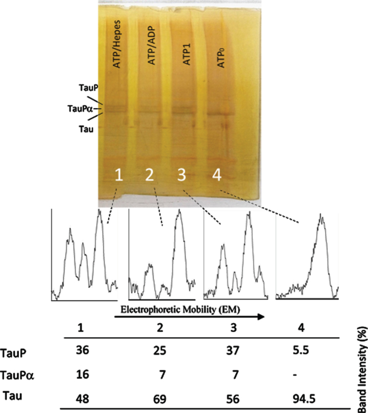
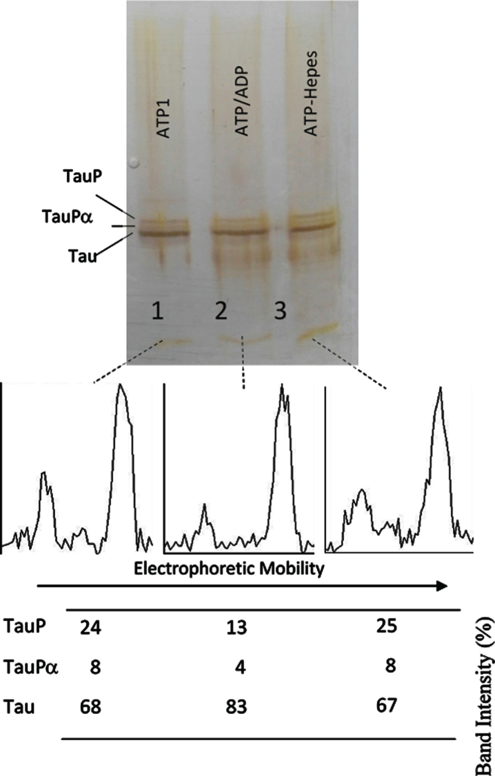
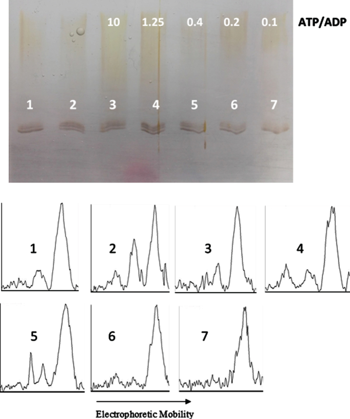
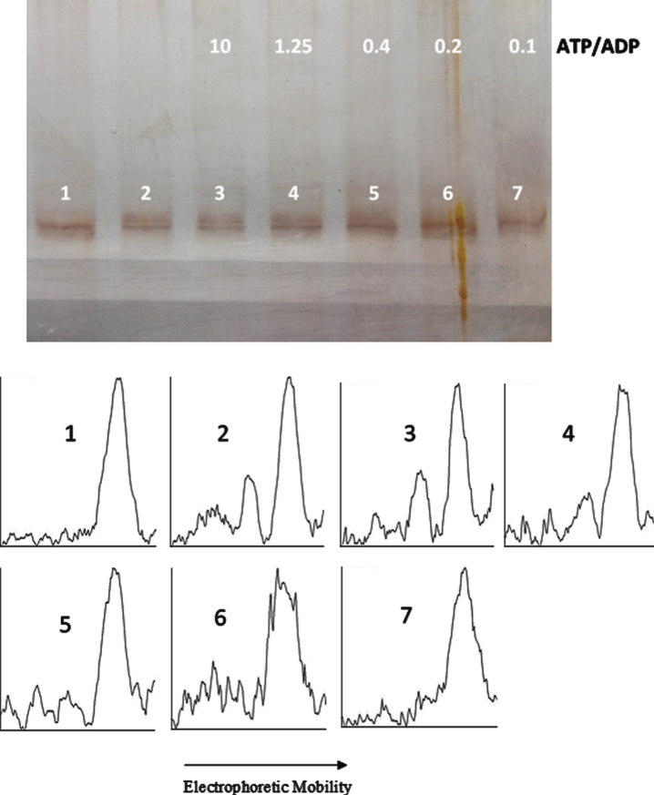
Background: Tau is a microtubule associated protein that regulates the stability of microtubules and the microtubule-dependent axonal transport. Its hyperphosphorylated form is one of the hallmarks of Alzheimer's disease and other tauopathies and the major component of the paired helical filaments that form the abnormal proteinaceous tangles found in these neurodegenerative diseases. It is generally accepted that the phosphorylation extent of tau is the result of an equilibrium in the activity of protein kinases and phosphatases. Disruption of the balance between both types of enzyme activities has been assumed to be at the origin of tau hyperphosphorylation and the subsequent toxicity and progress of the disease.

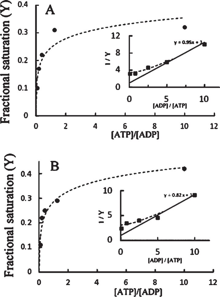
Objective: We explore the possibility that, beside the phosphatase action on phosphorylated tau, the catalytic subunit of PKA catalyzes both tau phosphorylation and also tau dephosphorylation, depending on the ATP/ADP ratio.

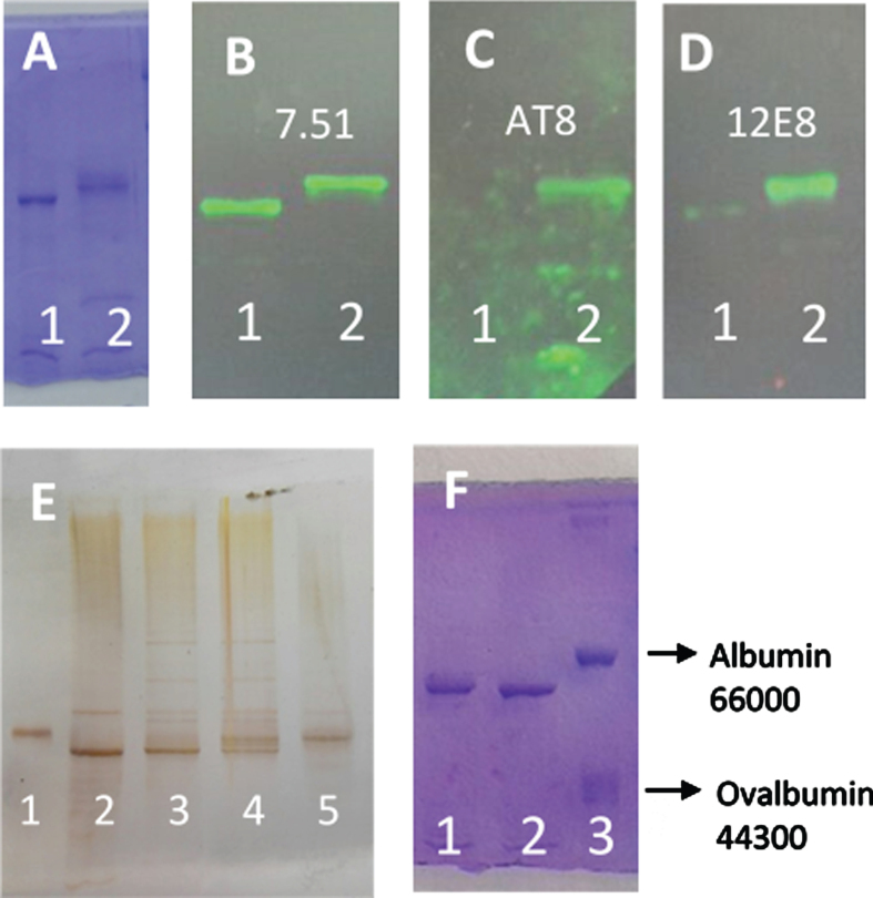
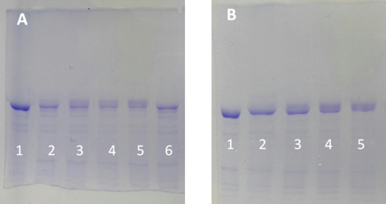
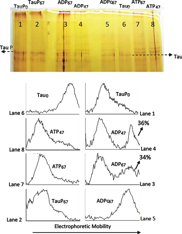
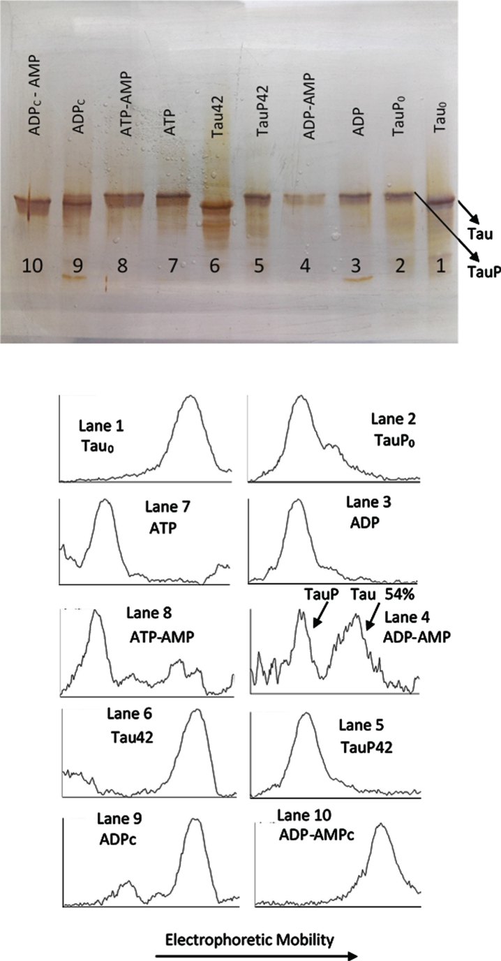
Methods: We use the shift in the relative electrophoretic mobility suffered by different phosphorylated forms of tau, as a sensor of the catalytic action of the enzyme.

Results: The results are in agreement with the long-known thermodynamic reversibility of the phosphorylation reaction (ATP + Protein = ADP+Phospho-Protein) catalyzed by PKA and many other protein kinases.

Conclusion: The results contribute to put the compartmentalized energy state of the neuron and the mitochondrial-functions disruption upstream of tau-related pathologies.

Keywords: Dephosphorylation; PKA; electrophoretic mobility; phosphorylation; tau protein; thermodynamic reversibility.


Conflict of interest statement

Authors’ disclosures available online (https://www.j-alz.com/manuscript-disclosures/20-1077r1).

Figures

Fig. 1
Fig. 1
SDS-PAGE (8% of polyacrylamide) of different forms of tau protein. A) Lane 1, tau protein (0.05 mg/ml) incubated for 4 h at 37°C in the presence of MgCl2 (10 mM), EGTA (0.25 mM), 2-mercaptoethanol (0.25 mM), Hepes (5 mM), and NaCl (50 mM), pH 7. Lane 2, the same tau protein incubation as Lane 1, but in the presence of ATP (2 mM) and C-PKA (4 units). B-D) Tau protein incubated as in (A) showing the western blots using antibody 7.51 to total tau (B), AT8 to Ser 202 and Thr 205 (C), and 12E8 to Ser 262 and Ser 356 (D). E) Silver stained of different forms of tau protein. Lane 1, Albumin used as a control. Lane 2, tau protein. Lanes 3 and 4, tau protein (3.6 ng/μl) incubated with ATP (5 mM) and C-PKA for 2.5 h (lane 3) and 17 h (lane 4). Lane 5 shows the phosphorylated tau protein prepared as described in the Materials and Methods. The amounts of tau and albumin loaded in lanes 1 and 2 were approximately 70 and 60 ng respectively. F) shows tau protein (0.05 mg/ml) incubated for 1 h with ATP 0.1 mM in the absence (lane 2) or the presence of C-PKA (lane 1). Lane 3 shows a mixture of bovine serum albumin and ovalbumin used as controls.
Fig. 2
Fig. 2
Electrophoretic mobility retard of tau protein as a function of the ATP concentration and as a function of time. A) SDS-PAGE (8% of polyacrylamide) of tau protein (0.05 mg/ml, in a buffer composed of 10 mM Hepes, 0.1 M NaCl, 10 mM MgCl2, 0.25 mM EGTA, and 0.25 mM 2-mercaptoethanol 10 mM at pH 7) in the absence of ATP and C-PKA (Lane 1). The rest of the lanes correspond to the same tau protein at 0.05 mg/ml incubated at 37°C for 2 h in the presence of the same buffer as in lane 1 but in the presence of 3.2 units of C-PKA and ATP 0.01 mM (lane 2), ATP 0.05 mM (lane 3), ATP 0.1 mM (lane 4), ATP 0.5 mM (lane 5), and no ATP (lane 6). B) Time dependence of the mobility retard. Tau protein as in (A), incubated with 0.1 mM of ATP at time 0 (lane 1), 30 min (lane 2), 1.5 h (lane 3), 3 h (lane 4), and 4 h (lane 5).
Fig. 3
Fig. 3
Silver stained of tau and phosphorylated tau separated by SDS-PAGE (8% acrylamide). Lanes 5–8: Tau protein (2.4 μg/ml final concentration) was diluted in a buffer composed of 31 μl of 10 mM Hepes, 0.15 M KCl, 0.25 mM 2-mercaptoethanol, 15 mM MgCl2, and 0.1 mg/ml Ampicillin at pH 6 adjusted with KOH; plus 10 μl of 10 mM Hepes, 0.15 M NaCl at pH 7; plus 5 μl of a solution of 250 mM potassium phosphate 10 mM and 0.1 mg/ml of Ampicillin at pH 7, containing 10 mM of P1, P5-Di(adenosine-5’) pentaphosphate. This solution was incubated at 20°C in the presence of 25 mM of AMP and 2.5 mM ATP for 47 h (lane 8) and 67 h (lane 7) or 2.5 mM ADP for 67 h (lane 5) and 4 units of C-PKA (Promega); lane 6 is a control in the absence of C-PKA, without incubation. Lanes 1–4: Phosphorylated tau (TauP) (1.8 μg/ml, final concentration) was diluted similarly to tau and incubated at 20°C with 2.5 mM ADP and 4 units of C-PKA for 47 h (lane 4) and 67 h (lane 3); dephosphorylation percentages of TauP are indicated. Lane 1 is a control in the absence of C-PKA, without incubation. Lane 2 is a control of TauP incubated for 67 h in the presence of ADP but in the absence of C-PKA. Tau and TauP samples were boiled before use.
Fig. 4
Fig. 4
Silver stained of tau and phosphorylated tau separated by SDS-PAGE (8% acrylamide). Lanes 1, 6–10: Tau protein (3 μg/ml final concentration) was diluted in a buffer composed of 22.5 μl of 10 mM Hepes, 0.15 M KCl, 0.25 mM 2-mercaptoethanol, 15 mM MgCl2, and 0.1 mg/ml Ampicillin at pH 7 plus 2.5 μl of a solution of 250 mM potassium phosphate 10 mM and 0.1 mg/ml of Ampicillin at pH 7, containing 10 mM of P1, P5-Di(adenosine-5’) pentaphosphate. This solution was incubated with C-PKA, at 20°C for 42 h, in the presence of 2.5 mM ATP plus the presence (Lane 8) or absence (Lane 7) of 25 mM of AMP and in presence of 2.5 mM ADP plus the presence (Lane 10) or absence (Lane 9) of 25 mM of AMP. Lanes 1 and 6 are controls of tau in the absence of C-PKA and nucleotides, incubated for 0 h (Lane 1) and 42 h (Lane 2). Lanes 2-5: Phosphorylated tau (TauP) (2.7 μg/ml, final concentration) was diluted in the same buffer solution as described for tau and incubated with C-PKA, at 20°C for 42 h, in the presence of 2.5 mM ADP plus the presence (Lane 4) or absence (Lane 3) of 25 mM of AMP; dephosphorylation percentage of TauP is indicated. Lanes 2 and 5 are controls of TauP in the presence of 2.5 mM ADP but in the absence of C-PKA, incubated for 0 h (Lane 2) and 42 h (Lane 5). 5 units of C-PKA from Promega were used, when added. Both tau and TauP solutions were boiled before used.
Fig. 5
Fig. 5
Successive incubation of tau protein with ATP and ADP. The mixture of tau protein and C-PKA prepared as described in Materials and Method (Tau-C-PKA) was diluted 1/10 in a buffer composed of 75 μl of buffer Hepes containing 10 mM Hepes and 0.1 M NaCl, at pH 7 and 50 μl of phosphate buffer containing 250 mM phosphate and 0.1 mg/ml Ampicillin at pH 7. This reaction mixture contained 15 mM MgCl2, 0.25 mM EGTA, 50 mM P1, P5-Di(adenosine-5’)pentaphosphate, 0.25 mM 2-mercaptoethanol and ATP 0.1 mM. Lane 4 corresponds to tau protein in this reaction mixture at time 0 of incubation (ATP0). After 2 h of incubation at 30°C, in the presence of ATPMg2+, the reaction mixture was divided in three parts: the first one (ATP1) was loaded in the gel (lane 3); the second one was incubated at 30°C for two additional hours in the presence of 1.1 mM ADP(ATP/ADP) (lane 2); the third one was incubated for the same time, at 30°C, in the presence of Hepes buffer, (ATP/Hepes) (lane 1). Scanning of the electrophoresis bands was carried out after applying a filter to improve the photo quality. The table at the bottom of the figure shows the results of peak percentages resulting from the scanning. TauP represent the band displaying the lowest mobility, and TauPα represents the intermediate band usually observed at short times of incubation. Scanning was done after applying a filter to improve the photo quality.
Fig. 6
Fig. 6
Successive incubation of tau protein with ATP and ADP. The mixture of tau protein and C-PKA prepared as described in Materials and Method (Tau-C-PKA) was diluted 1/10 in a buffer composed of 75 μl of Hepes buffer containing 10 mM Hepes and 0.1 M NaCl, at pH 7 and 50 μl of phosphate buffer containing 250 mM phosphate and 0.1 mg/ml Ampicillin at pH 7. This reaction mixture contained 15 mM MgCl2, 0.25 mM EGTA, 50 mM P1, P5-Di(adenosine- 5’)pentaphosphate, 0.25 mM 2-mercaptoethanol, 0.05 mg/ml DNA, and ATP 0.1 mM. After 2 h of incubation at 30°C, in the presence of ATPMg2+, the reaction mixture was divided in three parts: the first one (25 μl) (ATP1) was loaded in the gel (lane 1); the second one (22.5 μl) was incubated at 30°C for 30 min after adding 2.5 ml of 10 mM ADP (ATP/ADP) (lane 2); the third one (22.5 μl) was incubated for the same time, at 30°C, after adding 2.5 μl of Hepes buffer containing 10 mM Hepes, 0.1 M NaCl, 0.1 mg/ml DNA, and 100 μM P1, P5-Di(adenosine- 5’)pentaphosphate at pH 7 (lane 3). The Table at the bottom of the figure shows the results of peak percentages after the scanning of the bands. TauP represent the band displaying the lowest mobility, and TauPα represents the intermediate band usually observed at short times of incubation. The rolling ball radius used for subtraction of background was of 5 pixels.
Fig. 7
Fig. 7
ATP/ADP ratio-dependency of tau protein phosphorylation as probed by electrophoretic mobility. The mixture of tau protein and C-PKA, prepared as described in Materials and Method (Tau-C-PKA) was diluted 1/11.5 in a buffer composed of 15 μl of 10 mM Hepes and 0.1 M NaCl at pH 7, and 10 μl of phosphate buffer containing 250 mM phosphate and 0.1 mg/ml Ampicillin at pH 7. This reaction mixture contained 15 mM MgCl2, 0.25 mM EGTA, 0.25 mM 2-mercaptoethanol, ATP 0.1 mM and the following ADP concentrations (mM): 0 (lane 2), 0.01 (lane 3), 0.08 (lane 4), 0.25 (lane 5), 0.5 (lane 6), and 1 (lane 7). Lane 1 is a control in the absence of nucleotides. All samples were incubated at 20°C for 22 h. Scanning results are included in Table 1. The rolling ball radius used for subtraction of background was of 11 pixels.
Fig. 8
Fig. 8
ATP/ADP ratio-dependency of tau protein phosphorylation as probed by electrophoretic mobility. The mixture of tau protein and C-PKA prepared as described in Materials and Method (Tau-C-PKA) was diluted 1/10 in a buffer composed of 35 μl of 10 mM Hepes and 0.1 M NaCl at pH 7, and 23 μl of phosphate buffer containing 250 mM phosphate and 0.1 mg/ml Ampicillin at pH 7. This reaction mixture contained 13 mM MgCl2, 0.22 mM EGTA, 0.25 mM 2-mercaptoethanol, 0.87 mM ATP and the following ADP concentrations (mM): 0 (lane 2), 0.087 (lane 3), 0.7 (lane 4), 2.2 (lane 5), 4.4 (lane 6), and 8.7 (lane 7). Lane 1 is a control in the absence of nucleotides. All samples were incubated at 20°C for 22 h. The rolling ball radius used for subtraction of background was of 30 pixels.
Fig. 9
Fig. 9
Fractional saturation of phosphorylation sites on tau, following the data of Table 1. The fractional saturation as a function of the [ATP]/[ADP] ratio, keeping [ATP] constant at 0.1 mM (part A) or 0.87 mM (part B). The insets show the inverse plots corresponding to equation (5).

References

    1. Weingarten MD, Lockwood AH, Hwo SY, Kirschner MW (1975) A protein factor essential for microtubule assembly. Proc Natl Acad Sci U S A 72, 1858–1862. - PMC - PubMed
    1. Grundke-Iqbal I, Iqbal K, Tung YC, Quinlan M, Wisniewski HM, Binder LI (1986) Abnormal phosphorylation of the microtubule-associated protein (tau) in Alzheimer cytoskeletal pathology. Proc Natl Acad Sci U S A 83, 4913–4917. - PMC - PubMed
    1. Drubin DG, Kirschner MW (1986) Tau protein function in living cells. J Cell Biol 103, 2739–2746. - PMC - PubMed
    1. Ebneth A, Godemann R, Stamer K, Illenberger S, Trinczek B, Mandelkow E (1998) Overexpression of tau protein inhibits kinesin-dependent trafficking of vesicles, mitochondria, and endoplasmic reticulum: Implications for Alzheimer’s disease. J Cell Biol 143, 777–794. - PMC - PubMed
    1. Ávila J, Jiménez JS, Sayas CL, Bolós M, Zabala JC, Rivas G, Hernández F (2016) Tau structures. Front Aging Neurosci 8, 262. - PMC - PubMed

Publication types

Substances