. 2021 Apr;41(3):515-525.
doi: 10.1007/s10875-020-00920-5. Epub 2021 Jan 2.

SARS-CoV-2-Induced ARDS Associates with MDSC Expansion, Lymphocyte Dysfunction, and Arginine Shortage

Affiliations

SARS-CoV-2-Induced ARDS Associates with MDSC Expansion, Lymphocyte Dysfunction, and Arginine Shortage

Florian Reizine et al. J Clin Immunol. 2021 Apr.

Abstract

Purpose: The SARS-CoV-2 infection can lead to a severe acute respiratory distress syndrome (ARDS) with prolonged mechanical ventilation and high mortality rate. Interestingly, COVID-19-associated ARDS share biological and clinical features with sepsis-associated immunosuppression since lymphopenia and acquired infections associated with late mortality are frequently encountered. Mechanisms responsible for COVID-19-associated lymphopenia need to be explored since they could be responsible for delayed virus clearance and increased mortality rate among intensive care unit (ICU) patients.

Methods: A series of 26 clinically annotated COVID-19 patients were analyzed by thorough phenotypic and functional investigations at days 0, 4, and 7 after ICU admission.

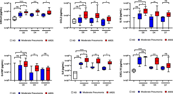
Results: We revealed that, in the absence of any difference in demographic parameters nor medical history between the two groups, ARDS patients presented with an increased number of myeloid-derived suppressor cells (MDSC) and a decreased number of CD8pos effector memory cell compared to patients hospitalized for COVID-19 moderate pneumonia. Interestingly, COVID-19-related MDSC expansion was directly correlated to lymphopenia and enhanced arginase activity. Lastly, T cell proliferative capacity in vitro was significantly reduced among COVID-19 patients and could be restored through arginine supplementation.

Conclusions: The present study reports a critical role for MDSC in COVID-19-associated ARDS. Our findings open the possibility of arginine supplementation as an adjuvant therapy for these ICU patients, aiming to reduce immunosuppression and help virus clearance, thereby decreasing the duration of mechanical ventilation, nosocomial infection acquisition, and mortality.

Keywords: ARDS; Arginine; Covid-19; Cross infection; Immunosuppression; Lymphocytes; MDSC.


Conflict of interest statement

The authors declare that they have no competing interests

Figures

Fig. 1
Fig. 1
CT scan from a patient hospitalized for SARS-CoV-2 induced ARDS at admission (A) and 7 days after (C). CT scan from a patient hospitalized for SARS-CoV-2-induced moderate pneumonia at admission (B) and 9 days after (D)
Fig. 2
Fig. 2
SARS-CoV-2-induced acute respiratory distress syndrome (ARDS) is associated with lymphopenia and an accumulation of circulating myeloid-derived suppressor cells (MDSC) leading to a higher susceptibility to nosocomial infections. A Blood count from 13 patients hospitalized for SARS-CoV-2 moderate pneumoniae (MP) and 13 patients hospitalized for SARS-CoV-2 ARDS (ARDS) 24 h after their admission (D0), 4 days after (D4), and 7 days after (D7) and lymphocytes subsets defined by flow cytometry from 13 healthy donors (HD), 13 patients hospitalized for SARS-CoV-2 MP, and 13 patients hospitalized for SARS-CoV-2 ARDS. B CD4 and CD8 effector memory (EM) T cell numeration by flow cytometry. C Peripheral monocytic-MDSC (M-MDSC) and granulocytic-MDSC (G-MDSC) recruitment among ARDS patients, moderate COVID cases, and HD. D Two groups were defined according to the presence or absence of a nosocomial infection. Lymphocyte count, M-MDSC, and G-MDSC recruitment according to the acquisition of nosocomial infection. Nosocomial infections as defined by the Centers for Disease Control and Prevention were screened among patients hospitalized for a SARS-CoV-2 infection over 28 days after their admission. Box and whiskers plot features are as follows: central line in the box is the median, bottom line of the box is first quartile (25%), and top line of box is third quartile (75%). Bottom of whiskers is minimum value; top of whiskers is maximum value. Groups were compared using Kruskal Wallis test with Dunn’s multiple comparison test (A, B, and C) or Mann-Whitney U test (A and D) as appropriate. Pearson correlation coefficients (rho) and P values are indicated for each correlation (C). *P < 0.05; **P < 0.01; ***P < 0.001
Fig. 3
Fig. 3
SARS-CoV2 ARDS induces a significant increase among inflammatory, chemoattractant, and immunosuppressive cytokines. MDSC recruiting (IL-6, CCL2, G-CSF), chemoattractant (CXCL9, CXCL10, CCL2), inflammatory (IL-6), and immunosuppressive (IL-10) cytokine assessments were performed among the ARDS patients (ARDS), the moderate COVID cases (MP), and the healthy donors (HD). Box and whiskers plot features are as follows: central line in the box is the median, bottom line of the box is first quartile (25%), and top line of box is third quartile (75%). Bottom of whiskers is minimum value; top of whiskers is maximum value. Groups were compared using Kruskal Wallis test with Dunn’s multiple comparison test. *P < 0.05; **P < 0.01; ***P < 0.001; ****P < 0.0001
Fig. 4
Fig. 4
SARS-CoV-2 ARDS induces a significant decrease in plasma arginine concentration; its supplementation restores the ability of T-cells to proliferate in-vitro. A Plasma arginine, ornithine, kynurenine, and tryptophan concentrations were measured by liquid chromatography coupled with tandem mass spectrometry among 13 healthy donors (HD), 13 patients hospitalized for SARS-CoV-2 moderate pneumonia (MP), and 13 patients hospitalized for SARS-CoV-2 ARDS (ARDS) 24 h after their admission (D0), 4 days after (D4), and 7 days after (D7). Arginase activity was calculated using the ornithine/arginine ratio and IDO activity was calculated using the kynurenine/tryptophan ratio. B PBMC obtained at admission (D0), 4 days after admission (D4), and 7 days after (D7) from 14 patients hospitalized for a SARS-CoV-2 infection and from 7 HD were stimulated with anti-CD3/anti-CD28 monoclonal antibodies after CFSE labelling. The proportion of proliferated T-cells were determined by flow cytometry. C Proportion of apoptotic T cells was determined by flow cytometry using a Caspase-3 staining among 26 patients hospitalized for a SARS-CoV-2 infection at admission (D0), 4 days after (D4), and 7 days after (D7) and from 11 HD. Results are expressed by the percentage of Caspase3pos T cells. D PBMC obtained from 7 patients hospitalized for a SARS-CoV-2 ARDS were stimulated with anti-CD3/anti-CD28 monoclonal antibodies after CFSE labelling. Culture media were enriched with either L-arginine (L-Arg) or with control D-arginine (D-arg), IDO inhibitor (Coptisine) or vehicle (NT), PD-L1 biding antibody (aPD-L1), or its isotype (IgG). The proportion proliferated T cells were determined by flow cytometry. Box and whiskers plot features are as follows: central line in the box is the median, bottom line of the box is first quartile (25%), and top line of box is third quartile (75%). Bottom of whiskers is minimum value; top of whiskers is maximum value. Groups were compared using Kruskal Wallis test with Dunn’s multiple comparison test (A), Mann-Whitney U test (A, B, and C) or Wilcoxon test (D) as appropriate. Pearson correlation coefficients (rho) and P values are indicated for each correlation (a). *P < 0.05; **P < 0.01; ***P < 0.001

References

    1. Grasselli G, Zangrillo A, Zanella A, Antonelli M, Cabrini L, Castelli A, Cereda D, Coluccello A, Foti G, Fumagalli R, Iotti G, Latronico N, Lorini L, Merler S, Natalini G, Piatti A, Ranieri MV, Scandroglio AM, Storti E, Cecconi M, Pesenti A, for the COVID-19 Lombardy ICU Network Baseline characteristics and outcomes of 1591 patients infected with SARS-CoV-2 admitted to ICUs of the Lombardy region, Italy. JAMA. 2020;323:1574–1581. doi: 10.1001/jama.2020.5394. - DOI - PMC - PubMed
    1. Zhou F, Yu T, Du R, Fan G, Liu Y, Liu Z, Xiang J, Wang Y, Song B, Gu X, et al. Clinical course and risk factors for mortality of adult inpatients with COVID-19 in Wuhan, China: a retrospective cohort study. Lancet. 2020;395:1054–1062. doi: 10.1016/S0140-6736(20)30566-3. - DOI - PMC - PubMed
    1. Wu C, Chen X, Cai Y, Xia J, Zhou X, Xu S, Huang H, Zhang L, Zhou X, Du C, et al. Risk factors associated with acute respiratory distress syndrome and death in patients with coronavirus disease 2019 pneumonia in Wuhan, China. JAMA Intern Med. 2020;180:934–943. doi: 10.1001/jamainternmed.2020.0994. - DOI - PMC - PubMed
    1. Du RH, Liang LR, Yang CQ, Wang W, Cao TZ, Li M, et al. Predictors of mortality for patients with COVID-19 pneumonia caused by SARS-CoV-2: a prospective cohort study. Eur Respir J. 2020. - PMC - PubMed
    1. Tay MZ, Poh CM, Renia L, MacAry PA, Ng LFP. The trinity of COVID-19: immunity, inflammation and intervention. Nat Rev Immunol. 2020;20:363–374. doi: 10.1038/s41577-020-0311-8. - DOI - PMC - PubMed

Publication types