. 2021 Jan;48(1):1017-1023.
doi: 10.1007/s11033-020-06109-8. Epub 2021 Jan 2.

Anthracycline-induced cytotoxicity in the GL261 glioma model system

Affiliations

Anthracycline-induced cytotoxicity in the GL261 glioma model system

Amber M Tavener et al. Mol Biol Rep. 2021 Jan.

Abstract

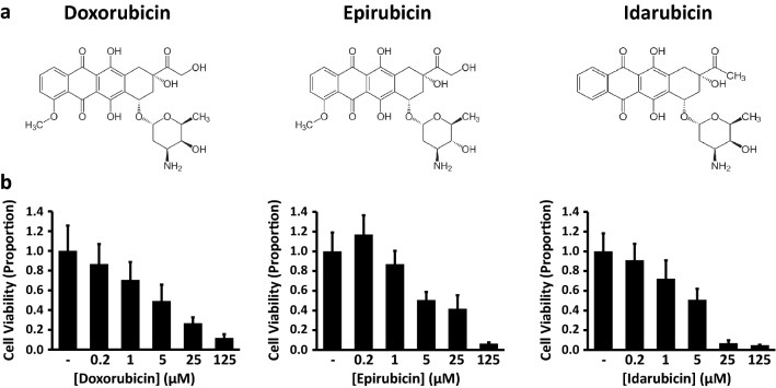
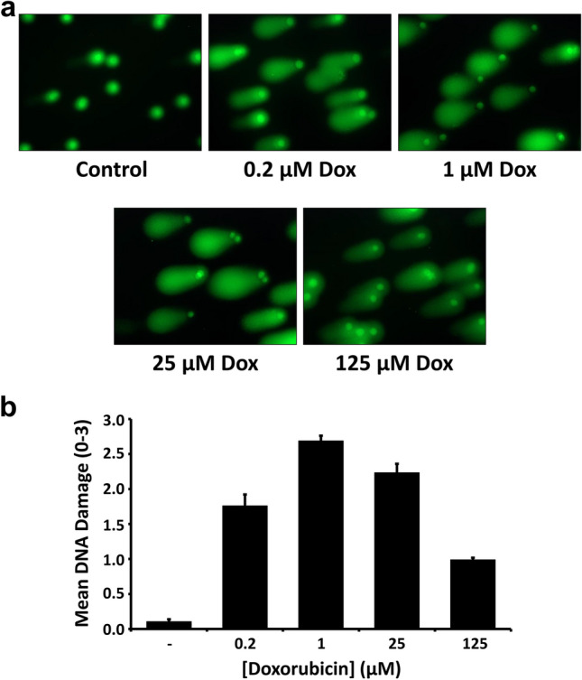
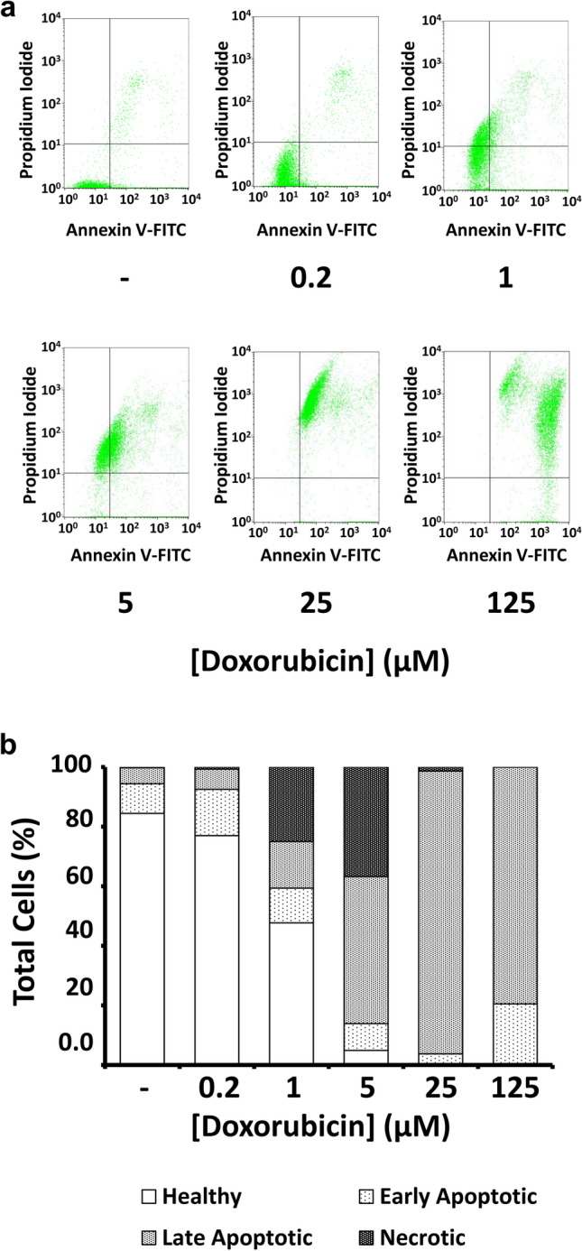
Glioblastoma (GBM) is a lethal astrocyte-derived tumor that is currently treated with a multi-modal approach of surgical resection, radiotherapy, and temozolomide-based chemotherapy. Alternatives to current therapies are urgently needed as its prognosis remains poor. Anthracyclines are a class of compounds that show great potential as GBM chemotherapeutic agents and are widely used to treat solid tumors outside the central nervous system. Here we investigate the cytotoxic effects of doxorubicin and other anthracyclines on GL261 glioma tumor cells in anticipation of novel anthracycline-based CNS therapies. Three methods were used to quantify dose-dependent effects of anthracyclines on adherent GL261 tumor cells, a murine cell-based model of GBM. MTT assays quantified anthracycline effects on cell viability, comet assays examined doxorubicin genotoxicity, and flow cytometry with Annexin V/PI staining characterized doxorubicin-induced apoptosis and necrosis. Dose-dependent reductions in GL261 cell viability were found in cells treated with doxorubicin (EC50 = 4.9 μM), epirubicin (EC50 = 5.9 μM), and idarubicin (EC50 = 4.4 μM). Comet assays showed DNA damage following doxorubicin treatments, peaking at concentrations of 1.0 μM and declining after 25 μM. Lastly, flow cytometric analysis of doxorubicin-treated cells showed dose-dependent induction of apoptosis (EC50 = 5.2 μM). Together, these results characterized the cytotoxic effects of anthracyclines on GL261 glioma cells. We found dose-dependent apoptotic induction; however at high concentrations we find that cell death is likely necrotic. Our results support the continued exploration of anthracyclines as compounds with significant potential for improved GBM treatments.

Keywords: Comet assay; Doxorubicin; Flow cytometry; GL261; Glioblastoma; MTT.


Conflict of interest statement

The authors have no competing interests to declare.

Figures

Fig. 1
Fig. 1
Anthracyclines decrease GL261 cell viability in a dose-dependent manner. (a) Chemical structures for doxorubicin, epirubicin, and idarubicin (b) Cell viability following 24-h treatments with vehicle control or test compounds at the indicated concentrations (MTT assay). Values indicate mean ± standard error (n = 7 for doxorubicin, epirubicin; n = 6 for idarubicin)
Fig. 2
Fig. 2
Doxorubicin treatment leads to a dose-dependent increase in DNA damage. (a) Photomicrographs (400X) depict SYBR® Gold fluorescent staining of GL261 cells following electrophoresis through low melting point agarose (Comet Assay). Prior to imaging, cells were treated for 15–17 h with vehicle control or doxorubicin at the indicated concentrations (b) Quantification of comet assay results. DNA Damage scores were calculated based on comet tail length, and results are reported as mean ± standard error (n = 5)
Fig. 3
Fig. 3
Doxorubicin treatment leads to apoptosis and necrosis in GL261 cells. (a) Flow cytometry analysis of GL261 cells treated for 15–17 h with doxorubicin, stained with Annexin V-FITC, and counterstained with propidium iodide (PI). Dot plots show positioning of quadrants distinguishing healthy cells (lower left; Annexin V- / PI -), early apoptotic cells (lower right; Annexin V+ / PI -), late apoptotic cells (upper right; Annexin V+ / PI+), and necrotic cells (upper left; Annexin V- / PI+). (b) Bar graph showing proportions of healthy, apoptotic, and necrotic cells after the indicated doxorubicin treatments

References

    1. Alexander BM, Cloughesy TF. Adult Glioblastoma. J Clin Oncol. 2017;35:2402–2409. doi: 10.1200/JCO.2017.73.0119. - DOI - PubMed
    1. Ostrom QT, Gittleman H, Fulop J, et al. CBTRUS statistical report: primary brain and central nervous system tumors diagnosed in the United States in 2008-2012. Neuro-Oncology. 2015;17:iv1–iv62. doi: 10.1093/neuonc/nov189. - DOI - PMC - PubMed
    1. Stavrovskaya AA, Shushanov SS, Rybalkina EY. Problems of glioblastoma multiforme drug resistance. Biochem. 2016;81:91–100. doi: 10.1134/S0006297916020036. - DOI - PubMed
    1. van Tellingen O, Yetkin-Arik B, de Gooijer MC, et al. Overcoming the blood–brain tumor barrier for effective glioblastoma treatment. Drug Resist Updat. 2015;19:1–12. doi: 10.1016/J.DRUP.2015.02.002. - DOI - PubMed
    1. Ringel F, Pape H, Sabel M, et al. Clinical benefit from resection of recurrent glioblastomas: results of a multicenter study including 503 patients with recurrent glioblastomas undergoing surgical resection. Neuro-Oncology. 2016;18:96–104. doi: 10.1093/neuonc/nov145. - DOI - PMC - PubMed

LinkOut - more resources