Review
. 2021 Jun:112:86-101.
doi: 10.1016/j.humpath.2020.12.009. Epub 2020 Dec 30.

Hepatocellular carcinoma: making sense of morphological heterogeneity, growth patterns, and subtypes

Affiliations
Review

Hepatocellular carcinoma: making sense of morphological heterogeneity, growth patterns, and subtypes

Michael S Torbenson. Hum Pathol. 2021 Jun.

Abstract

Hepatocellular carcinomas are not a homogenous group of tumors but have multiple layers of heterogeneity. This heterogeneity has been studied for many years with the goal to individualize care for patients and has led to the identification of numerous hepatocellular carcinoma subtypes, defined by morphology and or molecular methods. This article reviews both gross and histological levels of heterogeneity within hepatocellular carcinoma, with a focus on histological findings, reviewing how different levels of histological heterogeneity are used as building blocks to construct morphological hepatocellular carcinoma subtypes. The current best practice for defining a morphological subtype is outlined. Then, the definition for thirteen distinct hepatocellular carcinoma subtypes is reviewed. For each of these subtypes, unresolved issues regarding their definitions are highlighted, including recommendations for these problematic areas. Finally, three methods for improving the research on hepatocellular carcinoma subtypes are proposed: (1) Use a systemic, rigorous approach for defining hepatocellular carcinoma subtypes (four-point model); (2) Once definitions for a subtype are established, it should be followed in research studies, as this common denominator enhances the ability to compare results between studies; and (3) Studies of subtypes will be more effective when morphological and molecular results are used in synergistic and iterative study designs where the results of one approach are used to refine and sharpen the results of the other. These and related efforts to better understand heterogeneity within hepatocellular carcinoma are the most promising avenue for improving patient care by individualizing patient care.

Keywords: Clear cell; Fibrolamellar; Growth patterns; Hepatocellular carcinoma; Heterogeneity; Steatohepatitic; Subtypes.


Conflict of interest statement

Conflicts of interest: The author declares no conflict of interest

Figures
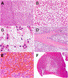
Figure 1.
Figure 1.. Cytoplasmic inclusions in tumor cells.
Panel A. Pale bodies are more common in fibrolamellar carcinoma than in conventional hepatocellular carcinoma but are not specific, as shown in this image of pale bodies in a conventional hepatocellular carcinoma. Several of the pale bodies are indicated by arrows. Panel B. Hyaline bodies are round to oval, densely pink inclusions. Several of the hyaline bodies are indicated by arrows. Panel C. Mallory Denk bodies are in tumor cells are similar to those seen in non-tumor cells, composed of ropy to chunky brightly eosinophilic material. Several Mallory Denk bodies are indicated by arrows.
Figure 2.
Figure 2.. Growth patterns in hepatocellular carcinomas.
These growth patterns can be seen in any subtype. Panel A. A solid growth pattern is shown. Panel B. A trabecular growth pattern is illustrated. Focal bile is also present. Panel C. This tumor has a pseudogland growth pattern. Panel D. A macrotrabecular growth pattern is seen.
Figure 3.
Figure 3.. Clonal progression.
This case shows a background steatohepatitic hepatocellular carcinoma (right side of image) with a nodule of poorly differentiated carcinoma that has lost the typical findings of steatohepatitic hepatocellular carcinoma (left side of image).
Figure 4.
Figure 4.. Most common subtypes of hepatocellular carcinoma.
Panel A. Steatohepatitic hepatocellular carcinomas show fat, inflammation, and often fibrosis. Panel B. Clear cell hepatocellular carcinomas have abundant clear cytoplasm. Most cases are well to moderately differentiated. Panel C. . Scirrhous hepatocellular carcinomas have abundant intratumoral fibrosis. They can closely mimic peripheral or small duct type cholangiocarcinoma. Panel D Macrotrabecular massive hepatocellular carcinoma. This hepatocellular carcinoma has small basophilic cells and a macrotrabecular growth pattern.
Figure 5.
Figure 5.. Less common subtypes of hepatocellular carcinoma.
Panel A. Fibrolamellar carcinoma. Intatumoral bands of fibrosis can be seen at low power. Panel B. Fibrolamellar carcinoma cells have abundant eosinophilic cytoplasm and prominent nucleoli. In the center top of the image, a tumor cell has abortive bile canaliculi located within the cytoplasm, a finding that is common but no specific for fibrolamellar carcinoma. Panel C. Chromophobe hepatocellular carcinoma. Scattered larger anaplastic nuclei are seen scattered in background of generally low grade nuclei—the sharp distinction of the two cell populations is captured in the term sudden anaplasia Panel D. Chromophobe hepatocellular carcinoma. Large pseudocysts are seen at low power.
Figure 6.
Figure 6.. Rare subtypes of hepatocellular carcinoma.
Panel A. Diffuse hepatocellular carcinoma. The tumor nodules are small and round, about the same size as an ordinary cirrhotic nodule. Panel B. Lymphocyte rich hepatocellular carcinoma. There is striking and diffuse lymphocytosis in the tumor sinusoids Panel C. GCSF producing hepatocellular carcinoma. The tumor sinusoids are markedly distended by neutrophils.
Figure 7.
Figure 7.. Mixed subtypes of hepatocellular carcinoma.
Panel A. Combined hepatocellular carcinoma-cholangiocarcinoma, with two distinct population of cells. Panel B. Carcinosarcoma, with two distinct populations of cells. Panel C. Mixed hepatocellular carcinoma-neuroendocrine carcinoma, with two distinct populations of cells. The small cell carcinoma is growing in small clusters in the tumor sinusoids, highlighted by arrows.

References

    1. Torbenson M, Washington K. Pathology of liver disease: advances in the last 50years. Hum Pathol 2019. - PubMed
    1. Kojiro M Pathology of Hepatocellular Carcinoma. Replika Presss Pvt. Ltd.: India, 2006. 174pp.
    1. Yeh CN, Lee WC, Jeng LB, Chen MF. Pedunculated hepatocellular carcinoma: clinicopathologic study of 18 surgically resected cases. World J Surg 2002;26:1133–8. - PubMed
    1. Nhieu JT, Renard CA, Wei Y, et al. Nuclear accumulation of mutated beta-catenin in hepatocellular carcinoma is associated with increased cell proliferation. Am J Pathol 1999;155:703–10. - PMC - PubMed
    1. Kitao A, Matsui O, Yoneda N, et al. Hepatocellular Carcinoma with beta-Catenin Mutation: Imaging and Pathologic Characteristics. Radiology 2015;275:708–17. - PubMed

Publication types

MeSH terms