

Reticular Pseudodrusen Characteristics and Associations in the Carotenoids in Age-Related Eye Disease Study 2 (CAREDS2), an Ancillary Study of the Women's Health Initiative
- PMID: 33387684
- PMCID: PMC8243566
- DOI: 10.1016/j.oret.2020.12.019
Reticular Pseudodrusen Characteristics and Associations in the Carotenoids in Age-Related Eye Disease Study 2 (CAREDS2), an Ancillary Study of the Women's Health Initiative
Abstract
Purpose: To determine the prevalence and morphologic features of reticular pseudodrusen (RPD) and their association with participant demographics and age-related macular degeneration (AMD) status in the Carotenoids in Age-Related Eye Disease Study 2 (CAREDS2) sample, an ancillary study of the Women's Health Initiative Observational Study.
Design: Cross-sectional, multicenter, natural history study.
Participants: Nine hundred and twenty-seven eyes from 466 postmenopausal women 69 to 101 years of age.
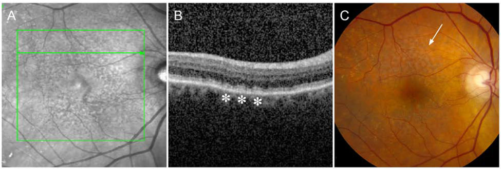
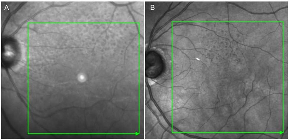
Methods: Multimodal imaging, including spectral-domain (SD) OCT and infrared reflectance (IR), were used to identify RPD characteristics, including location (within or outside the 6-mm diameter circle centered at the macula), presence of peripapillary RPD, pattern of RPD, and RPD area. Age-related macular degeneration features from SD OCT, IR, and color photographs also were assessed and AMD severity was categorized.
Main outcome measures: Reticular pseudodrusen prevalence using SD OCT and IR imaging and AMD status.
Results: Reticular pseudodrusen were present in 130 eyes (14% of eyes, 16% of participants), with increasing prevalence with age: 7% in those younger than 78 years, 14% in those 78 to 83 years of age, and 30% in those older than 83 years. Using clinical classification of AMD with color photography, RPD were seen in 2.4% of eyes with no AMD or aging changes, 11.5% in early AMD, 25.1% in intermediate AMD, and 51.1% in late AMD. Mean RPD area was 17.4 mm2 (standard deviation, 14.7 mm2). Ribbon morphologic RPD (53%) was more common than dot morphologic RPD (36%). Reticular pseudodrusen mostly were located both within and outside the 6-mm circle with primarily superior retinal distribution. Reticular pseudodrusen were visualized with corresponding color fundus photography in only 38 eyes (4% of total eyes). Participants with and without RPD had a visual acuity±standard error of 77.9 ± 1.4 letters and 81.3 ± 0.4 letters, respectively (P = 0.02).
Conclusions: The prevalence of RPD in CAREDS2 increased with age and was associated with AMD severity. Reticular pseudodrusen were detected in eyes without other features of AMD and could represent an earlier disease state. Multimodal imaging with SD OCT and IR has significantly greater sensitivity for visualizing RPD than color fundus photography.
Keywords: Age-related macular degeneration; Carotenoids in Age-Related Eye Disease Study 2; Optical coherence tomography; Reticular pseudodrusen.
Copyright © 2020 American Academy of Ophthalmology. Published by Elsevier Inc. All rights reserved.
Conflict of interest statement
Figures
Comment in
-
Reticular Pseudodrusen: Detecting a Common High-Risk Feature in Age-Related Macular Degeneration.Ophthalmol Retina. 2021 Aug;5(8):719-720. doi: 10.1016/j.oret.2021.05.005. Ophthalmol Retina. 2021. PMID: 34364530 No abstract available.
Similar articles
-
Local and Global Associations of Reticular Pseudodrusen in Age-Related Macular Degeneration.Ophthalmol Retina. 2024 Jul;8(7):646-656. doi: 10.1016/j.oret.2024.01.016. Epub 2024 Jan 24. Ophthalmol Retina. 2024. PMID: 38278174
-
Clinical and Genetic Characteristics of Japanese Patients with Age-Related Macular Degeneration and Pseudodrusen.Ophthalmology. 2016 Oct;123(10):2205-12. doi: 10.1016/j.ophtha.2016.06.052. Epub 2016 Aug 9. Ophthalmology. 2016. PMID: 27521170
-
Multimodal Imaging of Reticular Pseudodrusen in a Population-Based Setting: The Alienor Study.Invest Ophthalmol Vis Sci. 2016 Jun 1;57(7):3058-65. doi: 10.1167/iovs.16-19487. Invest Ophthalmol Vis Sci. 2016. PMID: 27367498
-
Perspectives on reticular pseudodrusen in age-related macular degeneration.Surv Ophthalmol. 2016 Sep-Oct;61(5):521-37. doi: 10.1016/j.survophthal.2016.02.005. Epub 2016 Mar 17. Surv Ophthalmol. 2016. PMID: 26994868 Review.
-
Reticular pseudodrusen in age-related macular degeneration.Optom Vis Sci. 2014 Aug;91(8):854-9. doi: 10.1097/OPX.0000000000000287. Optom Vis Sci. 2014. PMID: 24950032 Review.
Cited by
-
Reticular Pseudodrusen: The Third Macular Risk Feature for Progression to Late Age-Related Macular Degeneration: Age-Related Eye Disease Study 2 Report 30.Ophthalmology. 2022 Oct;129(10):1107-1119. doi: 10.1016/j.ophtha.2022.05.021. Epub 2022 May 31. Ophthalmology. 2022. PMID: 35660417 Free PMC article.
-
A Deep Learning Framework for the Detection and Quantification of Reticular Pseudodrusen and Drusen on Optical Coherence Tomography.Transl Vis Sci Technol. 2022 Dec 1;11(12):3. doi: 10.1167/tvst.11.12.3. Transl Vis Sci Technol. 2022. PMID: 36458946 Free PMC article.
-
Age-Related Macular Degeneration Masquerade: A Review of Pentosan Polysulfate Maculopathy and Implications for Clinical Practice.Asia Pac J Ophthalmol (Phila). 2022 Mar-Apr 01;11(2):100-110. doi: 10.1097/APO.0000000000000504. Asia Pac J Ophthalmol (Phila). 2022. PMID: 35533330 Free PMC article. Review.
-
Subretinal drusenoid deposits: An update.Taiwan J Ophthalmol. 2022 May 26;12(2):138-146. doi: 10.4103/tjo.tjo_18_22. eCollection 2022 Apr-Jun. Taiwan J Ophthalmol. 2022. PMID: 35813798 Free PMC article. Review.
-
Spatial Analysis Reveals Vascular Changes in Retinal and Choroidal Vessel Perfusion in Intermediate AMD With Reticular Pseudodrusen.Invest Ophthalmol Vis Sci. 2024 Feb 1;65(2):33. doi: 10.1167/iovs.65.2.33. Invest Ophthalmol Vis Sci. 2024. PMID: 38386332 Free PMC article.
References
Publication types
MeSH terms
Substances
Grants and funding
LinkOut - more resources
Full Text Sources
Medical
