. 2021 Feb:554:83-88.
doi: 10.1016/j.virol.2020.12.001. Epub 2020 Dec 16.

Dexamethasone inhibits SARS-CoV-2 spike pseudotyped virus viropexis by binding to ACE2

Affiliations

Dexamethasone inhibits SARS-CoV-2 spike pseudotyped virus viropexis by binding to ACE2

Yongjing Zhang et al. Virology. 2021 Feb.

Abstract

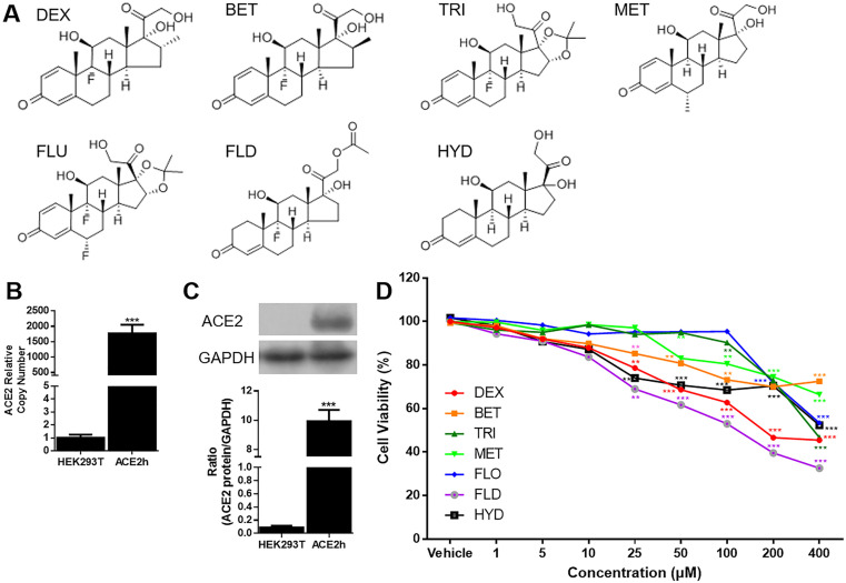
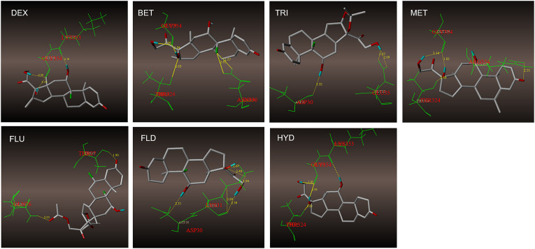
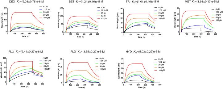
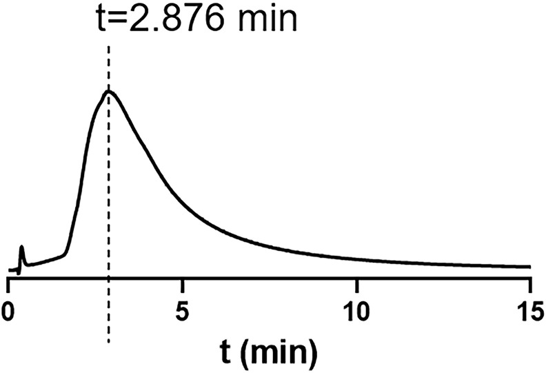
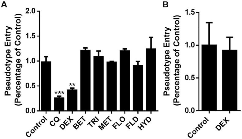
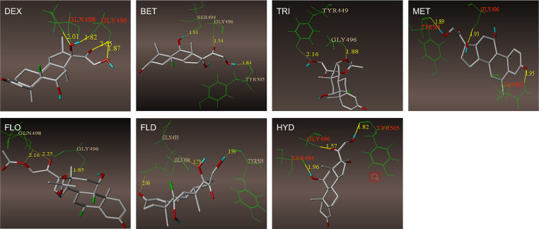
The SARS-CoV-2 outbreak, began in late 2019, has caused a worldwide pandemic and shows no signs of slowing. Glucocorticoids (GCs), including dexamethasone (DEX), have been widely used as effective anti-inflammatory and immunosuppressant drugs. In this study, seven GCs had no obvious effect on cell viability of angiotensin converting enzyme 2 (ACE2) high expressed HEK293T cells when concentrations were under 10 μM. Molecular docking results revealed that DEX occupied with active binding site of ACE2 of SARS-CoV-2 spike protein. Surface plasmon resonance (SPR) results showed that KD value between DEX and ACE2 was (9.03 ± 0.78) e-6 M. Cell membrane chromatography (CMC) results uncovered that DEX had a chromatographic retention. DEX was found out to inhibiting the viropexis into ACE2h cells using SARS-CoV-2 spike pseudotyped virus. Therefore, DEX inhibits the entrance of SARS-CoV-2 spike pseudotyped virus into cell by binding to ACE2.

Keywords: ACE2; Dexamethasone; SARS-CoV-2.


Conflict of interest statement

The authors declare that they have no known competing financial interests or personal relationships that could have appeared to influence the work reported in this paper.

Figures

Fig. 1
Fig. 1
Structure and cytotoxicity of GCs on ACE2h cells. A. Structure of 7 GCs; B. ACE2 mRNA was overexpressed in ACE2h cells compared to HEK293T cells. The experiments were repeat three times. Data are presented as mean ± S.D.; C. ACE2 was overexpressed in ACE2h cells compared to HEK293T cells. The experiments were repeat three times. Data are presented as mean ± S.D. (***p < 0.001, compared with HEK293T); (***p < 0.001, compared with HEK293T); D. Cytotoxicity of GCs on ACE2h cells under concentration from 0 to 400 μM. The experiments were repeat three times. Data are presented as mean ± S.D. (*p < 0.05, **p < 0.01, ***p < 0.001 compared to vehicle).
Fig. 2
Fig. 2
The binding sites and hydrogen bonds of 7 GCs with ACE2. A. GCs bind with ACE2 by several amino residues. DEX, TRI, MET, FLD and HYD bind to active binding sites. B. GCs bind with RBD by several amino residues. DEX, and FLO bind to active binding sites.
Fig. 3
Fig. 3
Interaction between GCs and ACE2. GCs have a certain ability to combine with ACE2 detected by SPR method. DEX showed better affinity with ACE2 than other 6 GCs, the KD value is (9.03 ± 0.78)e−6 M.
Fig. 4
Fig. 4
DEX was retained on the ACE2h-CMC column while other GCs were not. The retention time of DEX was 2.876 min.
Fig. 5
Fig. 5
Effect of GCs on the entrance of SARS-CoV-2 spike pseudotyped virus into ACE2h cells. A. DEX inhibit the entrance of SARS-CoV-2 spike pseudotyped virus into ACE2h cells; B. DEX had no effect on pseudovirus gene expression. The experiments were repeated three times. Data are presented as mean ± S.D. **p < 0.01, ***p < 0.001, compared with group 0.
figs1
figs1

References

    1. Arabi Y.M., Mandourah Y., Al-Hameed F., Sindi A.A., Almekhlafi G.A., Hussein M.A., Saudi Critical Care Trial G. Corticosteroid therapy for critically ill patients with Middle East respiratory syndrome. Am. J. Respir. Crit. Care Med. 2018;197(6):757–767. doi: 10.1164/rccm.201706-1172OC. - DOI - PubMed
    1. Ayroldi E., Cannarile L., Migliorati G., Nocentini G., Delfino D.V., Riccardi C. Mechanisms of the anti-inflammatory effects of glucocorticoids: genomic and nongenomic interference with MAPK signaling pathways. Faseb. J. 2012;26(12):4805–4820. doi: 10.1096/fj.12-216382. - DOI - PubMed
    1. Chen R.C., Tang X.P., Tan S.Y., Liang B.L., Wan Z.Y., Fang J.Q., Zhong N. Treatment of severe acute respiratory syndrome with glucosteroids: the Guangzhou experience. Chest. 2006;129(6):1441–1452. doi: 10.1378/chest.129.6.1441. - DOI - PMC - PubMed
    1. Colson P., Rolain J.M., Lagier J.C., Brouqui P., Raoult D. Chloroquine and hydroxychloroquine as available weapons to fight COVID-19. Int. J. Antimicrob. Agents. 2020;55(4):105932. doi: 10.1016/j.ijantimicag.2020.105932. - DOI - PMC - PubMed
    1. Coutinho A.E., Chapman K.E. The anti-inflammatory and immunosuppressive effects of glucocorticoids, recent developments and mechanistic insights. Mol. Cell. Endocrinol. 2011;335(1):2–13. doi: 10.1016/j.mce.2010.04.005. - DOI - PMC - PubMed

Publication types

MeSH terms