Review
. 2021 Jan 2;19(1):2.
doi: 10.1186/s12957-020-02098-x.

Heterotopic ossification in lymph node metastasis after rectal cancer resection: a case report and literature review

Affiliations
Review

Heterotopic ossification in lymph node metastasis after rectal cancer resection: a case report and literature review

Hideki Nagano et al. World J Surg Oncol. .

Abstract

Background: Heterotopic ossification (HO) is the formation of osseous tissue outside the skeleton. HO in malignant tumors of the digestive tract is extremely rare, as is ossification in metastatic lesions from HO-negative digestive tract tumors. Regarding the pathogenesis of HO, two theories have been proposed. The first is that the osteoblastic metaplasia of tumor cells (driven by the epithelial-mesenchymal transition, EMT) results in HO, and the second is that factors secreted by cancer cells lead to the metaplasia of stromal pluripotent cells into osteoblasts. However, the osteogenic mechanisms remain unclear.

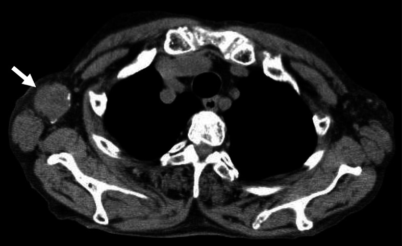
Case presentation: An 83-year-old Japanese woman underwent low anterior rectal resection for rectal cancer before presentation at our institution, in June 2018. The final diagnosis was stage IIB rectal adenocarcinoma (T4aN0M0). Histological examination did not reveal HO in the primary tumor. Thirteen months after the operation, a solitary metastatic lesion in the brain 20 mm in size and a solitary metastatic lesion in a right axillary lymph node 20 mm in size were diagnosed. The patient was treated with gamma-knife therapy for the brain metastasis. One month later, she was referred to our institution. She underwent lymph node resection. Histological examination revealed that most portions of the affected lymph node were occupied by metastatic tumor cells and that central necrosis and four small ossified lesions without an osteoblast-like cell rim were present in the peripheral region. Immunohistochemical analysis showed tumor cells positive for BMP-2, osteonectin, osteocalcin, AE1/AE3, TGF-β1, Gli2, Smad2/3, and CDX2 and negative for nestin, CD56, and CK7.

Conclusion: This is the first English case report of HO in a metachronous metastatic lymph node after the curative resection of HO-negative rectal cancer. Unlike HO lesions in past reports, the HO lesion did not show peripheral osteoblast-like cells, and the immunohistochemical findings indicated that the present case resulted from the EMT.

Keywords: BMP-2; Epithelial-mesenchymal transition; Gli2; Heterotopic ossification; Lymph node; Osteoblast-like cell; Rectal cancer; TGF-β1.


Conflict of interest statement

The authors have no competing interests to declare.

Figures

Fig. 1
Fig. 1
Findings of CT performed in October 2019. A solitary swollen lymph node measuring 37 × 33 mm in size with small high-density spots in the peripheral region was observed in the right axillary region (arrow)
Fig. 2
Fig. 2
a Gross appearance of resected rectal cancer. The resected specimen contains a polypoid tumor accompanied by an ulcer with a fungating polypoid component, measuring 48 × 46 mm. b Microscopy of the primary tumor. The primary tumor was mainly composed of moderately differentiated adenocarcinoma. c In a portion of the tumor, poorly differentiated carcinoma components were found. d Scattered lymphatic invasion was found (immunostaining by D2-40)
Fig. 3
Fig. 3
Microscopic findings of the axillary lymph node. a Metastatic tumor cells contained mucin as indicated by D-PAS staining; however, mucin retention in the stroma was not observed. b The tumor cells were also found positive for mucin by alcian blue staining. c Necrosis was observed in the central region of the node, and the metastasized tumor cells were directly adjacent. d In the peripheral region of the lymph node, four small ossified lesions were found, and the metastasized tumor cells were directly adjacent to ossified nests (arrow) (× 40). e Osteocytes were observed inside the osseous matrix; however, osteoblast-like cells were not found around the HO lesion (× 100). The upper right is a magnified view of the tumor cells beside the HO lesion
Fig. 4
Fig. 4
Findings of Azan staining and immunohistochemical staining of the axillary lymph node. a Azan staining showed that the metastatic tumor lesion was collagen rich (× 40). b Immunohistochemical staining using an antibody for chromogranin A showed negative staining in the metastatic tumor lesion as well as in the primary lesion. c The tumor cells also showed negative staining for synaptophysin. d The primary tumor cells showed positive staining for CDX2 in the nucleus (× 200). e The tumor cells in the lymph node also showed positive staining for CDX2 (× 200). f, g The tumor cells in the lymph node were negative for osteoblast marker CD56 (f) and breast cancer marker CK7 (g). h Cells positive for the macrophage cell marker CD68 were observed around the osseous lesion. i CD68-positive cells were also seen in the primary tumor. j The tumor cells showed BMP-2 overexpression in the lymph node (× 400). k, l BMP-2 expression was weakly positive in the cell membrane on the lumen side of the primary tumor (k: × 400) and adjacent normal mucosa (l: × 400). m, n Osteonectin expression was strongly positive (m: × 200) and osteocalcin expression was weakly positive (n: × 200) in the tumor cells. o, p The tumor cells expressed cytokeratin (AE1/AE3) (o: × 200) and TGF-β1 (p: × 200; *HO tissue). In the HO lesion, cytokeratin-positive osteocytes were observed (o: arrows). q, r Gli2 (q: × 200) and pSmad2/3 (r: × 200) showed overexpression in the nucleus of the tumor cells (*HO tissue). The upper right of q and r are magnified views (× 400). s The tumor cells showed no nestin expression (× 200). t Fibroblasts surrounding the tumor cells showed alpha-smooth muscle actin (α-SMA) expression (× 100)
Fig. 5
Fig. 5
A schematic representation of the HO in the present case. Although the primary tumor did not produce BMP-2 nor show HO, the metastasized tumor cells acquired the ability to express BMP-2 and TGF-β1. TGF-β1 stimulated the expression of BMP-2 and Gli2, and Gli2 stimulated the expression of BMP-2. BMP-2 induced the production of bone material, such as osteonectin or osteocalcin. BMP-2 and TGF-β1 were secreted outside the tumor cells and transformed fibroblasts and, probably, pluripotent stem cells into osteocytes. In addition, Gli2 stimulated the Smad pathway in the nucleus and induced bone formation through the RUNX2 pathway. Chemotherapy might contribute to Gli2 overexpression by activating the Hedgehog pathway. These findings provide evidence that HO can occur independently, without the transformation of pluripotent stem cells into osteoblast-like cells, and that the present case resulted from osteogenesis via the EMT. In contrast, most previously reported cases resulted from the metaplasia of stromal cells into osteoblasts
Fig. 6
Fig. 6
CT findings of the paraffin block of the rectal cancer, regional lymph nodes, and axillary lymph node. a The specimen blocks were set on the CT. Two left lower blocks, outlined in red, are of axillary lymph node with ossification. b The yellow horizontal line of the left image indicates the scan level of the CT. The left-hand side of the right image shows the CT of the axillary lymph node, and a minute high-density spot was apparent in the left specimen (arrow); however, not such spot was observed in the primary tumor specimen

Similar articles

Cited by

References

    1. Richard Sheng PH, Brown R, Buryanek J. Heterotopic ossification in metastatic colorectal carcinoma: case report with morphoproteomic insights into the histogenesis. Ann Clin Lab Sci. 2014;44:1. - PubMed
    1. Goswami M. Rectal adenocarcinoma with heterotopic ossification in metastatic lymph nodes: an unusual case. Nat J Lab Med. 2017;6:PL01–PL02.
    1. Clark A. Heterotopic bone formation associated with adenocarcinoma in an abdominal scar. Br J Surg. 1935;22:889–890. doi: 10.1002/bjs.1800228825. - DOI
    1. An T, Grathwohl M, Frable WJ. Breast carcinoma with osseous metaplasia: an electron microscope study. Am J Clin Pathol. 1983;80:127–132. - PubMed
    1. Maeda M, Kanayama M, Uchida T. A case of hepatocellular carcinoma associated with ossification. Cancer. 1986;57:134–137. doi: 10.1002/1097-0142(19860101)57:1<134::AID-CNCR2820570127>3.0.CO;2-0. - DOI - PubMed