. 2021 Feb 10;29(2):222-235.e4.
doi: 10.1016/j.chom.2020.12.016. Epub 2020 Dec 26.

Induction of alarmin S100A8/A9 mediates activation of aberrant neutrophils in the pathogenesis of COVID-19

Affiliations

Induction of alarmin S100A8/A9 mediates activation of aberrant neutrophils in the pathogenesis of COVID-19

Qirui Guo et al. Cell Host Microbe. .

Abstract

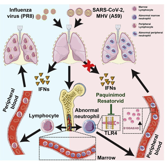
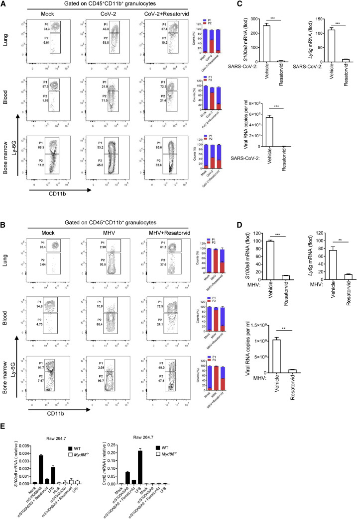
The severe acute respiratory syndrome coronavirus 2 (SARS-CoV-2) pandemic poses an unprecedented public health crisis. Evidence suggests that SARS-CoV-2 infection causes dysregulation of the immune system. However, the unique signature of early immune responses remains elusive. We characterized the transcriptome of rhesus macaques and mice infected with SARS-CoV-2. Alarmin S100A8 was robustly induced in SARS-CoV-2-infected animal models as well as in COVID-19 patients. Paquinimod, a specific inhibitor of S100A8/A9, could rescue the pneumonia with substantial reduction of viral loads in SARS-CoV-2-infected mice. Remarkably, Paquinimod treatment resulted in almost 100% survival in a lethal model of mouse coronavirus infection using the mouse hepatitis virus (MHV). A group of neutrophils that contributes to the uncontrolled pathological damage and onset of COVID-19 was dramatically induced by coronavirus infection. Paquinimod treatment could reduce these neutrophils and regain anti-viral responses, unveiling key roles of S100A8/A9 and aberrant neutrophils in the pathogenesis of COVID-19, highlighting new opportunities for therapeutic intervention.

Keywords: Paquinimod; S100A8/A9; SARS-CoV-2; aberrant neutrophils.


Conflict of interest statement

Declaration of interests The authors have no conflicts of interest to declare.

Figures

None
Graphical abstract
Figure 1
Figure 1
S100A8 and neutrophil-related gene expressions were significantly induced in the early stage of SARS-CoV-2 infection (A) A flow chart depicting the process of animal experiments with rhesus macaques. Rhesus macaques (3–4 years old) were challenged with 106 TCID50 SARS-CoV-2 virus by intratracheal routes. (B) Go analysis of the differences in rhesus macaques infected with SARS-CoV-2 in comparison with mock (fold change [FC] > 4 or < 0.25; p value < 0.05). (C) Analysis of neutrophil marker genes expression. n = 4. (D) Volcano plots show differentially expressed genes in rhesus macaques infected with SARS-CoV-2 at 3 and 5 dpi. S100A8 expression is significantly increased after SARS-CoV-2 infection. (E) Analysis of all known alarmins showing that S100A8 is the most significantly induced one. (F) qRT-PCR analysis for viral loads and the expression of S100A8 and S100A9 in the lungs of SARS-CoV-2-infected rhesus macaques at 3 and 5 dpi. n = 3. (G) Heatmap shows alarmins in the blood from rhesus macaques infected with SARS-CoV-2 at 5 dpi (left). The expression of neutrophil marker genes was analyzed (FC to mock, right). n = 4. (H) Heatmap depicting the expression of alarmins of the lung samples from healthy control and post-mortem lung samples from COVID-19 patients (left). The expression of neutrophil marker genes analyzed (FC to healthy control, right). Data from the lungs of COVID-19 patients and healthy control correspond to GEO: GSE147507. p < 0.05; ∗∗p < 0.01; ∗∗∗p < 0.001. Error bars, SD.
Figure 2
Figure 2
Anti-viral innate immune disorder and aberrant S100a8 expression induced by coronavirus but no other viruses (A) A flow chart depicting the process of animal experiments with mice. All the mice were inoculated intranasally with viruses. Virus doses: SARS-CoV-2 (105 TCID50), IAV (105 TCID50), EMCV (107 plaque-forming units [PFU]), HSV-1 (106 PFU), and MHV-A59 (105 PFU). (B) RNA-seq analysis of lungs from mice infected with SARS-CoV-2 or IAV. Go analysis was performed with the differentially expressed genes and compared with mock (FC > 4 or < 0.25; p value < 0.05). n = 3. (C) qRT-PCR analysis for the expression of S100a8, Ly6g, Ifnb, and Isg15 in the lungs of mice at different time points after IAV or SARS-CoV-2 infection. n = 3. (D) Heatmap depicting the expression differences of neutrophil marker genes in the lungs of mice infected with IAV or SARS-CoV-2. (E) qRT-PCR analysis for the expression of S100a8 and Ly6g in the blood and lungs of mice infected with different viruses. n = 3. (F) Post-infection survival curves of wild-type mice and Ifnar−/− mice infected with MHV. n = 10. (G) RNA-seq analysis of the lungs of Ifnar−/− mice infected with MHV at 5 dpi. Go and KEGG analysis were performed with the differentially expressed genes and compared with mock (FC > 4 or < 0.25; p value < 0.05). (H) qRT-PCR analysis for the expression of Ifnb, Isg15, S100a8, and Ly6g in the lungs of Ifnar−/− mice infected with IAV or MHV at 5 dpi. n = 3. p < 0.05; ∗∗p < 0.01; ∗∗∗p < 0.001. Error bars, SD.
Figure 3
Figure 3
A group of immature aberrant neutrophils emerged in coronavirus-infected mice (A) Immunohistochemical analysis of the location and expression of S100A8 in the lung tissue of mice infected with SARS-CoV-2 or MHV at 5 dpi. The S100A8+ cells in the lungs of mice infected with coronavirus were increased significantly. The red arrows indicate the S100A8+ cells. n = 5. Scale bars, 50 μm. (B) Flow cytometry analysis of neutrophils in lungs from mice infected with SARS-CoV-2 and MHV at 5 dpi. Control group means mice treated with vehicle. Gate P1 shows the conventional neutrophils (CD45+CD11b+Ly6Ghigh), and Gate P2 shows the pathologic aberrant neutrophils (CD45+CD11b+Ly6Gvariable). Aberrant neutrophils (P2) in the lungs of mice infected with coronavirus were significantly increased. n = 3. (C) Flow cytometry analysis of neutrophils in lungs of mice challenged with IAV, EMCV, HSV-1, and LPS at 5 dpi. The results showed that these treatments did not induce an increase in aberrant neutrophils. n = 3. (D) Flow cytometry analysis of neutrophils in bone marrow from mice infected with SARS-CoV-2 and MHV at 5 dpi. n = 3. (E) qRT-PCR-analyzed related gene expression of aberrant neutrophils in bone marrow of mice infected with MHV at 5 dpi and identified the differentiated types of aberrant neutrophils. n = 3. ∗∗∗p < 0.001. Error bars, SD.
Figure 4
Figure 4
Paquinimod rescues the mice infected by SARS-CoV-2 and MHV (A) A flow chart depicting the process of a drug rescue experiment. (B) Analysis of weight and survival rate of mice infected with SARS-CoV-2 and MHV after Paquinimod treatment. n = 6. (C) Analysis of the rescue effect of Paquinimod by H&E staining and pathology score of lung tissue in mice infected with SARS-CoV-2 or MHV at 5 dpi. Paquinimod treatment significantly prevented the bleeding and fibrosis in lung tissue. A number of pulmonary H&E staining images were randomly selected for pathological scoring. n = 10. Scale bars, 100 μm. (D) Immunohistochemical analysis of the S100A8+ cells in the lung tissue of mice infected with SARS-CoV-2 or MHV at 5 dpi after Paquinimod treatment. The S100A8+ cells in the lungs of mice infected with coronavirus were decreased significantly after Paquinimod treatment. The red arrows indicate the S100A8+ cells. n = 5. Scale bars, 50 μm. (E) qRT-PCR analysis for the expression of S100a8 and Ly6g in the lung of mice infected with SARS-CoV-2 at 5 dpi after Paquinimod treatment. n = 3. (F) qRT-PCR analysis of viral loads in the lungs of mice infected with coronavirus SARS-CoV-2 and MHV at 5 dpi after Paquinimod treatment. n = 3. (G) Flow cytometry analysis of neutrophils in lungs, blood, and bone marrow from mice infected with SARS-CoV-2 at 5 dpi after Paquinimod treatment. CD45+CD11b+Ly6Gvariable aberrant neutrophils (P2) in the mice infected with SARS-CoV-2 were significantly decreased by Paquinimod treatment. n = 3. (H) Heatmap depicting a decrease in B cell-related gene expression in the lungs of mice infected with SARS-CoV-2. (I) qRT-PCR analysis for the expression of B cell marker gene Cd19 in the peripheral blood of mice infected with SARS-CoV-2 after Paquinimod treatment. n ≥ 5. p < 0.05; ∗∗p < 0.01; ∗∗∗p < 0.001. Error bars, SD.
Figure 5
Figure 5
Blocking TLR4 signal can alleviate coronavirus fatal infection (A and B) Flow cytometry analysis of neutrophils in lungs, blood, and bone marrow from mice infected with SARS-CoV-2 (A) and MHV (B) at 5 dpi after Resatorvid treatment. Aberrant neutrophils (P2) in the mice infected with coronavirus were significantly decreased by Resatorvid treatment. n = 3. (C and D) qRT-PCR analysis for the expression of S100a8, Ly6g, and viral loads in the lungs of mice infected with SARS-CoV-2 (C) and MHV (D) at 5 dpi after Resatorvid treatment. n = 3. (E) qRT-PCR analysis for the effect of recombinant S100A8/A9 on S100a8 and Cxcl2 expression through TLR4 pathway. n = 3. p < 0.05; ∗∗p < 0.01; ∗∗∗p < 0.001. Error bars, SD.

Similar articles

Cited by

References

    1. Akira S., Uematsu S., Takeuchi O. Pathogen recognition and innate immunity. Cell. 2006;124:783–801. - PubMed
    1. Ali R.A., Gandhi A.A., Meng H., Yalavarthi S., Vreede A.P., Estes S.K., Palmer O.R., Bockenstedt P.L., Pinsky D.J., Greve J.M., et al. Adenosine receptor agonism protects against NETosis and thrombosis in antiphospholipid syndrome. Nat. Commun. 2019;10:1916. - PMC - PubMed
    1. Bao L., Deng W., Huang B., Gao H., Liu J., Ren L., Wei Q., Yu P., Xu Y., Qi F., et al. The pathogenicity of SARS-CoV-2 in hACE2 transgenic mice. Nature. 2020;583:830–833. - PubMed
    1. Bhattacharya M., Sharma A.R., Mallick B., Sharma G., Lee S.S., Chakraborty C. Immunoinformatics approach to understand molecular interaction between multi-epitopic regions of SARS-CoV-2 spike-protein with TLR4/MD-2 complex. Infect. Genet. Evol. 2020;85:104587. - PMC - PubMed
    1. Bianchi M.E. DAMPs, PAMPs and alarmins: all we need to know about danger. J. Leukoc. Biol. 2007;81:1–5. - PubMed

Publication types