. 2021 Apr 7;29(4):1395-1410.
doi: 10.1016/j.ymthe.2020.12.035. Epub 2021 Jan 1.

Telomerase therapy attenuates cardiotoxic effects of doxorubicin

Affiliations

Telomerase therapy attenuates cardiotoxic effects of doxorubicin

Shambhabi Chatterjee et al. Mol Ther. .

Abstract

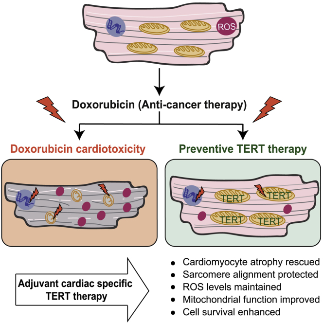
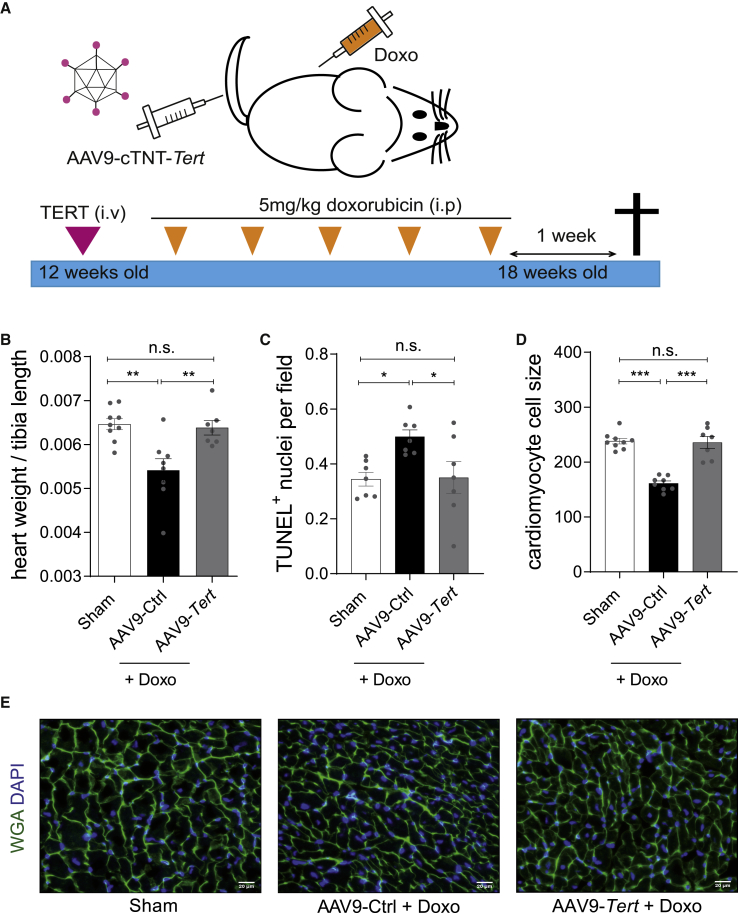
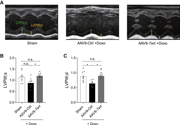
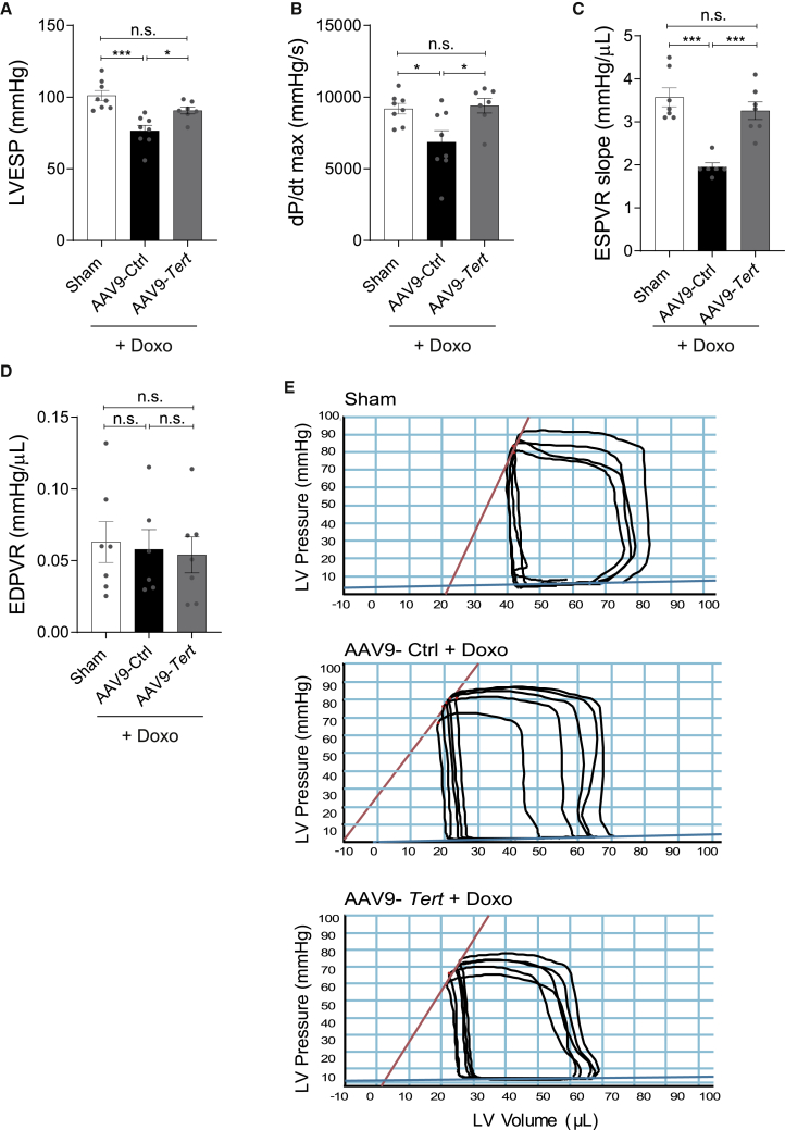
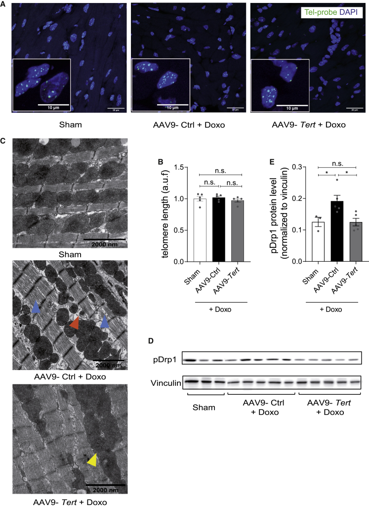
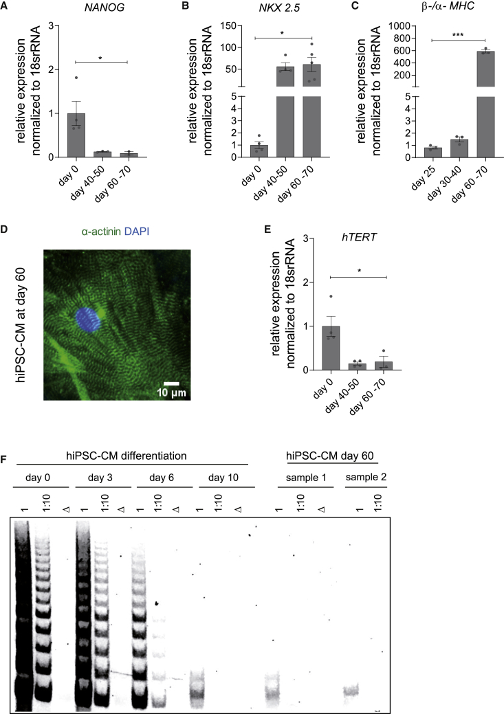
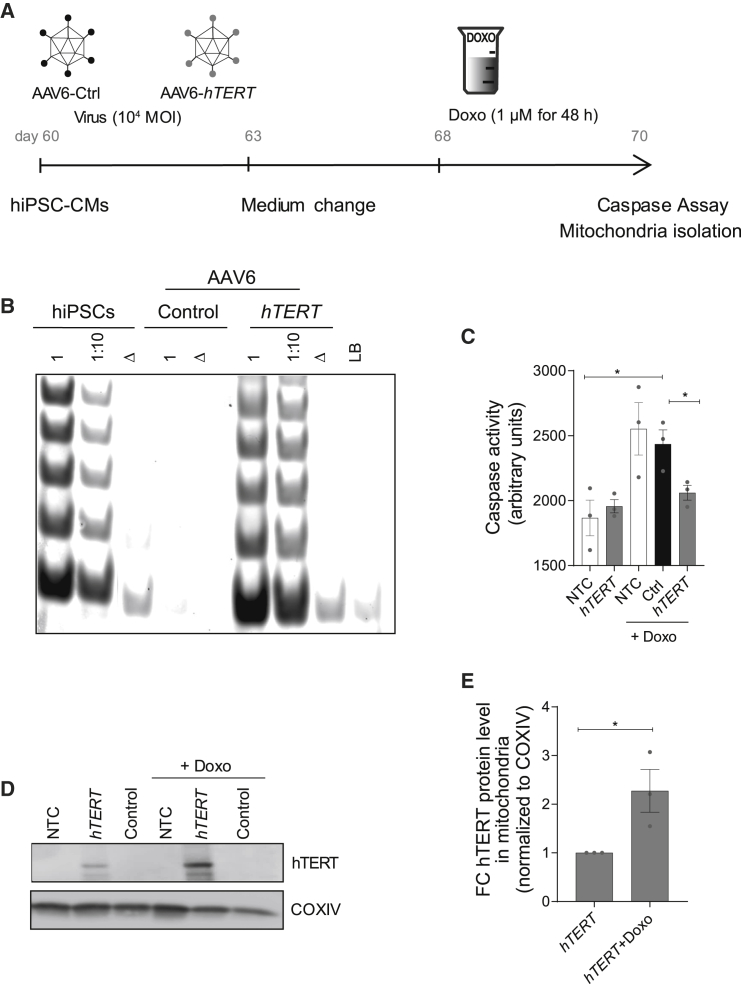
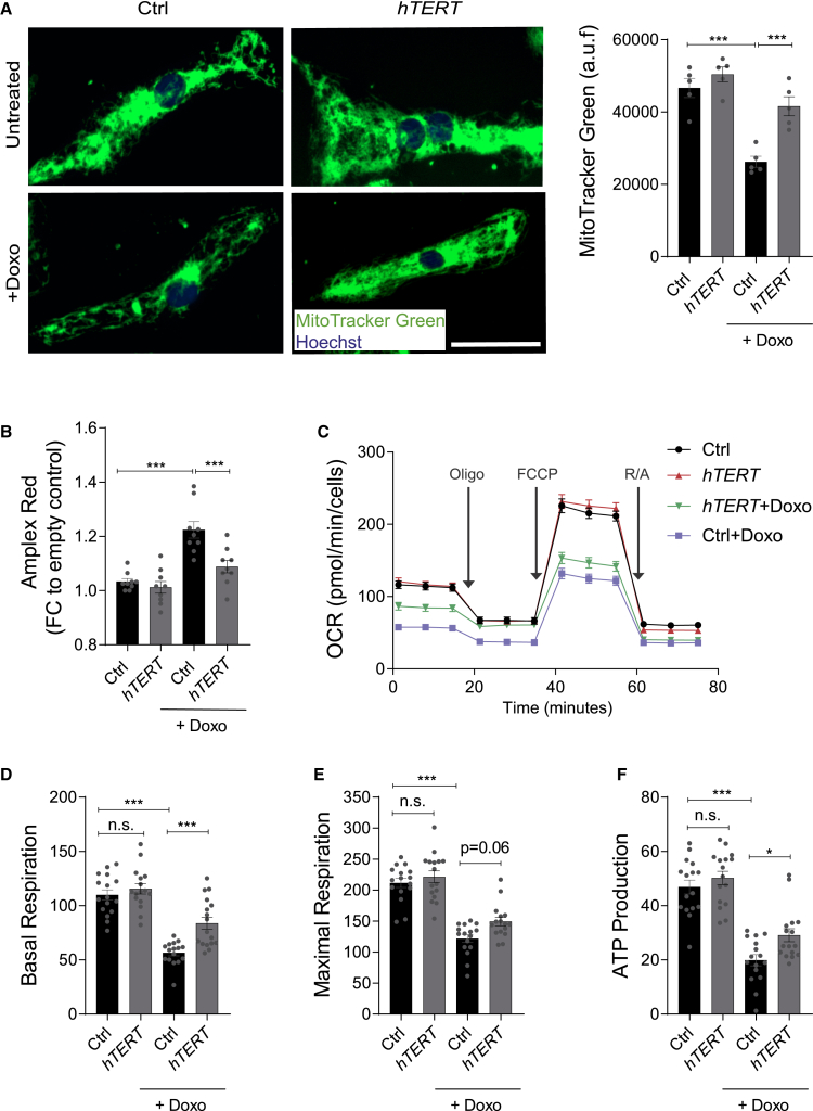
Doxorubicin is one of the most potent chemotherapeutic agents. However, its clinical use is restricted due to the severe risk of cardiotoxicity, partially attributed to elevated production of reactive oxygen species (ROS). Telomerase canonically maintains telomeres during cell division but is silenced in adult hearts. In non-dividing cells such as cardiomyocytes, telomerase confers pro-survival traits, likely owing to the detoxification of ROS. Therefore, we hypothesized that pharmacological overexpression of telomerase may be used as a therapeutic strategy for the prevention of doxorubicin-induced cardiotoxicity. We used adeno-associated virus (AAV)-mediated gene therapy for long-term expression of telomerase in in vitro and in vivo models of doxorubicin-induced cardiotoxicity. Overexpression of telomerase protected the heart from doxorubicin-mediated apoptosis and rescued cardiac function, which was accompanied by preserved cardiomyocyte size. At the mechanistic level, we observed altered mitochondrial morphology and dynamics in response to telomerase expression. Complementary in vitro experiments confirmed the anti-apoptotic effects of telomerase overexpression in human induced pluripotent stem cell-derived cardiomyocytes after doxorubicin treatment. Strikingly, elevated levels of telomerase translocated to the mitochondria upon doxorubicin treatment, which helped to maintain mitochondrial function. Thus, telomerase gene therapy could be a novel preventive strategy for cardiotoxicity by chemotherapy agents such as the anthracyclines.

Keywords: AAV gene therapy; ROS; anthracyclin; cancer; cardio-oncology; doxorubicin cardiotoxicity; heart failure; mitochondria; telomerase; telomere.


Conflict of interest statement

Declaration of interests S.B. and T.T. are co-founders and hold shares of Cardior Pharmaceuticals GmbH. T.T. filed and licensed patents on noncoding RNAs (outside of this paper). C. Bär. has filed and licensed patents on the therapeutic use of AAV9-mediated delivery of telomerase. The other authors declare no competing interests.

Figures

None
Graphical abstract
Figure 1
Figure 1
AAV9-Tert prevents doxorubicin-induced cardiac apoptosis and atrophy (A) The mouse model of doxorubicin-induced toxicity used to study the therapeutic effects of telomerase overexpression in vivo. Mice (12 weeks) were injected with AAV9-Tert (intravenously), and 5 mg/kg doxorubicin (intraperitoneally) was administered for 5 consecutive weeks. One week later, the animals were euthanized, and tissue samples were harvested for further analysis. (B) Decline in heart weight upon doxorubicin administration was preserved in mice injected with AAV9-Tert (n = 9/8/7 mice). (C) The AAV9-Tert therapy rescued cell death as measured using TUNEL assay (n = 7 mice in each group). (D) The AAV9-Tert therapy rescued CM cell size (n = 9/8/7 mice). The average cell size from each animal (n = 30 cells) has been plotted. (E) Representative images of WGA staining of transverse heart sections used to measure cell size. AAV9 particles were injected at a dose of 1 × 1012 vg/mouse. Doxo, doxorubicin. ∗p < 0.05; ∗∗p < 0.01; ∗∗∗p < 0.001; n.s., not significant; one-way ANOVA, Tukey multiple-comparisons test.
Figure 2
Figure 2
AAV9-mediated Tert overexpression rescues cardiac atrophy in doxorubicin-induced cardiotoxicity model (A) M-mode echocardiography of mice injected with Tert and then treated with saline (Sham) or injected with AAV9-Tert or control virus followed by doxorubicin treatment. Arrows indicate systole (green) and diastole (yellow) and highlight the difference. (B and C) Left ventricular posterior wall at end-systole and -diastole was assessed by echocardiography (n = 9/8/7 mice). AAV9 particles were injected at a dose of 1 × 1012 vg/mouse. LVPW, left ventricular posterior wall at end-systole (s) or -diastole (d). ∗p < 0.05; one-way ANOVA, Tukey multiple-comparisons test.
Figure 3
Figure 3
Effect of AAV9-mediated Tert overexpression on doxorubicin-induced systolic dysfunction (A–E) Measures of global systolic function (LVESP and dP/dT) (n = 8/8/7 mice) (A and B); load-independent systolic and diastolic parameters (ESPVR, EDPVR) (n = 7/6/7 mice) (C and D); and representative pressure-volume (PV) loops (E) obtained with a PV conductance catheter system at varying preload using transient vena cava occlusion, showing differences between mice injected with AAV9-Tert or control virus and then treated with saline or doxorubicin. AAV9 particles were injected at a dose of 1 × 1012 vg/mouse. LVESP, left ventricular end-systolic pressure; dP/dt max, ventricular contractility assessment; ESPVR, end-systolic pressure volume relationship; EDPVR, end-diastolic pressure volume relationship. ∗p < 0.05; ∗∗p < 0.01; ∗∗∗p < 0.001; one-way ANOVA, Tukey multiple-comparisons test.
Figure 4
Figure 4
AAV9-mediated Tert cardioprotection is conferred by non-canonical telomerase functions at the mitochondria (A and B) The telomere length in mouse heart does not increase upon overexpression of Tert as measured by qFISH (n = 5 mice/group). Scale bars indicate 20 μm, and, for insets, 10 μm. (C) Ultra-structure analysis using transmission electron microscopy indicated that the mitochondria undergo more fission post doxorubicin treatment (orange arrowhead), which was rescued partially by telomerase therapy (yellow arrowhead). Blue arrowhead indicates disrupted myofibril alignment. Scale bars indicate 2 μm. (D and E) Western blot depicting the levels of phosphorylated Drp1 (pDrp1), mitochondrial fission protein (n = 3/5/5 mice/group). AAV9 particles were injected at a dose of 1 × 1012 vg/mouse. a.u.f = arbitrary unit of fluorescence. ∗p < 0.05; one-way ANOVA, Tukey multiple-comparisons test.
Figure 5
Figure 5
Establishment of hiPSC-CMs as a translational research platform for AAV6-mediated hTERT overexpression (A–C) Expression of pluripotency marker (NANOG) (n = 4/3/2 independent differentiation experiments) (A), cardiac-specific lineage marker (NKX2.5) (n = 4/4/5 independent differentiation experiments) (B), and ratio of myosin heavy-chain isoforms (β-/α-MHC) (n = 3 independent differentiation experiments) (C) during differentiation and prolonged cultivation of hiPSC-CMs. (D) Purified hiPSC-CMs at day 60 exhibited well-organized sarcomere structures as seen by α-actinin immunostaining. (E) The mRNA expression of hTERT during CM differentiation protocol indicated downregulation by day 60 (n = 4/4/3 independent differentiation experiments). (F) Telomerase activity from cell lysates collected at various time points of the differentiation timeline showed a gradual decrease in TRAP activity by day 10 and undetectable by day 60. Δ indicates heat-inactivated cell lysate; 1 indicates undiluted cell lysate; 1:10 indicates cell lysate diluted by factor 10; scale bar indicates 10 μm. All data are mean fold change relative to control ± SEM. ∗p < 0.05; ∗∗∗p < 0.001; two-tailed unpaired t test with Welch’s correction.
Figure 6
Figure 6
AAV6-hTERT inhibits doxorubicin-induced apoptosis via regulating mitochondrial dynamics (A) Schematic of experimental strategy for investigating cardioprotection post doxorubicin-induced stress. Overview of experimental time course for 7 day virus transduction (MOI 104) and 48 h doxorubicin (1 μM) treatment in hiPSC-CMs. (B) Restoration of TRAP activity mediated via AAV6-hTERT transduction compared to AAV6 control. (C) Cell death measured by caspase activity before and after doxorubicin treatment in the presence and absence of AAV6-hTERT therapy indicated enhanced survival in the presence of hTERT overexpression. (D) Western blot indicating presence of TERT protein in the mitochondrial compartment, which further increases upon doxorubicin treatment. (E) Increase in hTERT protein levels within the mitochondria as measured from western blot. All data are mean relative to control ± SEM (n = 3 independent differentiation experiments). NTC, no virus treatment control; FC, fold change. ∗p < 0.05; ∗∗p < 0.01; ∗∗∗p < 0.001; two-tailed unpaired t test.
Figure 7
Figure 7
AAV6-hTERT protects from ROS and preserves mitochondrial metabolism post doxorubicin-induced cardiotoxicity (A) HiPSC-CMs overexpressing hTERT stained with MitoTracker Green FM in presence and absence of doxorubicin (1 μM, 48 h) and compared to control group. The fluorescence signal of MitoTracker Green FM from images on the left are quantified and represented on the right, which indicate mitochondrial content (n = 5 wells/group with 100,000 cells in each well from one differentiation experiment). Scale bar is 50 μm. (B) Bar graph represents the fold change of the extracellular ROS measured by Amplex Red assay before and after doxorubicin treatment in the presence and absence of AAV6-hTERT. The results indicate lower ROS levels after doxorubicin (1 μM) in the presence of hTERT. (n = 9 wells/group from triplicates of 3 independent differentiation experiments). (C) Analysis of hiPSC-CM mitochondrial metabolism using a Seahorse XFe96 Analyzer after transduction with AAV6 hTERT and in presence or absence of doxorubicin (1 μM). OCR was measured continuously at baseline and after addition of oligomycin (2 μM), FCCP (1 μM), and R/A (0.5 μM) (n = 16 wells/group with 50,000 cells in each well from one differentiation experiment). (D–F) The average levels of basal respiration, maximal respiration, and ATP production. The mitochondrial metabolism reduces after doxorubicin treatment, which is rescued by AAV6-hTERT therapy. All data are mean ± SEM. AAV6 transduction performed with 104 MOI. OCR, oxygen consumption rate; oligo = oligomycin; FCCP, carbonyl cyanide-4-phenylhydrazone; R/A, rotenone and antimycin A. ∗p < 0.05; ∗∗∗p < 0.001; one-way ANOVA, Tukey multiple-comparisons test.

Comment in

References

    1. Curigliano G., Cardinale D., Dent S., Criscitiello C., Aseyev O., Lenihan D., Cipolla C.M. Cardiotoxicity of anticancer treatments: Epidemiology, detection, and management. CA Cancer J. Clin. 2016;66:309–325. - PubMed
    1. Sturgeon K.M., Deng L., Bluethmann S.M., Zhou S., Trifiletti D.M., Jiang C., Kelly S.P., Zaorsky N.G. A population-based study of cardiovascular disease mortality risk in US cancer patients. Eur. Heart J. 2019;40:3889–3897. - PMC - PubMed
    1. Chatterjee S., Gupta S.K., Bär C., Thum T. Noncoding RNAs: potential regulators in cardioncology. Am. J. Physiol. Heart Circ. Physiol. 2019;316:H160–H168. - PMC - PubMed
    1. Haubner B.J., Adamowicz-Brice M., Khadayate S., Tiefenthaler V., Metzler B., Aitman T., Penninger J.M. Complete cardiac regeneration in a mouse model of myocardial infarction. Aging (Albany NY) 2012;4:966–977. - PMC - PubMed
    1. Haubner B.J., Schneider J., Schweigmann U., Schuetz T., Dichtl W., Velik-Salchner C., Stein J.I., Penninger J.M. Functional recovery of a human neonatal heart after severe myocardial infarction. Circ. Res. 2016;118:216–221. - PubMed

Publication types

MeSH terms