. 2021 Jul;70(7):1841-1851.
doi: 10.1007/s00262-020-02809-z. Epub 2021 Jan 3.

A study of the possible role of Fab-glycosylated IgG in tumor immunity

Affiliations

A study of the possible role of Fab-glycosylated IgG in tumor immunity

Qian Xu et al. Cancer Immunol Immunother. 2021 Jul.

Abstract

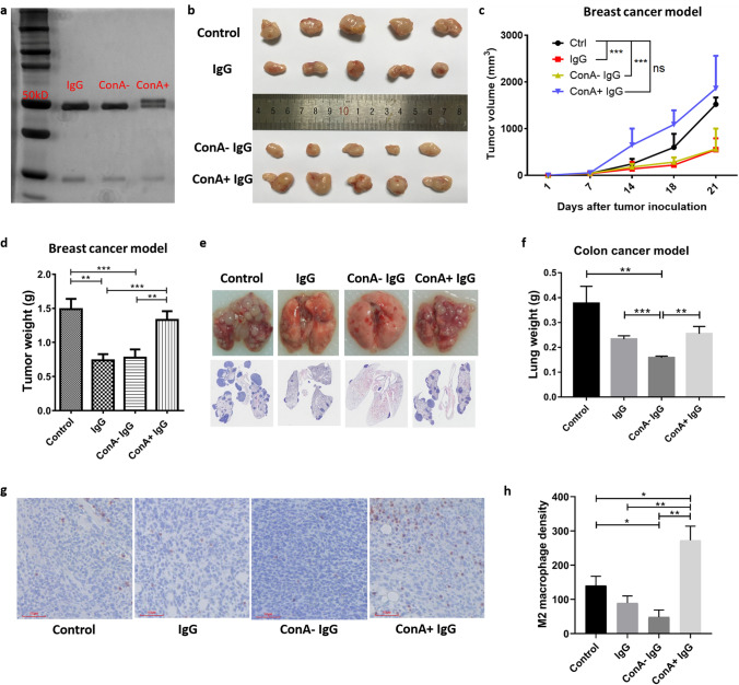
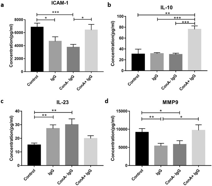
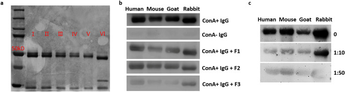
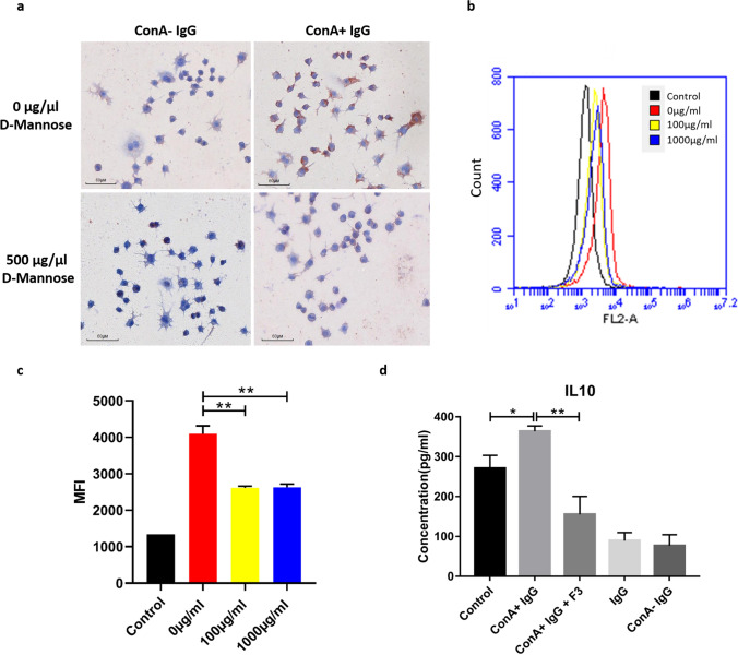
Previously we reported that administration of IgG could inhibit tumor progression in mouse models. At the same time, we also found that some IgGs have glycosylation modifications on their Fab fragments, which may have different biological functions than non-glycosylated IgG. In this study, we employed mouse tumor models to explore the roles of two different forms of IgG, i.e. Fab-glycosylated and Fab-non-glycosylated IgG, in tumor progression. The two types of IgGs were separated with ConA absorption which could react with glycan on the Fab arm but could not access glycan on the Fc fragment. In addition, we performed cytokine array, ELISA, western blotting, immunocytochemistry and other techniques to investigate the possible mechanisms of the actions of Fab-glycosylated IgG in the models. We found that Fab-glycosylated IgG, unlike Fab-non-glycosylated IgG, did not inhibit tumor growth and metastasis in the model. On the contrary, Fab-glycosylated IgG may bind to antigen-bound IgG molecules and macrophages through the glycosidic chain on the Fab fragment to affect antigen-antibody binding and macrophage polarization, which are likely to help tumor cells to evade the immune surveillance. A new mechanism of immune evasion with Fab-glycosylated IgG playing a significant role was proposed.

Keywords: Glycosylation; Immune evasion; Immunoglobulin G; Macrophage; Mouse tumor model.


Conflict of interest statement

The authors declare that they have no conflict of interest.

Figures

Fig. 1
Fig. 1
A diagram shows the experimental design of various groups of mice treated with different protocols
Fig. 2
Fig. 2
ConA+ IgG affects tumor progression by regulating TAMs polarization. The difference in molecular weight of ConA+ IgG and ConA− IgG was examined with electrophoresis and silver stain. ConA+ IgG had higher molecular weight bands above the 50 kDa heavy chain as extra N-glycans attached (a). IgG and ConA− IgG injection inhibited tumor growth in the breast cancer model (b–d) and colon cancer model (e, f), ConA+ IgG did not inhibit the growth of tumor but promoted tumor growth. The changes in tumor volume in four groups as time went on (c). CD206 immunostaining (g, h) revealed more M2 macrophages in ConA+ IgG-treated 4T1 tumor tissues than in other groups (n = 5, bar: 60 µm). All data were shown as the mean ± SEM (*p < 0.05, **p < 0.01, ***p < 0.001, ns p > 0.05)
Fig. 3
Fig. 3
Array analysis of the sera of tumor-bearing mice. The sera of mice treated with PBS (control), IgG, ConA− IgG and ConA+ IgG were measured with Mouse XL Cytokine Array analysis (a). 12 cytokines with obvious differences in expression were selected for further analysis (b). The expressions of cytokines associated with angiogenesis were increased in the ConA+ IgG group (c), which suggested that ConA+ IgG could promote tumor angiogenesis. In addition, the expressions of tumor-associated cytokines except IL-23 were increased in the ConA+ IgG groups (d), also suggested that ConA+ IgG played a role in tumor progression. The graphs (c, d) show the relative fold changes of protein concentration with significant difference upon IgG treatment. Pretreatment control was normalized to 1
Fig. 4
Fig. 4
ELISA analysis of the sera of tumor-bearing mouse. The sera of mice treated with PBS (control), IgG, ConA− IgG and ConA+ IgG were measured by ICAM-1 (a), IL-10 (b), IL-23 (c), and MMP9 (d) ELISA kit (*p < 0.05, **p < 0.01, ***p < 0.001). The results showed that the expressions of ICAM-1 and MMP9 were reduced in the IgG group and the ConA− IgG group, and the expression of IL-23 was increased in the IgG group and the ConA− IgG group. The expression of IL-10 was increased significantly in the ConA+ IgG group
Fig. 5
Fig. 5
ConA+ IgG reacts to IgG molecules from different species. ConA+ IgG treated with endoglycosidase was examined with electrophoresis and silver stain (a): ConA+ IgG (I), ConA+ IgG treated with reaction buffer (control, II), ConA+ IgG treated with endoglycosidase F1 (III), ConA+ IgG treated with endoglycosidase F2 (IV), ConA+ IgG treated with endoglycosidase F3 (V), ConA+ IgG treated with PNGase F (VI). It showed that endoglycosidase F3 removed most of the glycosidic chains attached to ConA+ IgG. ConA+ IgG, ConA− IgG, and ConA+ IgG treated with endoglycosidase F1, F2, F3 were used as the primary antibodies. The results showed that ConA+ IgG, but not ConA− IgG, reacted to IgG molecules from different species. As endoglycosidase F3 can remove the glycosidic chain of ConA+ IgG, the reaction between ConA+ IgG and IgG molecules from different species was the weakest (b). The reaction between ConA+ IgG and IgG molecules from different species was weakened after ConA+ IgG was neutralized with a large number of IgG molecules (c), indicating that ConA+ IgG reacted with IgG molecules specifically
Fig. 6
Fig. 6
ConA+ IgG binds to macrophages via mannose receptors to promote tumor progression. ConA+ IgG as a primary antibody reacted strongly with mouse macrophages, but the reaction was weakened after the macrophages were blocked by mannose (a). RAW246.7 cells were incubated with 0 µg/ml, 100 µg/ml and 1000 µg/ml d-mannose for 2 h first. Then we used PE-labeled ConA+ IgG to measure cell fluorescence intensity with Flowcytometry. Mean fluorescence intensity (MFI) represents the average binding affinity of ConA+ IgG on RAW246.7 (1 × 104 cells), and the binding affinity of ConA+ IgG became lower after d-mannose blocking (b, c, **p < 0.01). ConA+ IgG promoted the secretion of IL-10 by mouse macrophages, but this promotion was weakened after the ConA+ IgG was treated with endoglycosidase F3 to remove the glycosidic chain (d, *p < 0.05, **p < 0.01)

Similar articles

Cited by

References

    1. Poljak RJ. Three-dimensional structure, function and genetic control of immunoglobulins. Nature. 1975;256(5516):373–376. doi: 10.1038/256373a0. - DOI - PubMed
    1. Youings A, Chang SC, Dwek RA, Scragg IG. Site-specific glycosylation of human immunoglobulin G is altered in four rheumatoid arthritis patients. Biochem J. 1996;314(Pt 2):621–630. doi: 10.1042/bj3140621. - DOI - PMC - PubMed
    1. Jefferis R. Glycosylation of recombinant antibody therapeutics. Biotechnol Prog. 2005;21(1):11–16. doi: 10.1021/bp040016j. - DOI - PubMed
    1. van de Bovenkamp FS, Hafkenscheid L, Rispens T, Rombouts Y. The emerging importance of IgG Fab glycosylation in immunity. J Immunol. 2016;196(4):1435–1441. doi: 10.4049/jimmunol.1502136. - DOI - PubMed
    1. Borel IM, Gentile T, Angelucci J, Margni RA, Binaghi RA. Asymmetrically glycosylated IgG isolated from non-immune human sera. Biochem Biophys Acta. 1989;990(2):162–164. doi: 10.1016/S0304-4165(89)80029-7. - DOI - PubMed

LinkOut - more resources