

Comprehensive evaluation of corneas from normal, forme fruste keratoconus and clinical keratoconus patients using morphological and biomechanical properties
- PMID: 33389426
- PMCID: PMC8035106
- DOI: 10.1007/s10792-020-01679-9
Comprehensive evaluation of corneas from normal, forme fruste keratoconus and clinical keratoconus patients using morphological and biomechanical properties
Abstract
Objective: To more comprehensively evaluate the ability of the parameters reflecting the morphological and biomechanical properties of the cornea to distinguish clinical keratoconus (CKC) and forme fruste keratoconus (FFKC) from normal.
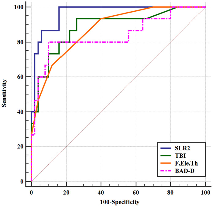
Methods: Normal eyes (n = 50), CKC (n = 45) and FFKC (n = 15) were analyzed using Pentacam, Corvis ST and ORA. Stepwise logistic regression of all parameters was performed to obtain the optimal combination model capable of distinguishing CKC, FFKC from normal, named SLR1 and SLR2, respectively. Receiver operating characteristic (ROC) curves were applied to determine the predictive accuracy of the parameters and the two combination models, as described by the area under the curve (AUC). AUCs were compared using the DeLong method.
Results: The SLR1 model included only the TBI output by Pentacam, while the SLR2 model included the morphological parameter F.Ele.Th and two parameters from the Corvis ST, HC DfA and SP-A1. The majority of the parameters had sufficient strength to differentiate the CKC from normal corneas, even the seven separate parameters and the SLR1 model had a discrimination efficiency of 100%. The predictive accuracy of the parameters was moderate for FFKC, and the SLR2 model (0.965) presented an excellent AUC, followed by TBI, F.Ele.Th and BAD-D.
Conclusion: The F.Ele.Th from Pentacam was the most sensitive morphological parameter for FFKC, and the combination of F.Ele.Th, HC DfA and SP-A1 made the diagnosis of FFKC more efficient. The CRF and CH output by ORA did not improve the combined diagnosis, despite the corneal combination of morphological and biomechanical properties that optimized the diagnosis of FFKC.
Keywords: Biomechanics; Forme fruste keratoconus; Keratoconus; Morphology.
Conflict of interest statement
The authors have no relevant financial or non-financial interests to disclose.
Figures
Similar articles
-
Progress of corneal morphological examination combined with biomechanical examination in preoperative screening for keratorefractive surgery.Indian J Ophthalmol. 2023 Jun;71(6):2369-2378. doi: 10.4103/ijo.IJO_1377_22. Indian J Ophthalmol. 2023. PMID: 37322646 Free PMC article. Review.
-
Comparison of the morphological and biomechanical characteristics of keratoconus, forme fruste keratoconus, and normal corneas.Semin Ophthalmol. 2021 Nov 17;36(8):671-678. doi: 10.1080/08820538.2021.1896752. Epub 2021 Mar 18. Semin Ophthalmol. 2021. PMID: 33734947
-
Application of a scheimpflug-based biomechanical analyser and tomography in the early detection of subclinical keratoconus in chinese patients.BMC Ophthalmol. 2021 Sep 20;21(1):339. doi: 10.1186/s12886-021-02102-2. BMC Ophthalmol. 2021. PMID: 34544392 Free PMC article.
-
An exploratory analysis of forme fruste keratoconus sensitivity diagnostic parameters.Int Ophthalmol. 2022 Aug;42(8):2473-2481. doi: 10.1007/s10792-022-02246-0. Epub 2022 Mar 5. Int Ophthalmol. 2022. PMID: 35247116
-
A Systematic Review of Subclinical Keratoconus and Forme Fruste Keratoconus.J Refract Surg. 2020 Apr 1;36(4):270-279. doi: 10.3928/1081597X-20200212-03. J Refract Surg. 2020. PMID: 32267959
Cited by
-
Progress of corneal morphological examination combined with biomechanical examination in preoperative screening for keratorefractive surgery.Indian J Ophthalmol. 2023 Jun;71(6):2369-2378. doi: 10.4103/ijo.IJO_1377_22. Indian J Ophthalmol. 2023. PMID: 37322646 Free PMC article. Review.
-
Development and validation of the VAE-NT index: a novel biomechanical parameter for distinguishing subclinical corneal abnormalities.Front Bioeng Biotechnol. 2025 Jul 16;13:1598546. doi: 10.3389/fbioe.2025.1598546. eCollection 2025. Front Bioeng Biotechnol. 2025. PMID: 40741532 Free PMC article.
-
Corneal biomechanics in early diagnosis of keratoconus using artificial intelligence.Graefes Arch Clin Exp Ophthalmol. 2024 Apr;262(4):1337-1349. doi: 10.1007/s00417-023-06307-7. Epub 2023 Nov 9. Graefes Arch Clin Exp Ophthalmol. 2024. PMID: 37943332 Review.
-
[Anisotropy and viscoelasticity of different corneal regions in rabbit corneal ectasia model].Sheng Wu Yi Xue Gong Cheng Xue Za Zhi. 2024 Feb 25;41(1):129-135. doi: 10.7507/1001-5515.202312022. Sheng Wu Yi Xue Gong Cheng Xue Za Zhi. 2024. PMID: 38403613 Free PMC article. Chinese.
-
Detection ability of corneal biomechanical parameters for early diagnosis of ectasia.Eye (Lond). 2023 Jun;37(8):1665-1672. doi: 10.1038/s41433-022-02218-9. Epub 2022 Aug 29. Eye (Lond). 2023. PMID: 36038724 Free PMC article.
References
-
- Muftuoglu O, Ayar O, Ozulken K, Ozyol E, Akıncı A. Posterior corneal elevation and back difference corneal elevation in diagnosing forme fruste keratoconus in the fellow eyes of unilateral keratoconus patients. J Cataract Refract Surg. 2013;39:1348–1357. doi: 10.1016/j.jcrs.2013.03.023. - DOI - PubMed
MeSH terms
Grants and funding
LinkOut - more resources
Full Text Sources
Other Literature Sources
Research Materials
