. 2021 Jun;17(3):981-998.
doi: 10.1007/s12015-020-10104-8. Epub 2021 Jan 3.

Human Amnion-Derived Mesenchymal Stromal Cells in Cirrhotic Patients with Refractory Ascites: A Possible Anti-Inflammatory Therapy for Preventing Spontaneous Bacterial Peritonitis

Affiliations

Human Amnion-Derived Mesenchymal Stromal Cells in Cirrhotic Patients with Refractory Ascites: A Possible Anti-Inflammatory Therapy for Preventing Spontaneous Bacterial Peritonitis

Mariangela Pampalone et al. Stem Cell Rev Rep. 2021 Jun.

Abstract

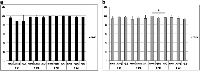
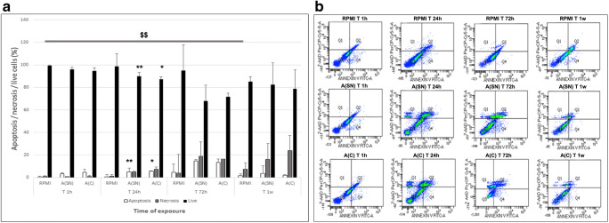
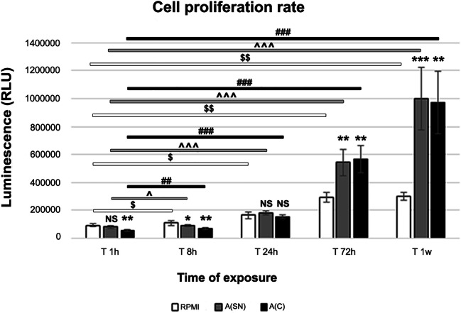
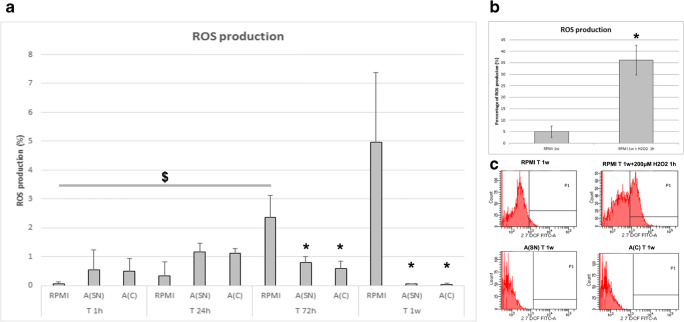
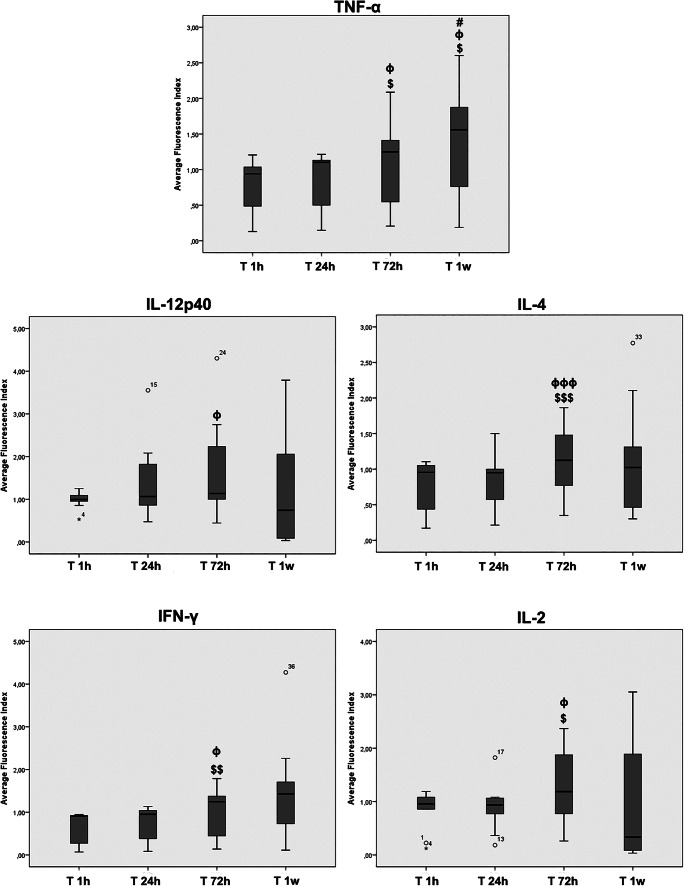
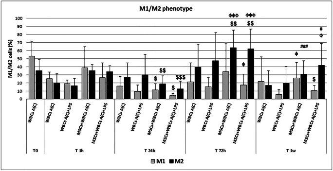
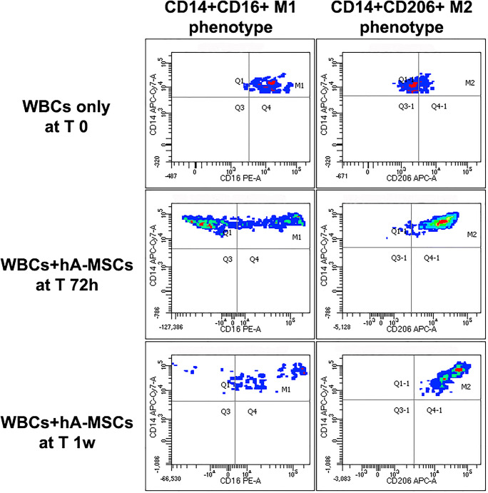
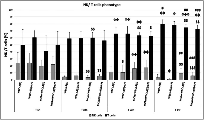
Cirrhosis is associated with dysregulated immune cell activation and immune dysfunction. These conditions modify gut flora, facilitate bacterial translocation, and increase susceptibility to bacterial peritonitis and consequent systemic infections by dramatically affecting long-term patient survival. Human amnion-derived mesenchymal stromal cells (hA-MSCs) exert immunomodulatory potential benefit, and have the ability to modulate their actions, especially in situations requiring immune activation through mechanisms not fully understood. In this study, we aimed to investigate, in vitro, the immunostimulant or immunosuppressive effects of hA-MSCs on cellular components of ascitic fluid obtained from cirrhotic patients with refractory ascites. We found that hA-MSCs viability is not affected by ascitic fluid and, interestingly, hA-MSCs diminished the pro-inflammatory cytokine production, and promoted anti-inflammatory M2 macrophage polarization. Moreover, we found that there was no simultaneous significant decrease in the M1-like component, allowing a continual phagocytosis activity of macrophages and NK cells to restore a physiological condition. These data highlight the plasticity of hA-MSCs' immunomodulatory capacity, and pave the way to further understanding their role in conditions such as spontaneous bacterial peritonitis.

Keywords: Anti-inflammatory therapy; Ascites; Cirrhosis; Human amnion-derived mesenchymal stromal cells; M2 polarization; Perinatal tissues; Placenta; Spontaneous bacterial peritonitis.


Conflict of interest statement

The authors declare no conflicts of interest.

Figures

Fig. 1
Fig. 1
Graph showing that hA-MSCs cultured in standard medium (RPMI) and AF (SN and C) maintained the expression of classical mesenchymal cell surface markers, such as CD90 (a) and CD73 (b) after 1 h, 24 h, 72 h, and 1 week, with a very slight decrease in CD73 expression after 72 h of A(C) compared with RPMI at the same time point (* p < 0.05). Also CD90 decreased after 1 week in RPMI compared with 72 h in the same condition. Values are expressed as means of percentages ± SD
Fig. 2
Fig. 2
a Graph showing the effects of AF apoptotic and necrotic events on hA-MSCs after 1 h, 24 h, 72 h, and 1 week of exposure, compared with standard culture in RPMI. A statistically significant increase of apoptosis (white) was seen after 24 h of exposure to A(SN) (** p < 0.01) and A(C) (* p < 0.05), with a concomitant decrease of live cells (black) in both A(SN) and A(C) (** p < 0.01, and * p < 0.05, respectively). However, the resistance to stressing events and decrease of apoptosis are visible as a trend after 1 week, while cells grown in RPMI showed a significant increase of necrosis (grey) ($$ p < 0.01) compared with 1 h. Values were expressed as means of percentages and SD. b Representative panel of flow cytometry analyses dot plot showing the possible adaptation of hA-MSCs to AF after long exposure
Fig. 3
Fig. 3
Graph showing proliferation rate of hA-MSCs cultured in complete standard medium (RPMI, white) and in both supernatant, A(SN) in grey, and complete, A(C) in black, ascitic fluid for 1, 8, 24, and 72 h, and 1 week, and expressed as relative luminescence units (RLU). Considering the time point of 1 h as our “time zero”, the cells grown in A(C) were immediately impaired after acute exposure at 1 h (* p < 0.01), while no significance was found when grown in A(SN). After 8 h, the cells cultured in both A(SN) and A(C) reduced their proliferation (* p < 0.05 and ** p < 0.01, respectively). The cells then increased their vitality starting at 24 h, in which no significant differences were seen compared with RPMI in each condition. The positive influence of AF on cell growth was seen after 72 h, with a p < 0.01 (**) for both A(SN) and A(C), and after 1 week, with a p < 0.001 (***) for A(SN), and p < 0.01 (**) for A(C). The increase in cell proliferation, considering the same culture condition (RPMI, $, A(SN) ^, and A(C) #) during the different time point sets, was considered statistically significant for p < 0.05 ($, ^, #), p < 0.01 ($$, ^^, ##), and p < 0.001 ($$$, ^^^, ###) compared with the” time zero” starting point of 1 h of exposure. NS: not statistically significant. Values were expressed as means of percentages ± SD
Fig. 4
Fig. 4
a Graph showing the ROS production by hA-MSCs during culture with standard medium (RPMI) and AF (SN and C) for 1 h, 24 h, 72 h, and 1 week. Significant increase was shown only during culture with RPMI after 72 h ($ p < 0.05) and with a trend of continual increase even after 1 week. Interestingly, not only did ROS never significantly increase when cells were grown in both A(SN) and A(C), but their amount significantly decreased after 72 h in both A-SN and A-C (both with p < 0.05, *), and much more also after 1 week in A-SN and A-C (both with p < 0.05, *). Values are expressed as means of percentages and SD. b Positive control of ROS production by hA-MSCs at 1 week treated with 200 μM H2O2 for 1 h and c representative plots showing the decrease in ROS production in A-SN and A-C in presence of hA-MSCs after 1 week of culture
Fig. 5
Fig. 5
Box plots showing how the inflammatory cytokine release was influenced in ascites after 1 h, 24 h, 72 h, and 1 week in the presence of hA-MSCs. Acute exposure (T 24 h) did not reflect any significant difference compared with 1 h. Significant increases were seen for TNF-α, IL-12p40, IL-4, IFN-γ, and IL-2 after 72 h. Only TNF-α showed a statistically significant increase after 1 week. Statistics were obtained comparing with 1 h ($), 24 h (ϕ), and 72 h (#).Values were statistically significant at p < 0.05 ($, ϕ, #), p < 0.01 ($$, ϕϕ, ##), and p < 0.001 ($$$, ϕϕϕ, ###). Values are expressed as means of fluorescence index± SD
Fig. 6
Fig. 6
Graph of M1 (grey) and M2 (black) phenotype expressed by WBCs derived from the ascites of cirrhotic patients after paracentesis, A(C) T0, and after co-culture with hA-MSCs for the time point chosen: 1 h, 24 h, 72 h, and 1 week. After 24 h of co-culture, a significant decrease of both M1- and M2-like cells was observed, while a boosted reaction was obtained after 72 h of co-culture. After 1 week, the macrophage composition was restored, and was comparable to 1 h. Statistics were obtained comparing with 1 h ($), 24 h (ϕ), and 72 h (#).Values were statistically significant when p < 0.05 ($, ϕ, #), p < 0.01 ($$, ϕϕ, ##), and p < 0.001 ($$$, ϕϕϕ, ###), and expressed as means of percentages and SD
Fig. 7
Fig. 7
A representative dot plot showing CD14 + CD16+ M1- and M2-like macrophages from total WBCs in post-paracentesis (T 0) AF, in which M1-like cells are prevalent compared with M2-like cells after 72 h of co-culture with hA-MSCs, when the high increase of M2 macrophages was observed, and after 1 week of co-culture with hA-MSCs, in which M2-like cells increased more than M1, even though the latter were still present
Fig. 8
Fig. 8
Graph of NK (grey) and T cells (black) phenotypes expressed by WBCs derived from the ascites of cirrhotic patients after co-culture with hA-MSCs for the time point chosen: 1 h, 24 h, 72 h, and 1 week. The co-culture with hA-MSCs significantly determined a decrease of NK and increase of T cells after long exposure (72 h and 1 week). Statistics were obtained compared with 1 h ($), 24 h (ϕ), and 72 h (#). Values were statistically significant when p < 0.05 ($, ϕ, #), p < 0.01 ($$, ϕϕ, ##), and p < 0.001 ($$$, ϕϕϕ, ###), and expressed as means of percentages and SD
Fig. 9
Fig. 9
Representative dot plots showing CD16 + CD56 + NK and CD3 + T cells in WBCs after only 1 h of experiment (T 1 h), in which T cells and NK co-exist with a prevalence of T cells. After 72 h of co-culture with hA-MSCs and after 1 week of co-culture with hA-MSCs in which T cells are highly increased compared with NK

Similar articles

Cited by

References

    1. Marcellin P, Kutala BK. Liver diseases: A major, neglected global public health problem requiring urgent actions and large-scale screening. Liver international: official journal of the International Association for the Study of the Liver. 2018;38(Suppl 1):2–6. doi: 10.1111/liv.13682. - DOI - PubMed
    1. Maurice J, Pinzani M. The stratification of cirrhosis. Hepatology research: the official journal of the Japan Society of Hepatology. 2020;50(5):535–541. doi: 10.1111/hepr.13493. - DOI - PubMed
    1. Northup PG, Intagliata NM, Shah NL, Pelletier SJ, Berg CL, Argo CK. Excess mortality on the liver transplant waiting list: Unintended policy consequences and model for end-stage liver disease (MELD) inflation. Hepatology (Baltimore, Md.) 2015;61(1):285–291. doi: 10.1002/hep.27283. - DOI - PubMed
    1. Gracia-Sancho J, Marrone G, Fernández-Iglesias A. Hepatic microcirculation and mechanisms of portal hypertension. Nature reviews. Gastroenterology & hepatology. 2019;16(4):221–234. doi: 10.1038/s41575-018-0097-3. - DOI - PubMed
    1. Sauerbruch, T., Schierwagen, R., Trebicka, J. (2018). Managing portal hypertension in patients with liver cirrhosis. F1000Res. May 2;7. Pii: F1000 faculty Rev-533. 10.12688/f1000research.13943.1. eCollection 2018. Review. - PMC - PubMed

Publication types

Substances

LinkOut - more resources