. 2021 Mar;30(3):613-623.
doi: 10.1002/pro.4020. Epub 2021 Jan 7.

A systematic analysis of the beta hairpin motif in the Protein Data Bank

Affiliations

A systematic analysis of the beta hairpin motif in the Protein Data Bank

Cory D DuPai et al. Protein Sci. 2021 Mar.

Abstract

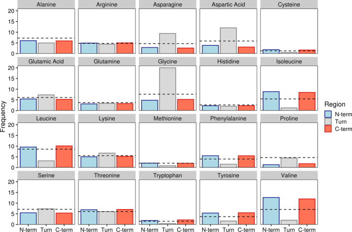
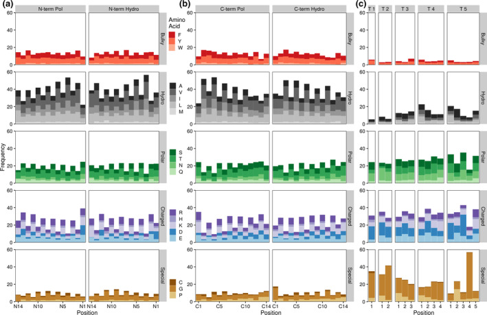
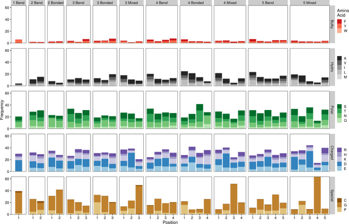
The beta hairpin motif is a ubiquitous protein structural motif that can be found in molecules across the tree of life. This motif, which is also popular in synthetically designed proteins and peptides, is known for its stability and adaptability to broad functions. Here, we systematically probe all 49,000 unique beta hairpin substructures contained within the Protein Data Bank (PDB) to uncover key characteristics correlated with stable beta hairpin structure, including amino acid biases and enriched interstrand contacts. We find that position specific amino acid preferences, while seen throughout the beta hairpin structure, are most evident within the turn region, where they depend on subtle turn dynamics associated with turn length and secondary structure. We also establish a set of broad design principles, such as the inclusion of aspartic acid residues at a specific position and the careful consideration of desired secondary structure when selecting residues for the turn region, that can be applied to the generation of libraries encoding proteins or peptides containing beta hairpin structures.

Keywords: PDB; beta hairpin; computational biology; protein design.


Figures

FIGURE 1
FIGURE 1
General beta hairpin structure. Beta hairpins consist of two anti‐parallel beta strands (grey arrows) linked with a flexible turn region (grey line). Beta strands typically have amphipathic characteristics conferred by alternating hydrophobic and hydrophilic residues. Triangles represent beta strand side amino acid side chains, with red indicating hydrophobic and blue indicating hydrophilic residues. Dashed triangles indicate side chains oriented away from the viewer while solid triangles indicate side chains oriented toward the viewer
FIGURE 2
FIGURE 2
Amino acid frequencies by beta hairpin secondary structure region. Bars indicate average amino acid frequencies for each amino acid within a given region of all beta hairpins. The black dashed line indicates background amino acid frequencies for all sites in all proteins containing the beta hairpin motif. N‐term and C‐term refer to the N‐ and C‐terminal beta strands while turn denotes the turn region
FIGURE 3
FIGURE 3
Amino acid frequencies by beta hairpin residue position. Bars indicate average amino acid frequencies for each amino acid at a given position across all beta hairpin structures. (a) and (b) N‐term and C‐term refer to the N‐ and C‐terminal beta strands. Pol refers to beta strands containing a polar face adjacent to the turn region, Hydro denotes a hydrophobic face at this position. Beta strand residues are numbered from the turn region, with residue 1 representing the residue closest to the turn. (c) T # denotes a turn region of a given length (e.g., T 3 indicates a three residue turn region). Turn residues are numbered from N‐terminal (residue 1) to C‐terminal
FIGURE 4
FIGURE 4
Amino acid frequencies by beta hairpin turn residue position, length, and type. Bars indicate average amino acid frequencies for each amino acid at a given position across all beta hairpin turn structures. Bonded refers to turn regions containing only hydrogen bonded turn residues, bend indicates turn regions with coil and/or bend residues, and mixed indicates a mixture or hydrogen bonded and coil and/or bend residues. Numbers preceding the turn type indicate the length of the turn region. Residue positions are numbered as in Figure 3. Frequencies are only shown for turn types with at least 100 representative structures in our dataset
FIGURE 5
FIGURE 5
Grouped differences in observed versus expected residue contacts. Dots represent individual contacting pairs with red, labeled dots indicating contacts that are enriched or depleted at least two‐fold versus expected values. Residues are grouped as follows: Special refers to cysteine, proline, and glycine; Hydrophobic refers to valine, leucine, isoleucine, methionine, alanine, tryptophan, tyrosine, phenylalanine; Polar refers to glutamine, threonine, serine, and asparagine; Charged refers to arginine, histidine, lysine, aspartic acid, and glutamic acid

References

    1. Watkins AM, Arora PS. Anatomy of β‐strands at protein‐protein interfaces. ACS Chem Biol. 2014;9:1747–1754. - PMC - PubMed
    1. Robinson JA. β‐Hairpin peptidomimetics: Design, structures and biological activities. Acc Chem Res. 2008;41:1278–1288. - PubMed
    1. Chakraborty K, Shivakumar P, Raghothama S, Varadarajan R. NMR structural analysis of a peptide mimic of the bridging sheet of HIV‐1 gp120 in methanol and water. Biochem J. 2005;390:573–581. - PMC - PubMed
    1. Batalha IL, Lychko I, Branco RJF, Iranzo O, Roque ACA. β‐Hairpins as peptidomimetics of human phosphoprotein‐binding domains. Org Biomol Chem. 2019;17:3996–4004. - PubMed
    1. Remmert M, Biegert A, Linke D, Lupas AN, Söding J. Evolution of outer membrane β‐barrels from an ancestral ββ hairpin. Mol Biol Evol. 2010;27:1348–1358. - PubMed

Publication types

LinkOut - more resources