Primary intracranial leiomyosarcoma presenting with frontal bone mass: a case report
- PMID: 33389983
- PMCID: PMC7785836
- DOI: 10.3857/roj.2020.00577
Primary intracranial leiomyosarcoma presenting with frontal bone mass: a case report
Abstract
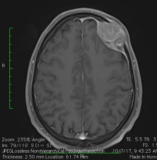
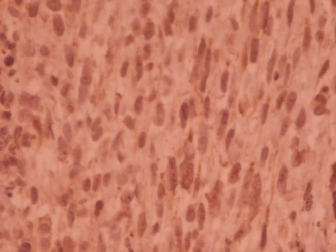
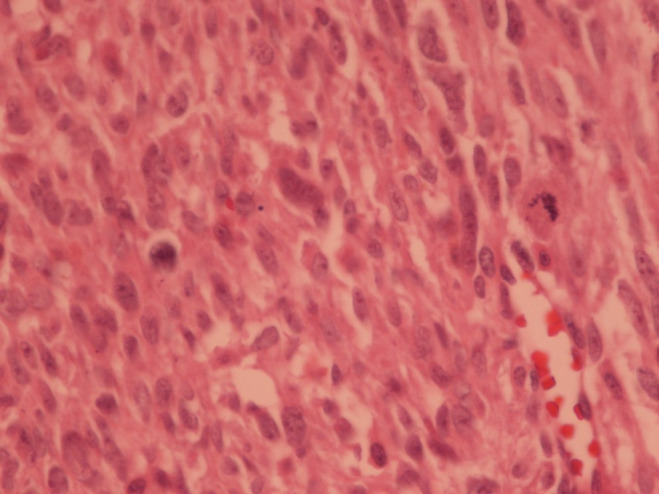
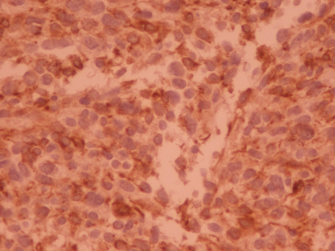
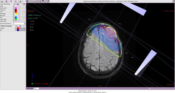
Primary intracranial mesenchymal neoplasms are rare tumors. These tumors are usually metastatic disease from other primary sites. We presented a 31-year-old man with a 6-month history of gradually enlarging frontal mass and positional headache. There was no other symptom demonstrating other organs' involvement. The patient underwent an uncomplicated craniotomy with clear surgical margins. The pathology review and the immunohistochemistry staining confirmed leiomyosarcoma grade II. We prescribed radiation therapy with tumor dose of 60 Gy in 30 fractions with conformal treatment planning to the tumor bed. As this disease has a high potency for metastasis, we advised four courses of single agent doxorubicin chemotherapy 75 mg/m2 every 4 weeks starting one month after the end of radiotherapy. In the last follow-up visit 34 months later, the patient was disease free in physical exam and imaging findings.
Keywords: Brain neoplasms; Intracranial; Leiomyosarcoma.
Conflict of interest statement
No potential conflict of interest relevant to this article was reported.
Figures
Similar articles
-
Letter to the Editor: Depression As The First Symptom Of Frontal Lobe Grade 2 Malignant Glioma.Turk Psikiyatri Derg. 2022 Summer;33(2):143-145. doi: 10.5080/u25957. Turk Psikiyatri Derg. 2022. PMID: 35730515 English, Turkish.
-
Primary intracranial leiomyosarcoma: report of a case and review of the literature.Sarcoma. 2006;2006:52140. doi: 10.1155/SRCM/2006/52140. Epub 2006 Dec 24. Sarcoma. 2006. PMID: 17496995 Free PMC article.
-
Intracranial dural metastasis from uterine leiomyosarcoma with orbital extension.Neurol Sci. 2012 Oct;33(5):1173-7. doi: 10.1007/s10072-011-0877-9. Epub 2012 Jan 10. Neurol Sci. 2012. PMID: 22231469
-
Primary intracranial leiomyosarcoma in an immunocompetent patient: Case report and review of the literature.Clin Neurol Neurosurg. 2018 Feb;165:76-80. doi: 10.1016/j.clineuro.2017.12.014. Epub 2018 Jan 6. Clin Neurol Neurosurg. 2018. PMID: 29324399 Review.
-
[Leiomyosarcoma of the paranasal sinuses with intracranial involvement: report of a clinical case and review of the literature].Neurocirugia (Astur). 2001 Aug;12(4):331-7. doi: 10.1016/s1130-1473(01)70690-2. Neurocirugia (Astur). 2001. PMID: 11706678 Review. Spanish.
Cited by
-
Primary Intracranial Leiomyosarcoma Secondary to Glioblastoma: Case Report and Literature Review.Front Oncol. 2021 May 20;11:642683. doi: 10.3389/fonc.2021.642683. eCollection 2021. Front Oncol. 2021. PMID: 34094927 Free PMC article.
-
Giant primary intracranial multi-fossa leiomyosarcoma involving the frontal sinus, ethmoid air cells, anterior fossa, middle fossa, and intraventricular space: A case report and literature review.Surg Neurol Int. 2023 Oct 27;14:384. doi: 10.25259/SNI_647_2023. eCollection 2023. Surg Neurol Int. 2023. PMID: 37941634 Free PMC article.
References
-
- Torihashi K, Chin M, Yoshida K, Narumi O, Yamagata S. Primary intracranial leiomyosarcoma with intratumoral hemorrhage: case report and review of literature. World Neurosurg. 2018;116:169–73. - PubMed
-
- Saito A, Ninomiya A, Ishida T, et al. Intractable repeated intracerebral hemorrhage due to primary dural leiomyosarcoma: case report and literature review. World Neurosurg. 2019;122:116–22. - PubMed
Publication types
LinkOut - more resources
Full Text Sources
Research Materials