. 2021 Jan 4;40(1):4.
doi: 10.1186/s13046-020-01786-6.

Progranulin induces immune escape in breast cancer via up-regulating PD-L1 expression on tumor-associated macrophages (TAMs) and promoting CD8+ T cell exclusion

Affiliations

Progranulin induces immune escape in breast cancer via up-regulating PD-L1 expression on tumor-associated macrophages (TAMs) and promoting CD8+ T cell exclusion

Wenli Fang et al. J Exp Clin Cancer Res. .

Erratum in

Abstract

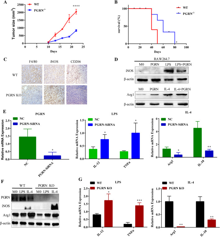
Background: Progranulin (PGRN), as a multifunctional growth factor, is overexpressed in multiple tumors, but the role of PGRN on tumor immunity is still unclear. Here, we studied the effect of PGRN on breast cancer tumor immunity and its possible molecular mechanism.

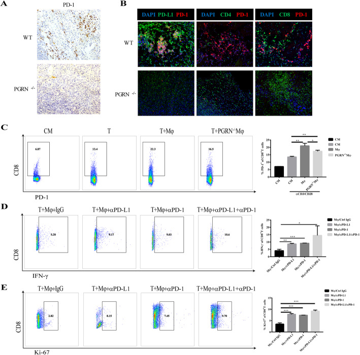
Methods: The changes of macrophage phenotypes after PGRN treatment were detected by western blot, quantitative polymerase chain reaction (PCR) and flow cytometry. Western blot was used to study the signal molecular mechanism of PGRN regulating this process. The number and localization of immune cells in Wild-type (WT) and PGRN-/- breast cancer tissues were analyzed by immunohistochemical staining and immunofluorescence techniques. The activation and proliferation of CD8+ T cells were measured by flow cytometry.

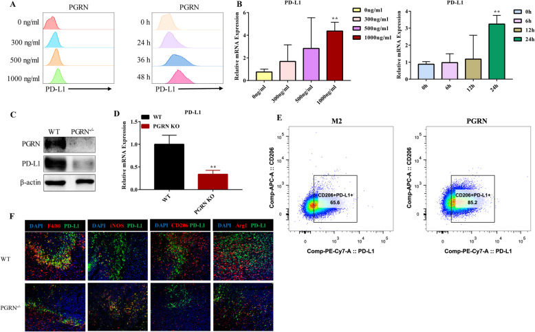
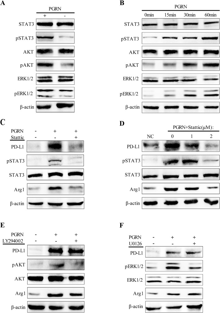
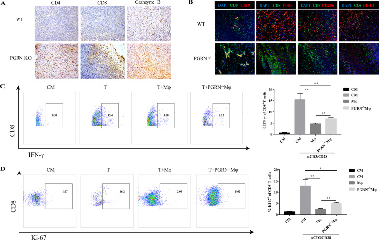
Results: After being treated with PGRN, the expressions of M2 markers and programmed death ligand 1 (PD-L1) on macrophages increased significantly. Signal transducer and activator of transcription 3 (STAT3) signaling pathway inhibitor Stattic significantly inhibited the expression of PD-L1 and M2 related markers induced by PGRN. In WT group, CD8 were co-localized with macrophages and PD-L1, but not tumor cells. The number of immune cells in PGRN-/- breast cancer tissue increased, and their infiltration into tumor parenchyma was also enhanced. Moreover, in the co-culture system, WT peritoneal macrophages not only reduced the ratio of activated CD8+ T cells but also reduced the proportion of proliferating CD8+ T cells. The addition of programmed death receptor 1 (PD-1) and PD-L1 neutralizing antibodies effectively reversed this effect and restored the immune function of CD8+ T cells.

Conclusion: These results demonstrate that PGRN promotes M2 polarization and PD-L1 expression by activating the STAT3 signaling pathway. Furthermore, through PD-1/PD-L1 interaction, PGRN can promote the breast tumor immune escape. Our research may provide new ideas and targets for clinical breast cancer immunotherapy.

Keywords: Breast cancer; PD-L1; Progranulin (PGRN); T cell; TAMs.


Conflict of interest statement

The authors declare that they have no competing interests.

Figures

Fig. 1
Fig. 1
PGRN promotes M2 polarization of macrophages. a-b. Breast cancer PY8119 cells were injected in situ into the fat pads of C57 wild-type mice and PGRN knock out mice (n = 5 per group). a. Tumor volume curve. b. Survival curve of mice. c. F4/80, iNOS and CD206 expression were detected by IHC in breast cancer tissue sections of WT and PGRN KO mice respectively. d-e. RAW264.7 macrophage cell line was treated with PGRN recombinant protein and LPS or IL-4. d. iNOS and Arg1 expression were examined by western blot. e. M1 markers (IL-12, TNF-α) and M2 markers (Arg1, IL-10) were tested by PCR. (F-G) WT and PGRN KO mouse peritoneal macrophages were treated with LPS or IL-4. f. Western blot was performed to analyze iNOS, and Arg1 expression. g. The differences in the expression of IL-12, TNF-α and Arg1, and IL-10 were measured by PCR. *p < 0.05; **p < 0.01; ***p < 0.001; ****p < 0.0001
Fig. 2
Fig. 2
PGRN up-regulates PD-L1 expression on TAMs. a-b M2 was treated with PGRN recombinant protein; then PD-L1 expression was measured by flow cytometry and PCR. c-d. IL-4 was used to induce WT, PGRN KO peritoneal macrophages into M2; then WB and PCR were used to detect the difference in PD-L1 expression between them. e. After being treated with PGRN, the proportion of CD206+ PD-L1+ cells in M2 were measured by flow cytometry. F. Immunofluorescence was performed to analyze colocalization of F4/80 (red), iNOS (red), CD206 (red), Arg1 (red) and PD-L1 (green) in WT and PGRN KO mice breast cancer sections, and the nucleus was stained with DAPI (blue). **p < 0.01
Fig. 3
Fig. 3
PGRN/STAT3 axis regulates TAMs polarization and up-regulates PD-L1 expression. a. After being treated with PGRN, western blot was used to detect STAT3/pSTAT3, AKT/pAKT and ERK1/2/pERK1/2 expression in M2. b. M2 was exposed to PGRN at a specified time point, and WB was used to detect the expression of downstream signaling proteins of PGRN. c-d. M2 was pretreated with STAT3 inhibitor Stattic, and then PGRN was added. Expression of PD-L1, STAT3/pSTAT3 and Arg1 was examined by Western blotting. e-f M2 was pretreated with AKT inhibitor LY294002 and ERK1/2 inhibitor U0126 respectively, and the expression changes of PD-L1, STAT3/pSTAT3 and Arg1 before and after PGRN stimulation were analyzed by Western blot
Fig. 4
Fig. 4
PGRN promotes CD8+ T cell exclusion and inhibits tumor immunity. a. The expression of CD4, CD8 and Granzyme B in WT and PGRN KO mice breast cancer tissue sections was tested by immunohistochemical staining. b. Colocalization of CK19 (red), F4/80 (red), CD206 (red), PD-L1 (red) and CD8 (green) in WT and PGRN KO mice breast cancer tissue sections was examined by immunofluorescence analysis, and the nucleus was stained with DAPI (blue). c-d WT and PGRN−/− peritoneal macrophages were co-cultured with mouse spleen lymphocytes activated by αCD3/CD28. Here CM stands for control medium, without αCD3/CD28 stimulation. c. Flow cytometry was used to measure the proportion of activated CD8+ T cells (IFN-γ+ CD8+ T cells) and (D) Ki-67+ CD8+ T cells. *p < 0.05; **p < 0.01
Fig. 5
Fig. 5
The interaction of PD-1/PD-L1 mediates the immunosuppressive function of PGRN in breast cancer. a. Expression of PD-1 in WT and PGRN KO mice breast cancer tissue sections was detected with immunohistochemical staining. b. PD-L1 (green), CD4 (green), CD8 (green) expression differences and co-localization with PD-1 (red) in WT and PGRN KO mice breast cancer tissue sections were examined by immunofluorescence, and the nucleus was stained with DAPI (blue). c. Mouse splenic lymphocytes activated with or without αCD3/CD28 antibody were co-cultured with WT or PGRN−/− peritoneal macrophages, and the frequency of PD-1+ CD8+ T cells was tested by flow cytometry. d-e Wild-type peritoneal macrophages were co-cultured with splenic lymphocytes preactivated by αCD3/CD28 antibody, and then anti-PD-1 or anti-PD-L1 neutralizing antibodies were added or not to the co-culture system. d. CD8+ T cell activation and (e) CD8+ T cell proliferation were detected by flow cytometry. *p < 0.05; **p < 0.01; ***p < 0.001

References

    1. Harbeck N, Gnant M. Breast cancer. Lancet. 2017;389(10074):1134–1150. - PubMed
    1. Spitzer MH, Carmi Y, Reticker-Flynn NE, Kwek SS, Madhireddy D, Martins MM, et al. Systemic immunity is required for effective Cancer immunotherapy. Cell. 2017;168(3):487–502. - PMC - PubMed
    1. Quigley DA, Kristensen V. Predicting prognosis and therapeutic response from interactions between lymphocytes and tumor cells. Mol Oncol. 2015;9(10):2054–2062. - PMC - PubMed
    1. Zhang J, Dang F, Ren J, Wei W. Biochemical aspects of PD-L1 regulation in Cancer immunotherapy. Trends Biochem Sci. 2018;43(12):1014–1032. - PMC - PubMed
    1. Dong P, Xiong Y, Yue J, Hanley SJB, Watari H. Tumor-intrinsic PD-L1 signaling in Cancer initiation, Development and Treatment: Beyond Immune Evasion. Front Oncol. 2018;8:386. - PMC - PubMed

MeSH terms