

Prefrontal Disinhibition in Social Fear: A Vital Action of Somatostatin Interneurons
- PMID: 33390908
- PMCID: PMC7773700
- DOI: 10.3389/fncel.2020.611732
Prefrontal Disinhibition in Social Fear: A Vital Action of Somatostatin Interneurons
Abstract
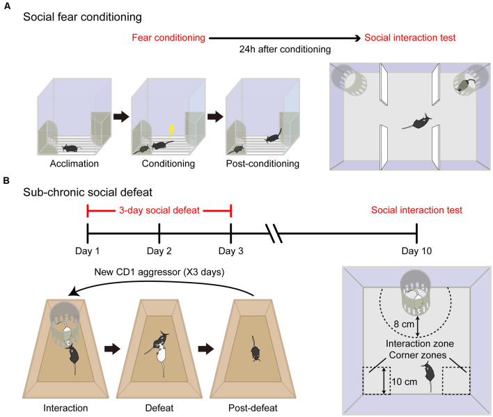
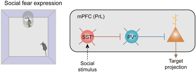
Social fear and avoidance of social partners and social situations represent the core behavioral symptom of Social Anxiety Disorder (SAD), a prevalent psychiatric disorder worldwide. The pathological mechanism of SAD remains elusive and there are no specific and satisfactory therapeutic options currently available. With the development of appropriate animal models, growing studies start to unravel neuronal circuit mechanisms underlying social fear, and underscore a fundamental role of the prefrontal cortex (PFC). Prefrontal cortical functions are implemented by a finely wired microcircuit composed of excitatory principal neurons (PNs) and diverse subtypes of inhibitory interneurons (INs). Disinhibition, defined as a break in inhibition via interactions between IN subtypes that enhances the output of excitatory PNs, has recently been discovered to serve as an efficient strategy in cortical information processing. Here, we review the rodent animal models of social fear, the prefrontal IN diversity, and their circuits with a particular emphasis on a novel disinhibitory microcircuit mediated by somatostatin-expressing INs in gating social fear behavior. The INs subtype distinct and microcircuit-based mechanism advances our understanding of the etiology of social fear and sheds light on developing future treatment of neuropsychiatric disorders associated with social fear.
Keywords: disinhibition; interneuron; prefrontal cortex; social anxiety disorder; social fear.
Copyright © 2020 Wang, Tian, Zeng and Xu.
Conflict of interest statement
The authors declare that the research was conducted in the absence of any commercial or financial relationships that could be construed as a potential conflict of interest.
Figures
Similar articles
-
A Disinhibitory Microcircuit Mediates Conditioned Social Fear in the Prefrontal Cortex.Neuron. 2019 May 8;102(3):668-682.e5. doi: 10.1016/j.neuron.2019.02.026. Epub 2019 Mar 18. Neuron. 2019. PMID: 30898376
-
Alcohol reduces the activity of somatostatin interneurons in the mouse prefrontal cortex: A neural basis for its disinhibitory effect?Neuropharmacology. 2021 May 1;188:108501. doi: 10.1016/j.neuropharm.2021.108501. Epub 2021 Feb 24. Neuropharmacology. 2021. PMID: 33636191 Free PMC article.
-
Inhibitory Gating of Basolateral Amygdala Inputs to the Prefrontal Cortex.J Neurosci. 2016 Sep 7;36(36):9391-406. doi: 10.1523/JNEUROSCI.0874-16.2016. J Neurosci. 2016. PMID: 27605614 Free PMC article.
-
PV Interneurons: Critical Regulators of E/I Balance for Prefrontal Cortex-Dependent Behavior and Psychiatric Disorders.Front Neural Circuits. 2018 May 16;12:37. doi: 10.3389/fncir.2018.00037. eCollection 2018. Front Neural Circuits. 2018. PMID: 29867371 Free PMC article. Review.
-
Cortical disinhibitory circuits: cell types, connectivity and function.Trends Neurosci. 2021 Aug;44(8):643-657. doi: 10.1016/j.tins.2021.04.009. Epub 2021 May 15. Trends Neurosci. 2021. PMID: 34006387 Review.
Cited by
-
Differences in mGluR5 Availability Depending on the Level of Social Avoidance in Drug-Naïve Young Patients with Major Depressive Disorder.Neuropsychiatr Dis Treat. 2022 Sep 12;18:2041-2053. doi: 10.2147/NDT.S379395. eCollection 2022. Neuropsychiatr Dis Treat. 2022. PMID: 36124236 Free PMC article.
-
Excitatory synapses and gap junctions cooperate to improve Pv neuronal burst firing and cortical social cognition in Shank2-mutant mice.Nat Commun. 2021 Aug 25;12(1):5116. doi: 10.1038/s41467-021-25356-2. Nat Commun. 2021. PMID: 34433814 Free PMC article.
-
The maternal brain is more flexible and responsive at rest: effective connectivity of the parental caregiving network in postpartum mothers.Sci Rep. 2023 Mar 23;13(1):4719. doi: 10.1038/s41598-023-31696-4. Sci Rep. 2023. PMID: 36959247 Free PMC article.
-
Development, Diversity, and Death of MGE-Derived Cortical Interneurons.Int J Mol Sci. 2021 Aug 27;22(17):9297. doi: 10.3390/ijms22179297. Int J Mol Sci. 2021. PMID: 34502208 Free PMC article. Review.
-
Neurochemical Alterations in Social Anxiety Disorder (SAD): A Systematic Review of Proton Magnetic Resonance Spectroscopic Studies.Int J Mol Sci. 2022 Apr 26;23(9):4754. doi: 10.3390/ijms23094754. Int J Mol Sci. 2022. PMID: 35563145 Free PMC article.
References
-
- American Psychiatric Publishing (2013). Diagnostic and Statistical Manual of Mental Disorders (DSM-5). Arlington: American Psychiatric Publishing .
Publication types
LinkOut - more resources
Full Text Sources
Miscellaneous