. 2020 Nov 30:11:585860.
doi: 10.3389/fphar.2020.585860. eCollection 2020.

Hydrogen Sulfide Attenuates High-Fat Diet-Induced Non-Alcoholic Fatty Liver Disease by Inhibiting Apoptosis and Promoting Autophagy via Reactive Oxygen Species/Phosphatidylinositol 3-Kinase/AKT/Mammalian Target of Rapamycin Signaling Pathway

Affiliations

Hydrogen Sulfide Attenuates High-Fat Diet-Induced Non-Alcoholic Fatty Liver Disease by Inhibiting Apoptosis and Promoting Autophagy via Reactive Oxygen Species/Phosphatidylinositol 3-Kinase/AKT/Mammalian Target of Rapamycin Signaling Pathway

Dongdong Wu et al. Front Pharmacol. .

Abstract

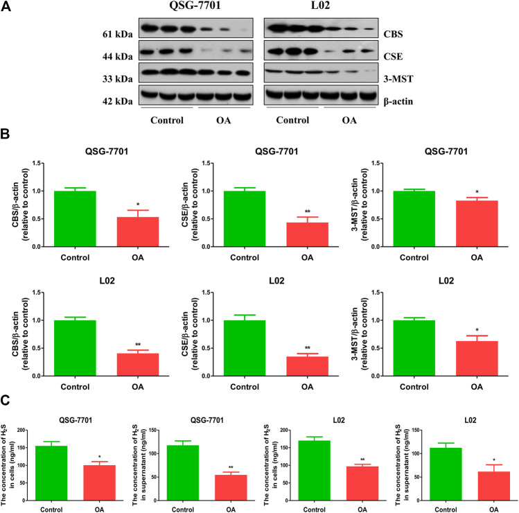
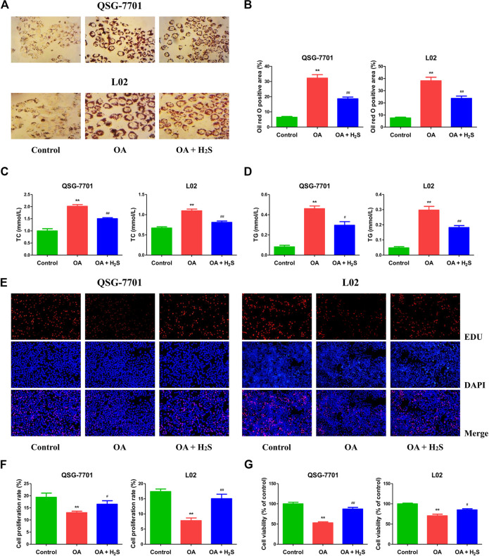
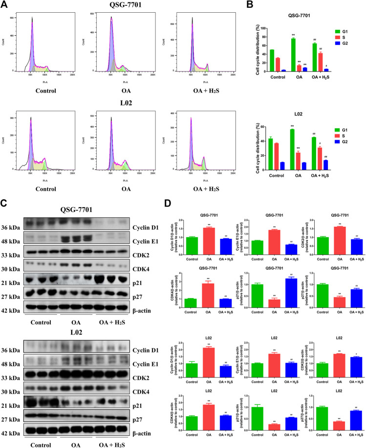
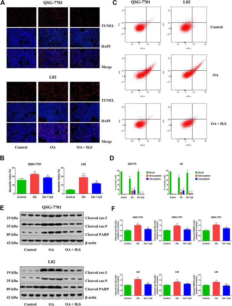
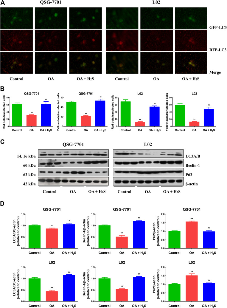
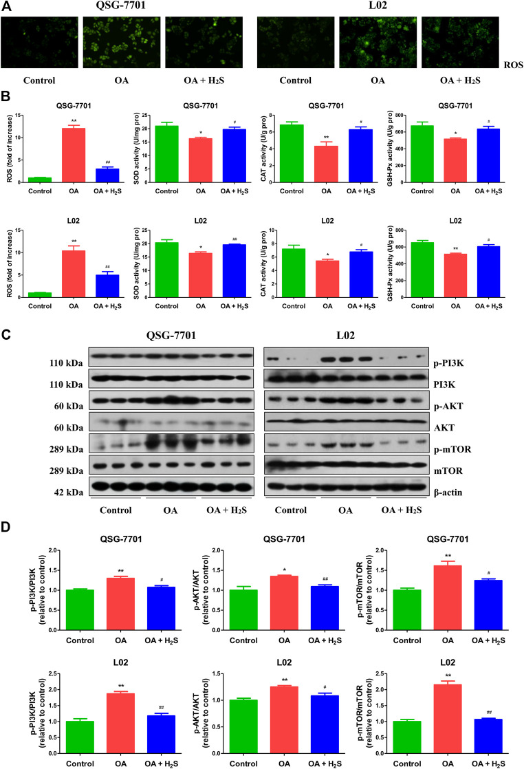
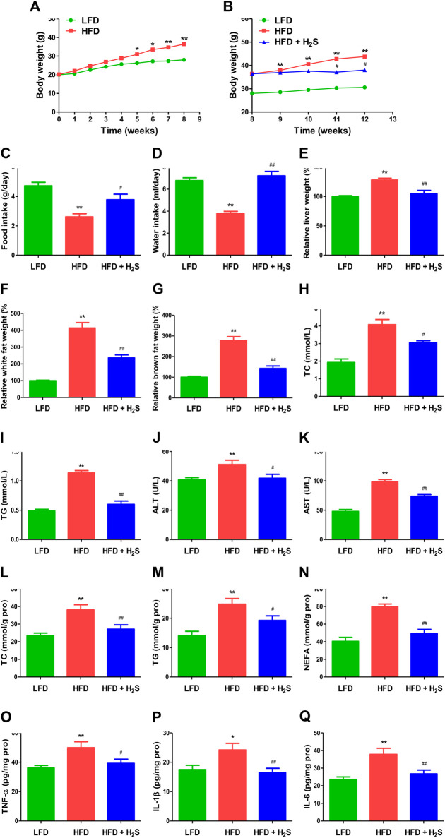
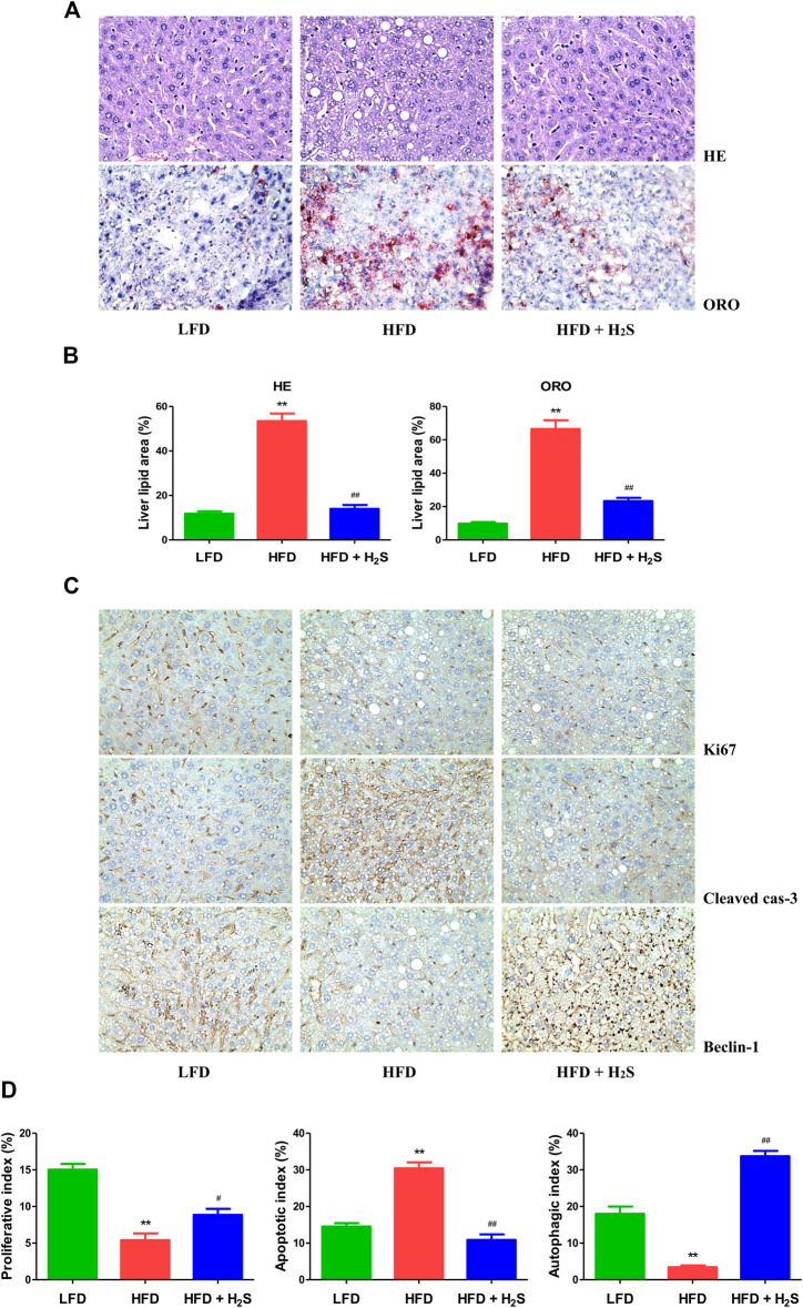
Non-alcoholic fatty liver disease (NAFLD) is a common chronic liver disease worldwide. Hydrogen sulfide (H2S) is involved in a wide range of physiological and pathological processes. Nevertheless, the mechanism of action of H2S in NAFLD development has not been fully clarified. Here, the reduced level of H2S was observed in liver cells treated with oleic acid (OA). Administration of H2S increased the proliferation of OA-treated cells. The results showed that H2S decreased apoptosis and promoted autophagy through reactive oxygen species (ROS)-mediated phosphatidylinositol 3-kinase (PI3K)/AKT/mammalian target of rapamycin (mTOR) cascade in OA-treated cells. In addition, administration of H2S relieved high-fat diet (HFD)-induced NAFLD via inhibition of apoptosis and promotion of autophagy. These findings suggest that H2S could ameliorate HFD-induced NAFLD by regulating apoptosis and autophagy through ROS/PI3K/AKT/mTOR signaling pathway. Novel H2S-releasing donors may have therapeutic potential for the treatment of NAFLD.

Keywords: apoptosis; autophagy; hydrogen sulfide; nonalcoholic fatty liver disease; signaling pathway.


Conflict of interest statement

The authors declare that the research was conducted in the absence of any commercial or financial relationships that could be constructed as a potential conflict of interest.

Figures

FIGURE 1
FIGURE 1
The levels of endogenous H2S in normal and OA-treated QSG-7701 and L02 cells and culture supernatant. (A) The protein levels of CSE, CBS, and 3-MST were examined by Western blot. β-actin was used as a loading control. (B) The densitometric quantification was performed, normalized to the level of β-actin. (C) The levels of H2S in normal and OA-treated liver cells and culture supernatant. The experiments were performed in triplicates. Data are presented as mean ± SEM; *p < 0.05, **p < 0.01 vs. control group.
FIGURE 2
FIGURE 2
The effect of H2S on the formation of lipid droplet and the growth of QSG-7701 and L02 cells treated with OA. (A) Lipid droplets in QSG-7701 and L02 cells were stained with ORO (original magnification, ×400). (B) The ORO stained area was measured. (C) The level of TC was detected. (D) The level of TG was detected. (E) The replication activity was determined by EdU assay (original magnification, ×200). (F) The proliferation rate was calculated. (G) The percentage of viable cells was calculated. The viability of control group was normalized to 100%. The experiments were performed in triplicates. Data are presented as mean ± SEM; **p < 0.01 vs. control group; # p < 0.05, ## p < 0.01 vs. OA group.
FIGURE 3
FIGURE 3
The effect of H2S on cell-cycle progression of QSG-7701 and L02 cells treated with OA. (A) The cell-cycle distribution was analyzed by flow cytometry. (B) Cell-cycle distribution was determined. (C) The expression levels of cyclin D1/E1, CDK2/4, and p21/p27 were detected by Western blot. β-actin was used as a loading control. (D) The densitometric quantification was performed, normalized to the level of β-actin. The experiments were performed in triplicates. Data are presented as mean ± SEM; **p < 0.01 vs. control group; # p < 0.05, ## p < 0.01 vs. OA group.
FIGURE 4
FIGURE 4
The effect of H2S on the apoptosis of QSG-7701 and L02 cells treated with OA. (A) The apoptotic levels were measured by TUNEL staining (original magnification, ×200). (B) The apoptotic index was calculated. (C) The apoptotic level was detected by flow cytometry. (D) The result of flow cytometry was determined. (E) The expression levels of cleaved cas-3, -9, and cleaved PARP were detected by Western blot. β-actin was used as a loading control. (F) The densitometric quantification was performed, normalized to the level of β-actin. The experiments were performed in triplicates. Data are presented as mean ± SEM; **p < 0.01 vs. control group; # p < 0.05, ## p < 0.01 vs. OA group.
FIGURE 5
FIGURE 5
The effect of H2S on the autophagy of QSG-7701 and L02 cells treated with OA. (A) The fluorescence microscopy was used to detect QSG-7701 and L02 cells transfected with GFP-RFP-LC3 plasmid (original magnification, ×1,000). (B) The ratios of red/yellow dots to the transfected cells were determined. (C) The expression levels of LC3A/B, P62, and beclin-1 were detected by Western blot. β-actin was used as a loading control. (D) The densitometric quantification was performed, normalized to the level of β-actin. The experiments were performed in triplicates. Data are presented as mean ± SEM; *p < 0.05, **p < 0.01 vs. control group; # p < 0.05, ## p < 0.01 vs. OA group.
FIGURE 6
FIGURE 6
The effect of H2S on ROS/PI3K/AKT/mTOR pathway in QSG-7701 and L02 cells treated with OA. (A) The ROS level was determined (original magnification, ×400). (B) The ROS levels and activities of SOD, CAT, and GSH-Px were detected. (C) The expression levels of PI3K, p-PI3K, AKT, p-AKT, mTOR, and p-mTOR were detected by Western blot. β-actin was used as a loading control. (D) The densitometric quantification was performed, normalized to the level of β-actin. The experiments were performed in triplicates. Data are presented as mean ± SEM; *p < 0.05, **p < 0.01 vs. control group; # p < 0.05, ## p < 0.01 vs. OA group.
FIGURE 7
FIGURE 7
The effect of H2S on NAFLD in HFD-fed mice. (A,B) The body weight was determined. (C,D) Food/water intake was measured. (E–G) Relative liver weight and relative white/brown fat weight were determined. (H–K) The plasmatic levels of TC, TG, ALT, and AST were measured. (L–Q) The levels of TC, TG, NEFA, TNF-α, IL-1β, and IL-6 were measured in the liver of mice. Data are presented as mean ± SEM (n = 6). *p < 0.05, **p < 0.01 vs. control group; # p < 0.05, ## p < 0.01 vs. OA group.
FIGURE 8
FIGURE 8
The effect of H2S on the proliferative, apoptotic, and autophagic activities in the liver of NAFLD mice. (A) Representive images stained with HE and ORO (original magnification, ×400). HFD induced vacuolar degeneration of hepatocytes, disruption of normal hepatic lobules, and inflammatory cells infiltration. H2S dramatically reduced hepatic lipid droplets and macrovesicular steatosis. (B) Liver lipid area was calculated in HE and ORO staining. (C) Representive images stained with Ki67, cleaved cas-3, and beclin-1 (original magnification, ×400). Ki67 and beclin-1 staining were reduced in HFD group, which were reversed by the administration of H2S. The results of cleaved cas-3 staining exhibited opposite trends. (D) The indexes of proliferation, apoptosis and autophagy were determined. Data are presented as mean ± SEM (n = 6). **p < 0.01 vs. control group; # p < 0.05, ## p < 0.01 vs. OA group.

References

    1. Ali A., Zhang Y., Fu M., Pei Y., Wu L., Wang R., et al. (2020). Cystathionine gamma-lyase/H2S system suppresses hepatic acetyl-CoA accumulation and nonalcoholic fatty liver disease in mice. Life Sci. 252, 117661 10.1016/j.lfs.2020.117661. | - DOI - PubMed
    1. Ashkenazi A., Fairbrother W. J., Leverson J. D., Souers A. J. (2017). From basic apoptosis discoveries to advanced selective BCL-2 family inhibitors. Nat. Rev. Drug Discov. 16, 273–284. 10.1038/nrd.2016.253. | - DOI - PubMed
    1. Chen L., Liu P., Feng X., Ma C. (2017). Salidroside suppressing LPS-induced myocardial injury by inhibiting ROS-mediated PI3K/Akt/mTOR pathway in vitro and in vivo . J. Cell. Mol. Med. 21, 3178–3189. 10.1111/jcmm.12871. | - DOI - PMC - PubMed
    1. Cheng Z., Garikipati V. N. S., Nickoloff E., Wang C., Polhemus D. J., Zhou J., et al. (2016). Restoration of hydrogen sulfide production in diabetic mice improves reparative function of bone marrow cells. Circulation 134, 1467–1483. 10.1161/circulationaha.116.022967. | - DOI - PMC - PubMed
    1. Cheng Z., Shen X., Jiang X., Shan H., Cimini M., Fang P., et al. (2018). Hyperhomocysteinemia potentiates diabetes-impaired EDHF-induced vascular relaxation: role of insufficient hydrogen sulfide. Redox Biol. 16, 215–225. 10.1016/j.redox.2018.02.006. | - DOI - PMC - PubMed