. 2020 Dec 18:11:620817.
doi: 10.3389/fneur.2020.620817. eCollection 2020.

Responses of Neurons in the Medullary Lateral Tegmental Field and Nucleus Tractus Solitarius to Vestibular Stimuli in Conscious Felines

Affiliations

Responses of Neurons in the Medullary Lateral Tegmental Field and Nucleus Tractus Solitarius to Vestibular Stimuli in Conscious Felines

John P Bielanin et al. Front Neurol. .

Abstract

Considerable evidence shows that the vestibular system contributes to adjusting sympathetic nervous system activity to maintain adequate blood pressure during movement and changes in posture. However, only a few prior experiments entailed recordings in conscious animals from brainstem neurons presumed to convey baroreceptor and vestibular inputs to neurons in the rostral ventrolateral medulla (RVLM) that provide inputs to sympathetic preganglionic neurons in the spinal cord. In this study, recordings were made in conscious felines from neurons in the medullary lateral tegmental field (LTF) and nucleus tractus solitarius (NTS) identified as regulating sympathetic nervous system activity by exhibiting changes in firing rate related to the cardiac cycle, or cardiac-related activity (CRA). Approximately 38% of LTF and NTS neurons responded to static 40° head up tilts with a change in firing rate (increase for 60% of the neurons, decrease for 40%) of ~50%. However, few of these neurons responded to 10° sinusoidal rotations in the pitch plane, in contrast to prior findings in decerebrate animals that the firing rates of both NTS and LTF neurons are modulated by small-amplitude body rotations. Thus, as previously demonstrated for RVLM neurons, in conscious animals NTS and LTF neurons only respond to large rotations that lead to changes in sympathetic nervous system activity. The similar responses to head-up rotations of LTF and NTS neurons with those documented for RVLM neurons suggest that LTF and NTS neurons are components of the vestibulo-sympathetic reflex pathway. However, a difference between NTS/LTF and RVLM neurons was variability in CRA over time. This variability was significantly greater for RVLM neurons, raising the hypothesis that the responsiveness of these neurons to baroreceptor input is adjusted based on the animal's vigilance and alertness.

Keywords: cardiovascular regulation; lateral tegmental field; nucleus tractus solitarius (NTS); reticular formation; rostral ventrolateral medulla; sympathetic nervous system; vestibular; vestibulo-sympathetic responses.


Conflict of interest statement

The authors declare that the research was conducted in the absence of any commercial or financial relationships that could be construed as a potential conflict of interest.

Figures

Figure 1
Figure 1
Example of data collected during one trial. The top trace shows table position, the second shows discriminated activity for one unit, the third shows raw unit activity recorded by a microelectrode, and the bottom trace is a recording of the electrocardiogram (ECG). Initially, animals were positioned 20° nose-down for a variable amount of time. A light cue was presented (onset indicated by arrow) that lasted for 2 s. Ten seconds after the light cue was initiated, animals were tilted 40° head-up (20° nose-down to 20° nose-up) and remained in that position for 30 s prior to returning to the prone position. To determine if the light cue had any effect on a unit's activity, firing rate during the period 5 s before light onset (Pre-) was compared to that during the period 5–10 s prior to tilt onset (Post-2).
Figure 2
Figure 2
(A–D) Cardiac-related activity (CRA) evident in two trials for an NTS neuron (A,B are for one trial and C,D are for the second trial). Histograms were triggered by event markers demarking ECG R-waves; bin width is 10 ms. (A,C) show intervals between R-waves, and (B,D) show intervals between the unit's action potentials. Unit activity peaked in periods between R-waves. (E,F) plot of counts during peak and trough periods for the LTF (E) and NTS (F) neurons classified as exhibiting CRA.
Figure 3
Figure 3
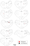
Locations of LTF and NTS neurons with CRA whose responses to 40° tilts were recorded during five or more trials. Red symbols designate units with significant differences in unit activity in the nose-down and nose-up position. Black symbols designate units without significant responses to tilts. The numbers above each section indicate the distance rostral to the obex in mm. LTF, lateral tegmental field; NTS, nucleus tractus solitarius; 5SN, spinal trigeminal nucleus; 5ST, spinal trigeminal tract; CUR, cuneate nucleus, rostral; CX, external cuneate nucleus; IO, inferior olivary nucleus; P, pyramid; VIN, inferior vestibular nucleus; RB, restiform body; VMN, medial vestibular nucleus; CD, dorsal cochlear nucleus.
Figure 4
Figure 4
Peak-to-trough ratio of activity related to the cardiac cycle for 8 LTF and 8 NTS units where multiple trials were conducted. Data for each unit is depicted by different shapes and colors. For most units, the peak-to-trough ratio varied throughout the course of recording although all units exhibited CRA during every trial.
Figure 5
Figure 5
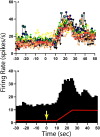
Change in activity for one NTS unit with CRA during 40° head-up tilt. (A) Unit activity during 1 s bins for 10 individual trials; each trial is demarked by lines of different colors as well as different symbols. (B) Histogram of averaged activity during 1 s bins for all trials. The red line designates table position; the transition from 20° nose-down to the 20° nose-up position (40° change in animal position) occurred 10 s after the light cue, which is demarked by a yellow arrow.
Figure 6
Figure 6
Percent change in firing rate when the animal was tilted nose-up (firing rate in nose-up position ÷ firing rate in the nose-down position) for the 12 units in the LTF and 8 units in the NTS with CRA whose activity was significantly modulated during 40° head-up tilts. Error bars indicate mean ± one standard deviation.
Figure 7
Figure 7
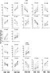
Firing rate during each trial for the 12 LTF and 8 NTS units with CRA whose activity was significantly modulated by 40° head-up tilt. Each panel depicts the firing rate for a single unit when the animal was positioned 20° nose-down (ND) and then subsequently positioned 20° nose-up (NU). P-values indicating the significance of the difference in firing rate in the ND and NU positions (paired t-test) are provided above each panel. The greatest significance level indicated is P < 0.01.
Figure 8
Figure 8
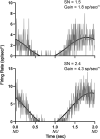
Averaged responses (~30 trials) of two LTF units with CRA to 10° sinusoidal rotations in the pitch plane at 0.5 Hz. Bin width was 4 ms (500 bins/trace). A sine wave fitted to responses is shown by black lines. The signal-to-noise (SN) ratio and gain for each unit's response is indicated. ND, nose-down; NU, nose-up.
Figure 9
Figure 9
Comparison of P-values (two-tailed t-test) for responses to head-up tilt (Tilt P) and responses to the light cue preceding tilts (Light P, pre-tilt vs. post-2 period indicated in Figure 1). Data for neurons with significant responses (P < 0.05) to head-up tilts are indicated by red symbols. Lines show the best-fit of the data with a linear regression analysis.
Figure 10
Figure 10
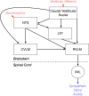
Brainstem regions that relay vestibular signals to the RVLM. This neural circuit diagram is based on the findings of both anatomical (–, –42) and electrophysiologic (21, 43) studies that mapped connections in the vestibulo-sympathetic pathway. Red text and arrows indicate afferent inputs, and blue text and arrows denote efferent (output) pathways from the central nervous system. CVLM, caudal ventrolateral medulla; IML, sympathetic intermediolateral cell column; LTF, lateral tegmental field; NTS, nucleus tractus solitarius; RVLM, rostral ventrolateral medulla.

Similar articles

Cited by

References

    1. Spyer KM. Annual review prize lecture - central nervous mechanisms contributing to cardiovascular control. J Physiol. (1994) 474:1–19. 10.1113/jphysiol.1994.sp019997 - DOI - PMC - PubMed
    1. Guyenet PG, Koshiya N, Huangfu D, Baraban SC, Stornetta RL, Li YW. Role of medulla oblongata in generation of sympathetic and vagal outflows. In: Holstege Bandler GR, Saper CB. editors. Emotional Motor System. Amsterdam: Elsevier; (1996). p. 127–44. - PubMed
    1. Dampney RA. Functional organization of central pathways regulating the cardiovascular system. Physiol Rev. (1994) 74:323–64. 10.1152/physrev.1994.74.2.323 - DOI - PubMed
    1. Barman SM, Yates BJ. Deciphering the neural control of sympathetic nerve activity: status report and directions for future research. Front Neurosci. (2017) 11:730. 10.3389/fnins.2017.00730 - DOI - PMC - PubMed
    1. Reis DJ, Ruggiero DA, Morrison SF. The C1 area of the rostral ventrolateral medulla oblongata a critical brainstem region for control of resting and reflex integration of arterial pressure. Am J Hypertens. (1989) 2:363S−74. 10.1093/ajh/2.12.363S - DOI - PubMed