. 2020 Dec 18:11:607957.
doi: 10.3389/fimmu.2020.607957. eCollection 2020.

Mycobacterium bovis Bacille-Calmette-Guérin Infection Aggravates Atherosclerosis

Affiliations

Mycobacterium bovis Bacille-Calmette-Guérin Infection Aggravates Atherosclerosis

Moises A Huaman et al. Front Immunol. .

Abstract

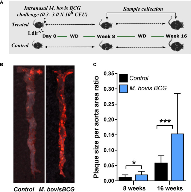
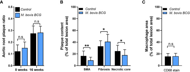
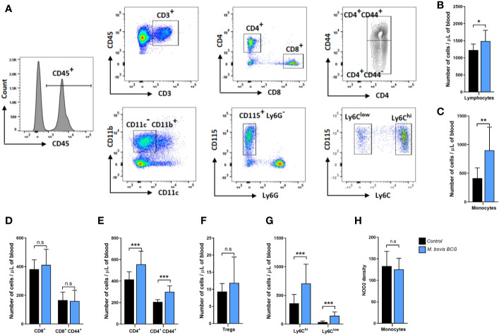
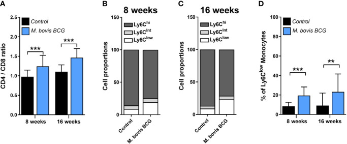
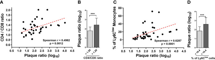
Tuberculosis has been associated with increased risk of atherosclerotic cardiovascular disease. To examine whether mycobacterial infection exacerbates atherosclerosis development in experimental conditions, we infected low-density lipoprotein receptor knockout (Ldlr-/-) mice with Mycobacterium bovis Bacille-Calmette-Guérin (BCG), an attenuated strain of the Mycobacterium tuberculosis complex. Twelve-week old male Ldlr-/- mice were infected with BCG (0.3-3.0x10⁶ colony-forming units) via the intranasal route. Mice were subsequently fed a western-type diet containing 21% fat and 0.2% cholesterol for up to 16 weeks. Age-matched uninfected Ldlr-/- mice fed with an identical diet served as controls. Atherosclerotic lesions in aorta were examined using Oil Red O staining. Changes induced by BCG infection on the immunophenotyping profile of circulating T lymphocytes and monocytes were assessed using flow cytometry. BCG infection increased atherosclerotic lesions in en face aorta after 8 weeks (plaque ratio; 0.021±0.01 vs. 0.013±0.01; p = 0.011) and 16 weeks (plaque ratio, 0.15±0.13 vs. 0.06±0.02; p = 0.003). No significant differences in plasma cholesterol or triglyceride levels were observed between infected and uninfected mice. Compared to uninfected mice, BCG infection increased systemic CD4/CD8 T cell ratio and the proportion of Ly6Clow non-classical monocytes at weeks 8 and 16. Aortic plaque ratios correlated with CD4/CD8 T cell ratios (Spearman's rho = 0.498; p = 0.001) and the proportion of Ly6Clow non-classical monocytes (Spearman's rho = 0.629; p < 0.001) at week 16. In conclusion, BCG infection expanded the proportion of CD4+ T cell and Ly6Clow monocytes, and aggravated atherosclerosis formation in the aortas of hyperlipidemic Ldlr-/- mice. Our results indicate that mycobacterial infection is capable of enhancing atherosclerosis development.

Keywords: Bacille-Calmette-Guérin; T cells; atherosclerosis; inflammation; monocytes; mycobacterium; tuberculosis.


Conflict of interest statement

CF has received research support to the University of Cincinnati from Gilead, Pfizer, BMS, ViiV, Janssen, and Merck. The remaining authors declare that the research was conducted in the absence of any commercial or financial relationships that could be construed as a potential conflict of interest.

Figures

Figure 1
Figure 1
M. bovis BCG increases en face aorta atherosclerosis by 8 and 16 weeks post-challenge. (A) Twelve-week old male Ldlr-/- mice were inoculated with M. bovis BCG (0.3–3.0x10⁶ CFU) via the intranasal route. Mice were fed a western-type diet for up to 16 weeks. Age-matched uninfected Ldlr -/- mice fed with an identical diet served as controls. (B) Atherosclerotic lesions in en face aorta were examined using Oil Red O staining at weeks 8 and 16. Data are representative of the aortae of one M. bovis BCG-infected mouse and one control mouse at 16 weeks. (C) Plaque burden was quantified by the plaque size per aorta ratio in M. bovis BCG-infected (blue) and control mice (black). Data are mean ± SD. n = 20 mice per group pooled from 2 independent experiments. Significance was determined by Student’s t-test. *p < 0.05; ***p < 0.001.
Figure 2
Figure 2
M. bovis BCG does not increase extent of aortic root atherosclerosis. Ldlr-/- mice were inoculated with M. bovis BCG (0.3–3.0x10⁶ CFU) via the intranasal route. Mice were fed a western-type diet for 8 to 16 weeks. Age-matched uninfected Ldlr -/- mice fed with an identical diet served as controls. (A) Plaque area per total area of aortic root ratios were quantified in M. bovis BCG-infected (blue) and control mice (black) at weeks 8 and 16. (B) Aortic root plaque composition of smooth muscle (using smooth muscle alpha actin staining; SMA), fibrosis content (using Syrius red staining), and necrotic core were quantified as percentage per total lesion area at week 16. (C) Macrophage content using CD68 staining was quantified as percentage per total lesion area at week 16. Data are mean ± SD. n = 20 mice per group pooled from 2 independent experiments. Significance was determined by Student’s t-test. *p < 0.05; **p < 0.01; n.s., non-significant (p > 0.05).
Figure 3
Figure 3
M. bovis BCG induces CD4+ T cell and monocyte activation by week 8. Flow cytometry analysis of blood lymphocytes and monocytes from M. bovis BCG-infected and control mice at 8 weeks. Ldlr-/- mice were inoculated with M. bovis BCG (0.3–3.0x10⁶ CFU) via the intranasal route. Mice were fed a western-type diet for 8 weeks. Age-matched uninfected Ldlr -/- mice fed with an identical diet served as controls. (A) Representative flow cytometry plots showing the gating strategy used for identifying subsets of T cells and monocytes. T cells (upper panel) were defined as CD45+ CD3+ cells and seperated into CD4+ and CD8+ T cell subsets. Expression of CD44 was further assessed within T cell subsets. Monocytes (lower panel) were defined as CD45+ CD3- CD11b+ CD11c- CD115+ Ly6G- cells. Monocyte subsets were defined based on Ly6C expression. (B–H) Number of lymphocytes (B), monocytes (C), T cell subsets (D–F), monocyte subsets (G) and monocyte NOD2 MFI (H) from M. bovis BCG-infected (blue) and control mice (black). Data are mean ± SD. n = 10 mice per group. Significance was determined by Student’s t-test. **p < 0.01; ***p < 0.001; n.s., non-significant (p > 0.05).
Figure 4
Figure 4
M. bovis BCG increases the CD4/C8 ratio and the proportion of Ly6Clow monocytes. Ldlr-/- mice were inoculated with M. bovis BCG (0.3–3.0x10⁶ CFU) via the intranasal route. Mice were fed a western-type diet for 8 to 16 weeks. Age-matched uninfected Ldlr -/- mice fed with an identical diet served as controls. (A) CD4/CD8 T cell ratios from M. bovis BCG and control mice at weeks 8 and 16. (B, C) Mean proportions of Ly6Clow, Ly6Cintermediate and Ly6Chigh monocyte subsets in blood on (B) week 8 and (C) week 16. (D) Percentage of Ly6Clow monocytes in blood. Data are means ± SD for A, D. Mean for B, C. n = 10 mice per group; pooled from 2 independent experiments. Significance was determined by Student’s t-test. **p < 0.01; ***p < 0.001.
Figure 5
Figure 5
The CD4/CD8 T cell ratio and the proportion of Ly6Clow monocytes correlate with the extent of aortic plaque by week 16. Ldlr-/- mice were inoculated with M. bovis BCG (0.3–3.0x10⁶ CFU) via the intranasal route. Mice were fed a western-type diet for 16 weeks. Age-matched uninfected Ldlr -/- mice fed with an identical diet served as controls. (A) Spearman correlation of blood CD4/CD8 T cell ratios with aorta plaque ratio. (B) Aorta plaque ratio in mice grouped as below or above CD4/CD8 ratio mean. (C) Spearman correlation of the proportion of Ly6Clow monocytes and aorta plaque ratio. (D) Aorta plaque ratio in mice grouped as below or above mean proportion of Ly6Clow monocytes in blood. n = 40; data are pooled from 2 independent experiments. Significance was determined by Spearman’s correlation test for (A, C). Significance was determined using Student’s t-test for (B, D). ***p < 0.001.

References

    1. World Health Organization Global tuberculosis report 2018. Geneva, Switzerland: World Health Organization; (2018).
    1. Houben RM, Dodd PJ. The Global Burden of Latent Tuberculosis Infection: A Re-estimation Using Mathematical Modelling. PloS Med (2016) 13(10):e1002152. 10.1371/journal.pmed.1002152 - DOI - PMC - PubMed
    1. Sheu JJ, Chiou HY, Kang JH, Chen YH, Lin HC. Tuberculosis and the risk of ischemic stroke: a 3-year follow-up study. Stroke (2010) 41(2):244–9. 10.1161/STROKEAHA.109.567735 - DOI - PubMed
    1. Chung WS, Lin CL, Hung CT, Chu YH, Sung FC, Kao CH, et al. Tuberculosis increases the subsequent risk of acute coronary syndrome: a nationwide population-based cohort study. Int J Tuberc Lung Dis (2014) 18(1):79–83. 10.5588/ijtld.13.0288 - DOI - PubMed
    1. Huaman MA, Kryscio RJ, Fichtenbaum CJ, Henson D, Salt E, Sterling TR, et al. Tuberculosis and risk of acute myocardial infarction: a propensity score-matched analysis. Epidemiol Infect (2017) 145(7):1363–7. 10.1017/S0950268817000279 - DOI - PMC - PubMed

Publication types

MeSH terms