

Synthesis and evaluation of 68Ga-labeled dimeric cNGR peptide for PET imaging of CD13 expression with ovarian cancer xenograft
- PMID: 33391421
- PMCID: PMC7738837
- DOI: 10.7150/jca.49628
Synthesis and evaluation of 68Ga-labeled dimeric cNGR peptide for PET imaging of CD13 expression with ovarian cancer xenograft
Abstract
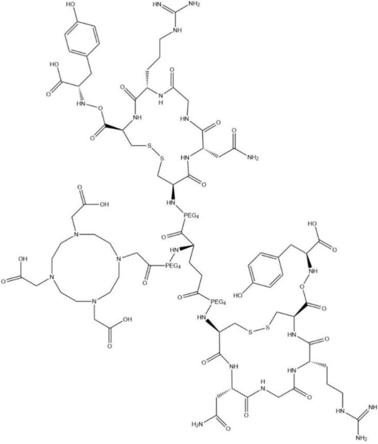
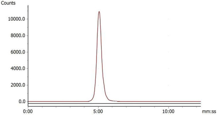
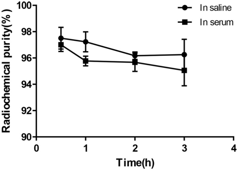
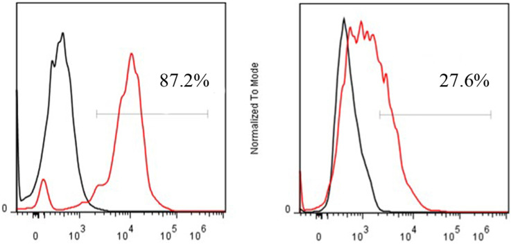
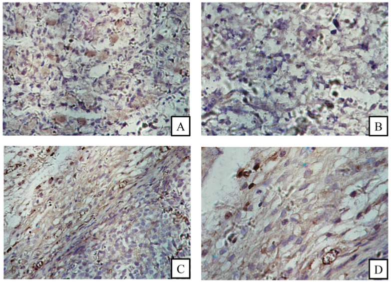
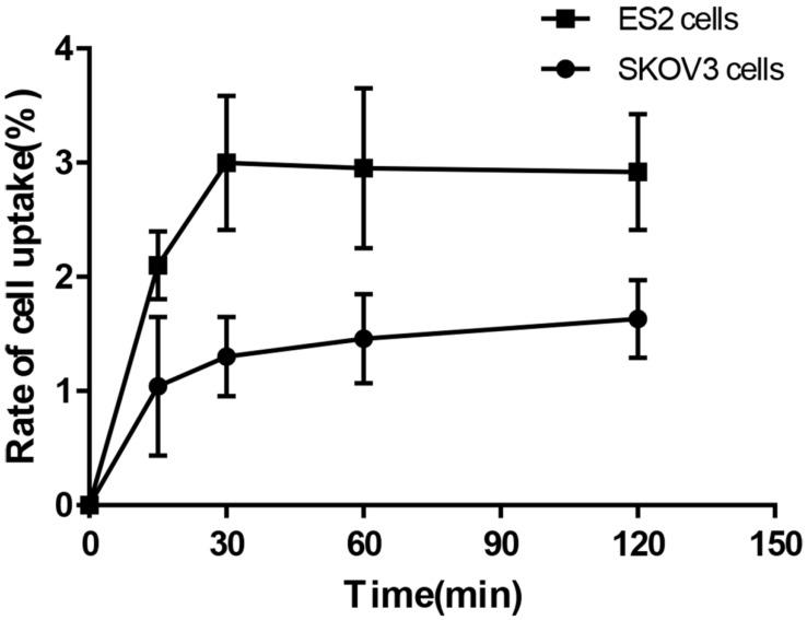
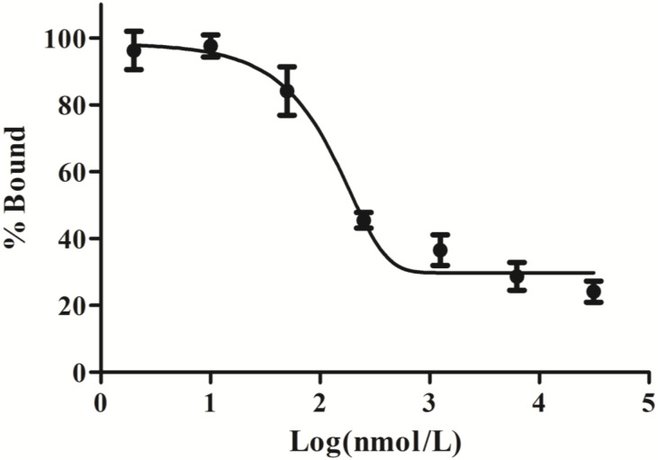
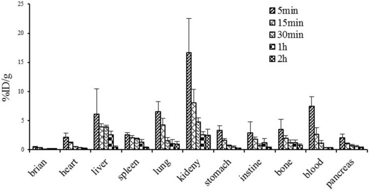
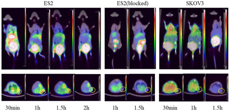
Introduction: Previous studies have shown that peptides containing the asparagine-glycine-arginine (NGR) sequence can specifically bind to CD13 (aminopeptidase N) receptor, a tumor neovascular biomarker that is over-expressed on the surface of angiogenic blood vessels and various tumor cells, and it plays an important role in angiogenesis and tumor progression. In the present study, we aimed to evaluate the efficacy of a gallium-68 (68Ga)-labeled dimeric cyclic NGR (cNGR) peptide as a new molecular probe that binds to CD13 in vitro and in vivo. Materials and Methods: A dimeric cNGR peptide conjugated with 1,4,7,10-tetraazacyclododecane-N,N',N'',N'''-tetraacetic acid (DOTA) was synthesized and labeled with 68Ga. In vitro uptake and binding analyses of the 68Ga- DOTA-c(NGR)2 were performed in two ovarian tumor cell lines, ES2 and SKOV3, which had different CD13 expression patterns. An in vivo biodistribution study was performed in normal mice, and micro positron emission tomography (PET) imaging was conducted in nude mice bearing ES2 and SKOV3 tumors. Results: 68Ga-DOTA-c(NGR)2 was prepared with high radiochemical purity (>95%), and it was stable both in saline at room temperature and in bovine serum at 37°C for 3 h. In vitro studies showed that the uptake of 68Ga-DOTA-c(NGR)2 in ES2 cells was higher compared with SKOV3 cells, and such uptake could be blocked by the cold DOTA-c(NGR)2. Biodistribution studies demonstrated that 68Ga-DOTA-c(NGR)2 was rapidly cleared from blood and mainly excreted from the kidney. MicroPET imaging of ES2 tumor xenografts showed the focal uptake of 68Ga-DOTA-c(NGR)2 in tumors from 1 to 1.5 h post-injection. The high-contrast tumor visualization occurred at 1 h, corresponding to the highest tumor/background ratio of 10.30±0.26. The CD13-specific tumor targeting of the 68Ga-DOTA-c(NGR)2 was further supported by the reduced uptake of the probe in ES2 tumors by co-injection of the unlabeled cold peptide. In SKOV3 tumor models, the tumor was not obviously visible under the same imaging conditions. Conclusions: 68Ga-DOTA-c(NGR)2 was easily synthesized, and it showed favorable CD13-specific targeting ability by in vitro data and microPET imaging with ovarian cancer xenografts. Collectively, 68Ga-DOTA-c(NGR)2 might be a potential PET imaging probe for non-invasive evaluation of the CD13 receptor expression in tumors.
Keywords: 68Ga labeling; CD13; NGR peptide; Tumor angiogenesis; microPET imaging.
© The author(s).
Conflict of interest statement
Competing Interests: The authors have declared that no competing interest exists.
Figures
Similar articles
-
68Ga-DOTA-NGR as a novel molecular probe for APN-positive tumor imaging using MicroPET.Nucl Med Biol. 2014 Mar;41(3):268-75. doi: 10.1016/j.nucmedbio.2013.12.008. Epub 2013 Dec 18. Nucl Med Biol. 2014. PMID: 24438818
-
Synthesis and evaluation of 64Cu-labeled monomeric and dimeric NGR peptides for MicroPET imaging of CD13 receptor expression.Mol Pharm. 2013 Jan 7;10(1):417-27. doi: 10.1021/mp3005676. Epub 2012 Dec 13. Mol Pharm. 2013. PMID: 23190134
-
Evaluation of 68Ga-labeled iNGR peptide with tumor-penetrating motif for microPET imaging of CD13-positive tumor xenografts.Tumour Biol. 2016 Sep;37(9):12123-12131. doi: 10.1007/s13277-016-5068-0. Epub 2016 May 24. Tumour Biol. 2016. PMID: 27220318
-
68Ga-Labeled DOTA-conjugated sCCK8[Phe2(p-CH2SO3H),Nle3,6], a sulfated cholecystokinin 8 (sCCK8) peptide derivative.2013 Jan 10 [updated 2013 Jan 31]. In: Molecular Imaging and Contrast Agent Database (MICAD) [Internet]. Bethesda (MD): National Center for Biotechnology Information (US); 2004–2013. 2013 Jan 10 [updated 2013 Jan 31]. In: Molecular Imaging and Contrast Agent Database (MICAD) [Internet]. Bethesda (MD): National Center for Biotechnology Information (US); 2004–2013. PMID: 23390663 Free Books & Documents. Review.
-
Radiolabeled NGR-Based Heterodimers for Angiogenesis Imaging: A Review of Preclinical Studies.Cancers (Basel). 2023 Sep 7;15(18):4459. doi: 10.3390/cancers15184459. Cancers (Basel). 2023. PMID: 37760428 Free PMC article. Review.
Cited by
-
NGR-Based Radiopharmaceuticals for Angiogenesis Imaging: A Preclinical Review.Int J Mol Sci. 2023 Aug 11;24(16):12675. doi: 10.3390/ijms241612675. Int J Mol Sci. 2023. PMID: 37628856 Free PMC article. Review.
-
NGR-modified curcumin nanovesicles reverse immunotherapy resistance in triple-negative breast cancer via TLR9 and mTOR pathway modulation.Cell Biol Toxicol. 2025 Jul 1;41(1):109. doi: 10.1007/s10565-025-10055-1. Cell Biol Toxicol. 2025. PMID: 40590998 Free PMC article.
-
Development and evaluation of albumin binder-conjugated heterodimeric radiopharmaceuticals targeting integrin αvβ3 and CD13 for cancer therapy.Eur J Nucl Med Mol Imaging. 2024 Sep;51(11):3334-3345. doi: 10.1007/s00259-024-06766-y. Epub 2024 May 24. Eur J Nucl Med Mol Imaging. 2024. PMID: 38787395
-
Recent Trends in Diagnostic Biomarkers of Tumor Microenvironment.Mol Imaging Biol. 2023 Jun;25(3):464-482. doi: 10.1007/s11307-022-01795-1. Epub 2022 Dec 14. Mol Imaging Biol. 2023. PMID: 36517729 Review.
-
Advances in Radionuclides and Radiolabelled Peptides for Cancer Therapeutics.Pharmaceutics. 2023 Mar 17;15(3):971. doi: 10.3390/pharmaceutics15030971. Pharmaceutics. 2023. PMID: 36986832 Free PMC article. Review.
References
-
- Tokuhara T, Hattori N, Ishida H. et al. Clinical significance of aminopeptidase N in non-small cell lung cancer. Clin Cancer Res. 2006;12(13):3971–8. - PubMed
-
- Hashida H, Takabayashi A, Kanai M. et al. Aminopeptidase N is involved in cell motility and angiogenesis: its clinical significance in human colon cancer. Gastroenterology. 2002;122(2):376–86. - PubMed
-
- Ishii K, Usui S, Sugimura Y. et al. Aminopeptidase N regulated by zinc in human prostate participates in tumor cell invasion. Int J Cancer. 2001;92(1):49–54. - PubMed
-
- Sjöström H, Norén O, Olsen J. Structure and function of aminopeptidase N. Adv Exp Med Biol. 2000;477:25–34. - PubMed
LinkOut - more resources
Full Text Sources
Other Literature Sources
Miscellaneous
