. 2021 Jan 1;11(2):700-714.
doi: 10.7150/thno.47800. eCollection 2021.

PLAGL2 promotes the proliferation and migration of gastric cancer cells via USP37-mediated deubiquitination of Snail1

Affiliations

PLAGL2 promotes the proliferation and migration of gastric cancer cells via USP37-mediated deubiquitination of Snail1

Liang Wu et al. Theranostics. .

Abstract

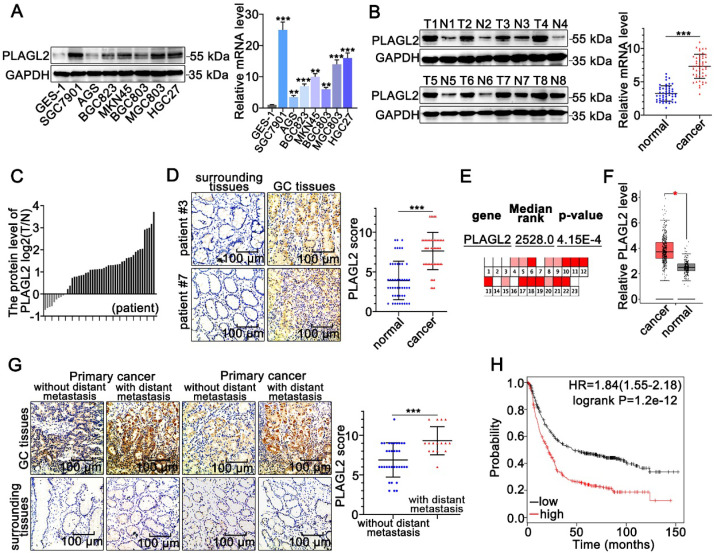
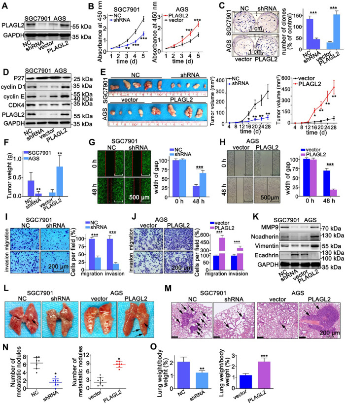
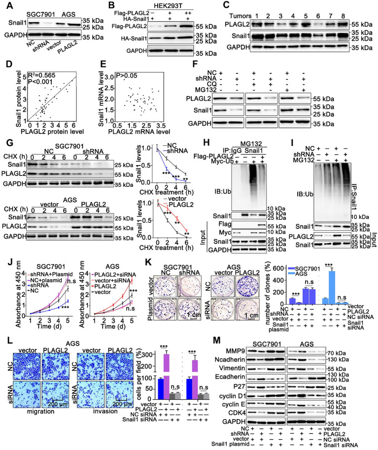
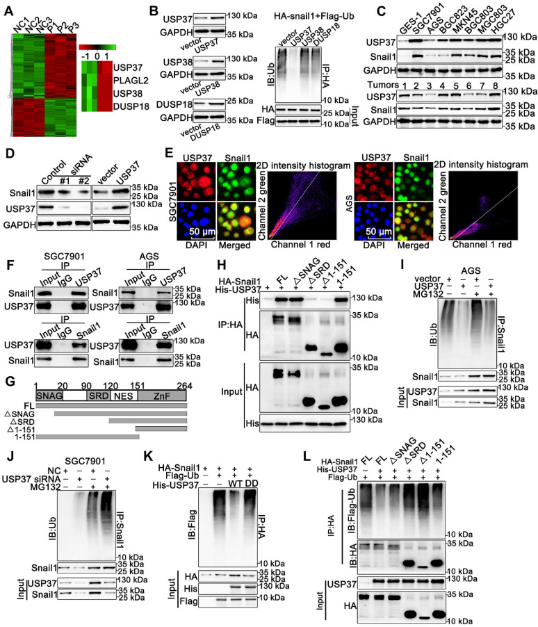
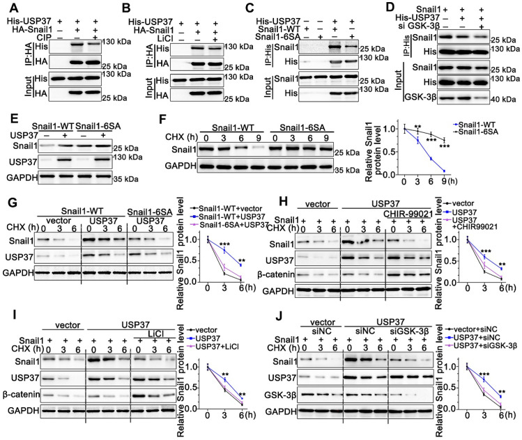
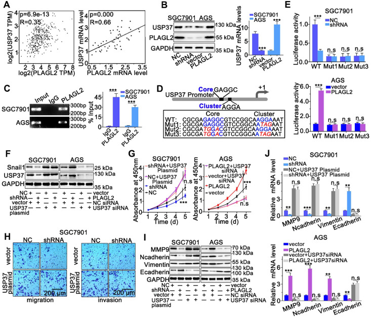
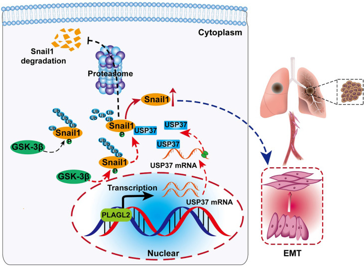
Rationale: PLAGL2 (pleomorphic adenoma gene like-2), a zinc finger PLAG transcription factor, is aberrantly expressed in several malignant tumors. However, the biological roles of PLAGL2 and its underlying mechanism in gastric cancer (GC) remain unclear. Methods: A series of experiments in vitro and in vivo were conducted to reveal the role of PLAGL2 in GC progression. Results: The data revealed that PLAGL2 promotes GC cell proliferation, migration, invasion, and EMT in vitro and in vivo. Mechanistically, we demonstrated the critical role of PLAGL2 in the stabilization of snail family transcriptional repressor 1 (Snail1) and promoting Snail1-mediated proliferation and migration of GC cells. PLAGL2 activated the transcription of deubiquitinase USP37, which then interacted with and deubiquitinated Snail1 protein directly. In addition, GSK-3β-dependent phosphorylation of Snail1 protein is essential for USP37-mediated Snail1 deubiquitination regulation. Conclusions: In general, PLAGL2 promotes the proliferation and migration of GC cells through USP37-mediated deubiquitination of Snail1 protein. This work provided potential therapeutic targets for GC treatment.

Keywords: GC; PLAGL2; Snail1; USP37; deubiquitination.


Conflict of interest statement

Competing Interests: The authors have declared that no competing interest exists.

Figures

Figure 1
Figure 1
PLAGL2 is highly expressed in GC. (A) WB and qRT-PCR analyses of PLAGL2 expression in human GC cell lines and GES-1. (B) WB and qRT-PCR analyses of PLAGL2 expression levels in clinical GC specimens. T, GC tissue; N, paired normal tissues. (C) Quantitative analysis of relative PLAGL2 expression in 49 paired GC tissues. (D)Representative IHC images of the PLAGL2 expression levels in paired GC tissues. Scale bars, 100 µm. (E) A meta-analysis of the PLAGL2 gene expression derived from the Oncomine database. (F) A box plot reflecting the PLAGL2 expression in GC specimens and normal specimens was obtained from the GEPIA database (G) Representative IHC images of the PLAGL2 expression levels in primary GC tumors without metastasis vs. GC tumors with metastasis. Scale bars, 100 µm. (H) Kaplan-Meier survival analysis of overall survival based on PLAGL2 expression in GC patients.
Figure 2
Figure 2
PLAGL2 promotes the proliferation, migration, and invasion of GC cells. (A) WB analysis of PLAGL2 expression in SGC7901 transfected with Lenti-shPLAGL2 and AGS transfected with Lenti-PLAGL2. (B-C) The cell proliferation was examined by CCK8 (B) and colony formation assays (C) in SGC7901 and AGS cells. Scale bars, 1cm. (D) WB analysis of the expression level of crucial cell cycle regulatory proteins. (E) Representative images of the corresponding xenograft. The growth curve of the tumors in the different groups. (F) The weight of tumors in different groups. (G-H) The cell migration was examined by the wound-healing assay. Pictures were taken at 0 and 48 h, respectively. Scale bars, 500μm. (I-J) The migration and invasion capacities of GC cells were also evaluated with Transwell assays. Scale bars, 200 µm. (K) WB analysis of the expression level of EMT-related proteins. (L) Representative images of visible lung metastases. The metastatic nodules were indicated with arrows. (M) Representative images of the corresponding HE staining. Scale bars, 200 µm. (N) Numbers of metastatic nodules. (O) The ratio of lung weight/body weight.
Figure 3
Figure 3
PLAGL2 stabilizes Snail1 protein by inhibiting its ubiquitination. (A) WB analysis of Snail1 expression of in PLAGL2 knockdown and overexpression cells. (B) WB analysis of exogenous Snail1 in HEK293T cells co-expressing PLAGL2 and Snail1. (C) WB analysis of protein levels of PLAGL2 and Snail1 in clinical GC specimens. (D) The correlation between PLAGL2 protein and Snail1 protein in GC tissues. (E) The correlation between PLAGL2 mRNA and Snail1 mRNA in GC tissues. (F) WB analysis of Snail1 level in PLAGL2 knockdown cells treated with MG132 and CQ. (G) CHX chase analysis of Snail1 protein half-life in PLAGL2 knockdown SGC7901 cell and PLAGL2 overexpression AGS cell. (H-I) Ubiquitination assays of endogenous Snail1 in the lysates from AGS cells transfected with Flag-PLAGL2 (H) or AGC7901 cells stably expressing PLAGL2 shRNA (I). (J-K) The stable PLAGL2 knockdown (SGC7901-shRNA) cell was transfected with Snail1 plasmid, and stable overexpression (AGS-PLAGL2) cell was transfected with the Snail1 siRNA. The role of Snail1 in PLAGL2-induced proliferation was examined by CCK8 (J) and colony formation assays (K). (L) Transwell assays detected the effect of Snail1 on PLAGL2-induced migration. Scale bars, 200 µm (l). (M) WB analysis of the expression level of EMT-related proteins and critical cell cycle regulatory proteins in cotransfected SGC7901 and AGS cells.
Figure 4
Figure 4
USP37 interacts with and deubiquitinates Snail1 directly. (A) Microarray analysis for mRNAs was performed with RNA extracted from SGC7901 NC and SGC7901 shRNA cells. (B) Ubiquitination assays of endogenous Snail1 in HEK293T cell, which was cotransfected with HA-Snail1, Flag-Ubi, and one of the three DUBs (USP37, USP38, and DUSP18). (C) WB analysis of protein levels of USP37 and Snail1 in clinical GC specimens and human GC cell lines. (D) WB analysis of protein levels of Snail1 and USP37 in SGC7901 cells transfected with two independent USP37 siRNAs and AGS cells expressing USP37 plasmid. (E) The representative images of IF staining of Snail1 (green) and USP37 (red) were shown in SGC7901 and AGS cells. Scale bars, 50 µm. (F) Reciprocal Co-IP and WB assays indicated the interaction between endogenous USP37 and Snail1 in AGS and SGC7901 cells. (G) Schematic diagram of the Snail1 full-length and deletion mutant plasmid. (H) HEK293T cells were cotransfected with USP37-His and the full length or truncation mutants of HA-Snail1. Cell lysates were immunoprecipitated with anti-HA antibody, and its production was analyzed by WB analysis with anti-His antibody (I-J) Ubiquitination assays of endogenous Snail1 in the lysates from AGS cells transfected with the USP37 plasmid (I) or AGC7901 cells transfected with the USP37 siRNA (J). (K) Ubiquitination assays of endogenous Snail1 in the lysates from HEK293T cells, which were cotransfected with the His-USP37-WT or His-USP37-DD, HA-Snail1 and Flag-Ub expressing plasmids. (L) Ubiquitination assays of endogenous Snail1 in the lysates from HEK293T cells, which were cotransfected with USP37-His and the full length or truncation mutants of HA-Snail1.
Figure 5
Figure 5
USP37 targets Snail1 for degradation in a GSK-3β phosphorylation-dependent manner. (A) Co-IP and western blot assays showed the mutual binding ability between USP37 and Snail1 in HEK293T cells treated either with or without CIP. (B) Co-IP and western blot assays showed the mutual binding ability between USP37 and Snail1 in HEK293T cells treated either with or without LiCl (C) Co-IP and western blot assays showed the mutual binding ability between USP37 and Snail1 in HEK293T cells cotransfected with His-USP37 and Wild-type Snail1 or Snail1-6SA mutant. (D) Co-IP and western blot assays showed the mutual binding ability between USP37 and Snail1 in HEK293T cells cotransfected either with or without si GSK3β (E) The WB analysis reflecting the expression regulation of USP37 on Wild-type Snail1 and Snail1-6SA mutants in HEK293T cells. (F) Pulse-chase assays of wild-type Snail1 or Snail1-6SA mutant in HEK293T cells. (G) Pulse-chase assays of Snail1 in HEK293T cells cotransfected with USP37 and Wild-type Snail1 or Snail1-6SA mutant. (H) Pulse-chase assays of Snail1 in HEK293T cells, which were transfected with His-USP37 plasmid and were treated either with or without LiCl. (I) Pulse-chase assays of Snail1 in HEK293T cells, which were transfected with His-USP37 plasmid and were treated either with or without CHIR-99021. (J) Pulse-chase assays of Snail1 in AGS cells, which were cotransfected with His-USP37 plasmid and siGSK-3β.
Figure 6
Figure 6
PLAGL2 modulates Snail1 stability by activating USP37 transcription. (A) A significant positive correlation between PLAGL2 mRNA and USP37 mRNA in GC tissues could be observed both in GEPIA database(left) and our data(right). (B) WB and qRT-PCR analyses of Snail1expression in PLAGL2 knockdown SGC7901 cell and PLAGL2 overexpression AGS cell. (C) The ChIP DNA was amplified by qRT-PCR, and then the products of qRT-PCR were electrophoresed on a 2% agarose gel. (D) The schematic representation of USP37 promoter. The sequences of wild-type and mutant PLAGL2 binding sites were indicated. (E) Luciferase activity assays were performed in PLAGL2 knockdown SGC7901 cell and PLAGL2 overexpression AGS cell, which were then transfected with wild type (USP37-WT) or mutant-type (USP37-mut) USP37 promoter-reporter plasmids. (F) WB of Snail1 protein level in SGC7901 cell cotransfected with Lenti-shPLAGL2 and USP37 plasmid and AGS cell cotransfected with Lenti-PLAGL2 and USP37 siRNA. (G-H) The stable PLAGL2 knockdown (SGC7901-shRNA) cell was transfected with USP37 plasmid, and stable overexpression (AGS-PLAGL2) cell was transfected with the USP37 siRNA. The role of USP37 in PLAGL2-induced proliferation was examined by CCK8 (G). Transwell assays detected the effect of USP37 on PLAGL2-induced migration. Scale bars, 200 µm (H). (I) WB analysis of the expression level of EMT-related proteins in cotransfected SGC7901 and AGS cells. (J) The qRT-PCR analysis of the expression level of EMT-related genes in cotransfected SGC7901 and AGS cells.
Figure 7
Figure 7
Schematic diagram of the molecular mechanism of PLAGL2 promoting proliferation and migration of GC cells. Schematic diagram of the molecular mechanism of PLAGL2 promoting proliferation and migration of GC cells. High expression of PLAGL2 transcriptionally activates USP37 expression to protect Snail1 from degradation by the proteasome.

References

    1. Bray F, Ferlay J, Soerjomataram I, Siegel RL, Torre LA, Jemal A. Global cancer statistics 2018: GLOBOCAN estimates of incidence and mortality worldwide for 36 cancers in 185 countries. CA Cancer J Clin. 2018;68(6):394–424. - PubMed
    1. Chaffer CL, Weinberg RA. A perspective on cancer cell metastasis. Science. 2011;331(6024):1559–64. - PubMed
    1. Landrette SF, Madera D, He F, Castilla LH. The transcription factor PlagL2 activates Mpl transcription and signaling in hematopoietic progenitor and leukemia cells. Leukemia. 2011;25(4):655–62. - PMC - PubMed
    1. Wu L, Yuan W, Chen J, Zhou Z, Shu Y, Ji J. et al. Increased miR-214 expression suppresses cell migration and proliferation in Hirschsprung disease by interacting with PLAGL2. Pediatr Res. 2019;86(4):460–70. - PMC - PubMed
    1. Wu L, Zhou Z, Han S, Chen J, Liu Z, Zhang X. et al. PLAGL2 promotes epithelial-mesenchymal transition and mediates colorectal cancer metastasis via beta-catenin-dependent regulation of ZEB1. Br J Cancer. 2020;122(4):578–89. - PMC - PubMed

Publication types

MeSH terms