. 2021 Jan 1;11(3):1129-1146.
doi: 10.7150/thno.49771. eCollection 2021.

ERK1/2 inhibition reduces vascular calcification by activating miR-126-3p-DKK1/LRP6 pathway

Affiliations

ERK1/2 inhibition reduces vascular calcification by activating miR-126-3p-DKK1/LRP6 pathway

Peng Zeng et al. Theranostics. .

Abstract

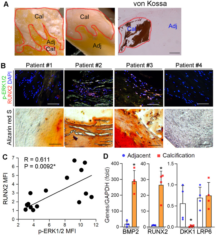
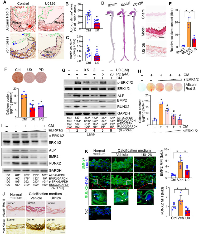
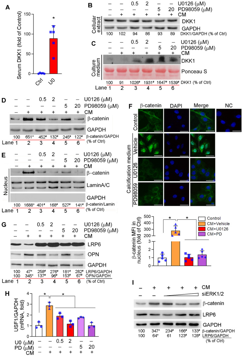
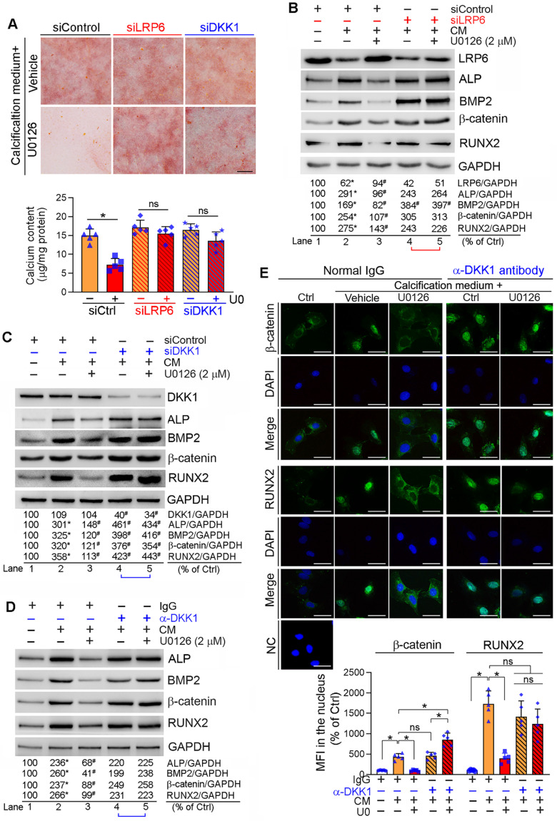
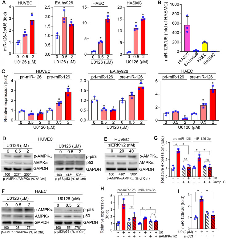
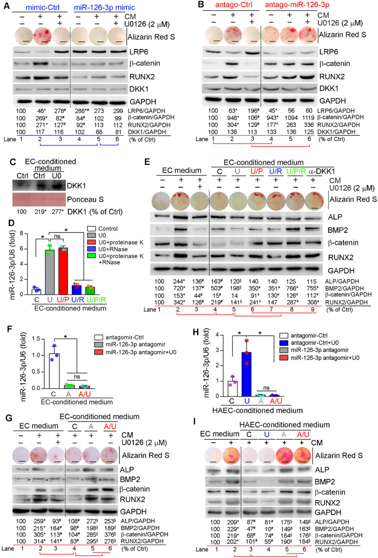
Rationale: Vascular microcalcification increases the risk of rupture of vulnerable atherosclerotic lesions. Inhibition of ERK1/2 reduces atherosclerosis in animal models while its role in vascular calcification and the underlying mechanisms remains incompletely understood. Methods: Levels of activated ERK1/2, DKK1, LRP6 and BMP2 in human calcific aortic valves were determined. ApoE deficient mice received ERK1/2 inhibitor (U0126) treatment, followed by determination of atherosclerosis, calcification and miR-126-3p production. C57BL/6J mice were used to determine the effect of U0126 on Vitamin D3 (VD3)-induced medial arterial calcification. HUVECs, HAECs and HASMCs were used to determine the effects of ERK1/2 inhibitor or siRNA on SMC calcification and the involved mechanisms. Results: We observed the calcification in human aortic valves was positively correlated to ERK1/2 activity. At cellular and animal levels, U0126 reduced intimal calcification in atherosclerotic lesions of high-fat diet-fed apoE deficient mice, medial arterial calcification in VD3-treated C57BL/6J mice, and calcification in cultured SMCs and arterial rings. The reduction of calcification was attributed to ERK1/2 inhibition-reduced expression of ALP, BMP2 and RUNX2 by activating DKK1 and LRP6 expression, and consequently inactivating both canonical and non-canonical Wnt signaling pathways in SMCs. Furthermore, we determined ERK1/2 inhibition activated miR-126-3p production by facilitating its maturation through activation of AMPKα-mediated p53 phosphorylation, and the activated miR-126-3p from ECs and SMCs played a key role in anti-vascular calcification actions of ERK1/2 inhibition. Conclusions: Our study demonstrates that activation of miR-126-3p production in ECs/SMCs and interactions between ECs and SMCs play an important role in reduction of vascular calcification by ERK1/2 inhibition.

Keywords: ECs; ERK1/2; Wnt signaling; miR-126-3p; vascular calcification.


Conflict of interest statement

Competing Interests: The authors have declared that no competing interest exists.

Figures

Figure 1
Figure 1
Activated ERK1/2 is positively correlated to calcification in human calcific aortic valves. (A) The resected aortic valve samples were collected from CAVD patients (n = 15). The sample was divided into two parts: calcification and adjacent areas. The calcification was initially confirmed by von Kossa staining. Bar: 200 µm. (B-C) the paraffin sections of aortic valves were prepared and used to determine calcification by Alizarin Red S staining and expression of p-ERK1/2 and RUNX2 by immunofluorescent staining. The images from 4 representative samples were presented. Bars: 200 µm. (D) the calcification and adjacent areas were collected from samples in Figure 1A and extracted total RNA, followed by determination of BMP2, DKK1, LRP6 and RUNX2 mRNA expression by qRT-PCR. *P < 0.05 (n = 4).
Figure 2
Figure 2
Inhibition of ERK1/2 by inhibitor/siRNA reduces calcification. (A) apoE-/- mice (15 mice/group) were fed HFD or HFD containing U0126 (3 mg/day/kg bodyweight) for 16 weeks. Aortic root cross sections were used to determine calcification formed within aortic root lesion area by Alizarin Red S (top panel) and von Kossa staining (bottom panel) (black dash lines: lesion area; green dash lines: aortic valve; black arrowheads and blue dash lines: calcification). Bars: 200 µm. (B) the percentage of calcium positive area in whole aortic lesion area was quantitatively analyzed based on images in the bottom panel of Figure 2A. *P < 0.05 (n = 15). (C) Total calcium of aorta was extracted and determined by an assay kit. *P < 0.05 (n = 15). (D-E) C57BL/6J mice (6 mice/group) were s.c. injected with 100 µL olive oil (Sham) or VD3 (5.5x10⁵ U/kg) dissolved in olive oil every day for three days, followed by feeding chow diet (Model) or chow diet containing U0126 (3 mg/kg bodyweight/day) (U0126) for 6 weeks. The whole artery and thoracic aorta (indicated by black arrows) cross sections were used to determine calcification by Alizarin Red S staining (D) and calcium quantitatively analysis (E), bars: 50 µm. (F-G) HASMCs were cultured in complete DMEM/F12 (1 : 1) medium or calcification medium (CM). Cells in CM were also treated with U0126 (U0) or PD98059 (PD) at the indicated concentrations (F: 2 µM U0126; 20 µM PD98059). After 7 days of calcification induction or plus treatment, cells were conducted Alizarin Red S staining and calcium quantitative assay (n = 5). Bars: 10 mm (F). Total cellular proteins were extracted and determined expression of p-ERK1/2, ERK1/2, ALP, BMP2 and RUNX2 by Western blot, *P < 0.05 vs. lane 1; #P < 0.05 vs. lane 2, n = 3 (G). (H-I) HASMCs were transfected with ERK1/2 siRNA (siERK1/2: 5, 20, 40 nM) for 24 h, followed by culture in complete DMEM/F12 (1 : 1) medium or CM for 7 days. Cells were then conducted Alizarin Red S staining and calcium quantitative assay (H, n = 5), Bars: 5 mm. Total cellular proteins were used to determine expression of p-ERK1/2, ERK1/2, ALP, BMP2 and RUNX2 by Western blot, *P < 0.05 vs. lane 1; #P < 0.05 vs. lane 2, n = 3 (I). (J-K) thoracic aorta was collected from apoE-/- mouse and cut into 5-mm long aortic rings. After cultured in complete DMEM/F12 medium, CM or CM plus U0126 (2 µM) for 14 days, rings were used to prepare 5-µm frozen sections. Calcium deposit and expression of BMP2 and RUNX2 in aortic rings were determined by Alizarin Red S and von Kossa staining (J), immunofluorescent staining (K) with quantitation of BMP2 and RUNX2 MFI, respectively. White arrow head indicated the nuclear RUNX2. Bars: 50 µm.
Figure 3
Figure 3
Regulation of canonical and non-canonical Wnt signaling pathways by ERK1/2 inhibition. (A) DKK1 levels in serum samples collected from mice in Figure 2A were determined by the “Antibody Arrays” method. The mean DKK1 level in control mice was defined as 1. *P < 0.05 (n = 5). (B-H) HASMCs were cultured in complete DMEM/F12 medium, CM or CM plus U0126/PD98059 at the indicated concentrations for 7 days, and then completed the following assays: expression of DKK1 (B), β-catenin (D), LRP6 and OPN (G) in total cellular proteins, DKK1 in concentrated HASMC-conditioned medium (C) and nuclear β-catenin (E) were determined by Western blot. *P < 0.05 vs. lane 1; #P < 0.05 vs. lane 2 (n = 3). Ponceau S staining was conducted as the loading control of the HASMC-conditioned medium. Expression of β-catenin in HASMCs was determined by immunofluorescent staining (F) with quantitation of β-catenin MFI in nucleus. Bars: 20 µm. Expression of USF1 mRNA was determined by qRT-PCR (H). *P < 0.05 (n = 3). (I) HASMCs were transfected with ERK1/2 siRNA (siERK1/2: 5, 20, 40 nM) for 24 h, followed by culture in complete DMEM/F12 (1 : 1) medium or CM for 7 days. Total cellular proteins were used to determine expression of β-catenin and LRP6 by Western blot, *P < 0.05 vs. Control; #P < 0.05 vs. CM control (n = 3).
Figure 4
Figure 4
Inhibition of DKK1 or LRP6 expression abolishes ERK1/2 inhibition-inhibited calcification in HASMCs. HASMCs were induced calcification by culture in CM or CM plus the following treatment for 7 days: (A-C) HASMCs were transfected with siRNA against human LRP6 (siLRP6) or DKK1 (siDKK1) for 24 h. Cells were then switched to complete DMEM/F12 medium, CM or CM plus U0126 (2 µM). The siRNA transfection was repeated once on day 3 of the calcification induction. Bars: 0.5 mm; (D-E) HASMCs were cultured in complete DMEM/F12 medium, CM or CM containing normal IgG or anti-DKK1 antibody or plus U0126 (2 µM) as indicated for 7 days. At the end of experiments, cells were used for the following assays: (A) calcium deposit by Alizarin Red S staining and calcium quantitative assay. *P < 0.05 vs. CM alone, n = 3; (B-D) expression of BMP2, β-catenin, RUNX2, ALP, DKK1 and LRP6 by Western blot. *P < 0.05 vs. lane 1; #P < 0.05 vs. lane 2 (n = 3). (E) Nuclear translocation of β-catenin and RUNX2 by immunofluorescent staining of HASMCs with quantitation of β-catenin and RUNX2 MFI. Bars: 20 µm. *P < 0.05; ns: not significant (n = 5).
Figure 5
Figure 5
Activation of miR-126-3p expression by ERK1/2 inhibition is mediated by activating AMPKα/p53 pathway. (A) HUVECs, EA.hy926 cells, HAECs and HASMCs were treated with U0126 at the indicated concentrations overnight, followed by determination of miR-126-3p expression by quantitative miR stem-loop RT-PCR technology. *P < 0.05, (n = 3); (B) total RNA extracted from un-treated HUVECs, EA.hy926 cells, HAECs or HASMCs was used to determine miR-126-3p expression by quantitative miR stem-loop RT-PCR technology. (C-D, F) HUVECs, EA.hy926 cells or HAECs were treated with U0126 at the indicated concentrations overnight. Expression of pri-miR-126 and pre-miR-126 was determined by qRT-PCR (C). Expression of AMPKα, p-AMPKα, p53 and p-p53 in HUVECs (D) or HAECs (F) were determined by Western blot. *P < 0.05 vs. control in the corresponding group (n = 3). (E) HUVECs were transfected with siERK1/2 at the indicated concentration for 24 h, followed by incubation in complete medium for another 48 h. Expression of AMPKα and p-AMPKα were determined by Western blot. *P < 0.05 (n = 3). (G) HUVECs were treated with U0126 (2 µM) or U0126 plus Compound C (Comp. C, 10 µM) overnight followed by determination of pre-miR-126 by qRT-PCR and miR-126-3p by quantitative miR stem-loop RT-PCR technology. *P < 0.05, ns: not significant (n = 3). (H-I) HUVECs were transfected with control, AMPKα1/2 or p53 siRNA for 24 h. Cells were then switched to complete EC medium and cultured for another 24 h, followed by U0126 (2 µM) treatment for 24 h. Expression of pre-miR-126-3p was determined by qRT-PCR and miR-126-3p by quantitative miR stem-loop RT-PCR technology. *P < 0.05 (n = 3).
Figure 6
Figure 6
Activated EC miR-126-3p mediates U0126-inhibited HASMC calcification. (A-B) HASMCs were transfected with miR-126-3p mimic or antagomir for 24 h. Cells were then switched to complete DMEM/F12 medium, CM or CM plus U0126 (2 µM). (C-D) HUVECs cultured in 100-mm dishes received treatment of vehicle or U0126 (U0126-treated) at 2 µM for 24 h, followed by washing with PBS twice. Cells were continued culture in serum-free EC medium for another 24 h, and the 24-h conditioned medium from vehicle or U0126-treated HUVECs was collected separately. The vehicle-treated EC conditioned medium was named as “C” medium. The U0126-treated EC conditioned medium was named as “U” medium. HASMCs in 6-well plate was cultured with DMEM/F12 medium plus the “C” or “U” EC-conditioned medium (1 : 1) for 24 h. After washed with PBS, the cells were cultured in serum-free DMEM/F12 medium for another 24 h. Then the medium was collected and used to determine DKK1 levels by Western blot (C). *P < 0.05 (n = 3). The “U” medium was also divided into 4 parts, treated with PBS, proteinase K, RNase or proteinase K plus RNase, and named as “U”, “U/P”, “U/R” or “U/P/R” medium separately. After inactivation of RNase and/or proteinase K by heating, miR-126-3p levels were determined by quantitative miR stem-loop RT-PCR technology (D). *P < 0.05; ns: not significant (n = 3). (E) HASMCs were cultured in complete DMEM/F12 medium, double-concentrated CM (containing 20 mM β-glycerol phosphate and 500 µM ascorbic acid) mixed with EC medium (1 : 1), EC medium plus U0126 (2 µM) (sample #1-3), the EC-conditioned medium collected from Figure 6D (1 : 1, sample #4-8), or EC medium (1 : 1) plus anti-DKK1 antibody (sample #9). (F, H) HUVECs or HAEC in 60-mm dishes were transfected with control antagomir or miR-126-3p antagomir followed by U0126 (2 µM) treatment. After 24 h of transfection or plus U0126 treatment, cells were switched to serum-free EC medium and continued culture for another 24 h. The 24-h EC-conditioned medium was collected, and named as “C”, “U”, “A” and “A/U” medium, respectively, followed by determination of miR-126-3p levels by quantitative miR stem-loop RT-PCR technology. *P < 0.05; ns: not significant (n = 3). (G) HASMCs were cultured in complete DMEM/F12 medium, double-concentrated CM mixed with EC medium (1 : 1) or EC medium plus U0126 (2 µM) (sample #1-3); or double-concentrated CM mixed with the HUVEC-conditioned medium (1 : 1) collected from Figure 6F (1 : 1, sample #4-6). I: HASMCs were cultured in complete DMEM/F12 medium, double-concentrated CM mixed with EC medium (1 : 1) (sample #1-2); or double-concentrated CM mixed with the HAEC-conditioned medium (1 : 1) collected from Figure 6H (1 : 1, sample #3-6). (A, B, E, G, I) all the medium or plus U0126 treatment were changed daily. After 7 days of treatment, cellular calcium deposit was determined by Alizarin Red S staining. Expression of LRP6, β-catenin, RUNX2, DKK1, BMP2 and ALP in total cellular proteins were determined by Western blot. A, B, E, G, I: *P < 0.05 vs. lane 1, #P < 0.05 vs. lane 2, §P < 0.05 vs. lane 4; D: ǂP < 0.05 vs. lane 5 (n = 3). Bars: 5 mm.

References

    1. Sage AP, Tintut Y, Demer LL. Regulatory mechanisms in vascular calcification. Nat Rev Cardiol. 2010;7:528–36. - PMC - PubMed
    1. O'Rourke RA, Brundage BH, Froelicher VF, Greenland P, Grundy SM, Hachamovitch R. et al. American College of Cardiology/American Heart Association Expert Consensus document on electron-beam computed tomography for the diagnosis and prognosis of coronary artery disease. Circulation. 2000;102:126–40. - PubMed
    1. Hoshino T, Chow LA, Hsu JJ, Perlowski AA, Abedin M, Tobis J. et al. Mechanical stress analysis of a rigid inclusion in distensible material: a model of atherosclerotic calcification and plaque vulnerability. Am J Physiol Heart Circ Physiol. 2009;297:H802–10. - PMC - PubMed
    1. Ehara S, Kobayashi Y, Yoshiyama M, Shimada K, Shimada Y, Fukuda D. et al. Spotty calcification typifies the culprit plaque in patients with acute myocardial infarction: an intravascular ultrasound study. Circulation. 2004;110:3424–9. - PubMed
    1. Budoff MJ, Shaw LJ, Liu ST, Weinstein SR, Mosler TP, Tseng PH. et al. Long-term prognosis associated with coronary calcification: observations from a registry of 25,253 patients. J Am Coll Cardiol. 2007;49:1860–70. - PubMed

Publication types

MeSH terms

Substances

Supplementary concepts