. 2021 Jan 1;11(3):1429-1445.
doi: 10.7150/thno.45351. eCollection 2021.

Breast cancer exosomes contribute to pre-metastatic niche formation and promote bone metastasis of tumor cells

Affiliations

Breast cancer exosomes contribute to pre-metastatic niche formation and promote bone metastasis of tumor cells

Xinxin Yuan et al. Theranostics. .

Abstract

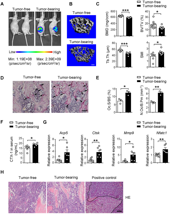
Rationale: Breast cancer preferentially develops osteolytic bone metastasis, which makes patients suffer from pain, fractures and spinal cord compression. Accumulating evidences have shown that exosomes play an irreplaceable role in pre-metastatic niche formation as a communication messenger. However, the function of exosomes secreted by breast cancer cells remains incompletely understood in bone metastasis of breast cancer. Methods: Mouse xenograft models and intravenous injection of exosomes were applied for analyzing the role of breast cancer cell-derived exosomes in vivo. Effects of exosomes secreted by the mildly metastatic MDA231 and its subline SCP28 with highly metastatic ability on osteoclasts formation were confirmed by TRAP staining, ELISA, microcomputed tomography, histomorphometric analyses, and pit formation assay. The candidate exosomal miRNAs for promoting osteoclastogenesis were globally screened by RNA-seq. qRT-PCR, western blot, confocal microscopy, and RNA interfering were performed to validate the function of exosomal miRNA. Results: Implantation of SCP28 tumor cells in situ leads to increased osteoclast activity and reduced bone density, which contributes to the formation of pre-metastatic niche for tumor cells. We found SCP28 cells-secreted exosomes are critical factors in promoting osteoclast differentiation and activation, which consequently accelerates bone lesion to reconstruct microenvironment for bone metastasis. Mechanistically, exosomal miR-21 derived from SCP28 cells facilitates osteoclastogenesis through regulating PDCD4 protein levels. Moreover, miR-21 level in serum exosomes of breast cancer patients with bone metastasis is significantly higher than that in other subpopulations. Conclusion: Our results indicate that breast cancer cell-derived exosomes play an important role in promoting breast cancer bone metastasis, which is associated with the formation of pre-metastatic niche via transferring miR-21 to osteoclasts. The data from patient samples further reflect the significance of miR-21 as a potential target for clinical diagnosis and treatment of breast cancer bone metastasis.

Keywords: bone metastasis; exosomes; miR-21; osteoclasts; pre-metastatic niche.


Conflict of interest statement

Competing Interests: The authors have declared that no competing interest exists.

Figures

Figure 1
Figure 1
Implantation of SCP28 breast cancer cells results in reduction of bone density. A. Representative bioluminescent imaging (BLI) of recipients with SCP28 cells (right) or control mice (left) at checkpoint of 5 weeks post implantation. B. Representative images showing three-dimensional trabecular architecture by micro-computed tomography (micro-CT) reconstruction in the distal femurs of tumor-free and tumor-bearing mice (n = 8 mice per group). Scale bar, 300 µm. C. Quantification of BMD, BV/TV, Tb.Th, SMI from statistical micro-CT data. (n = 8 mice per group). BMD, bone mineral density; BV/TV, bone volume/tissue volume ratio; Tb.Th, trabecular thickness; SMI, structure model index. D. Representative images of TRAP-positive cells in trabecular bone of tibias from tumor-free and tumor-bearing mice. Scale bar, 50 µm. E. Osteoclast surface/bone surface (Oc.S/BS) and osteoclast number/bone perimeter (N.Oc/B.Pm) of the proximal tibia from distinct mice in D were indicated (n = 3 in each group). F. ELISA analysis of serum cross linked C-telopeptide of type 1 collagen (CTX-1) (ng/mL) in tumor-free (n = 6) and tumor-bearing mice (n = 5). G. qRT-PCR analysis of the expression of osteoclast marker genes including Acp5, Ctsk, Mmp9, and Nfatc1 in tibias and femurs from tumor-free mice (n = 6) and tumor-bearing mice (n = 6). The relative expression of each target transcript (after normalization to the housekeeping Gapdh gene) in tumor-free mice was set as 1, and that in tumor-bearing mice was normalized, accordingly. H. Representative images of H&E staining of tibia section from tumor-free, tumor-bearing mice and mice with bone metastasis of SCP28 cells as a positive control at checkpoint of 5 weeks post implantation. T: tumor cell. Scale bar, 50 µm. Cumulative data are means ± SEM. * P < 0.05; ** P < 0.01; *** P < 0.001 (unpaired Student's t test). All data are from at least three independent experiments.
Figure 2
Figure 2
SCP28 cell-secreted exosomes serve as a critical role in bone homeostasis. A. Representative transmission electron microscopy (TEM) images of SCP28 cell-secreted exosomes. Scale bar, 25 nm. B. Nanoparticle tracking the size distribution of SCP28 cells-secreted exosomes (representative of five independent measurements). C. Detection of protein levels of Alix, HSP70, TSG101, TFIIB and LaminA/C in SCP28 cell-secreted exosomes and parental cell lysates by western blot. D. Representative BLI imaging showed the fluorescence signal distribution in multiple organs from mice injected with PBS or Dil-labeled exosomes derived from indicated cell lines. E. Representative images of TRAP staining in the proximal tibias of mice from indicated groups. Scale bar, 50 µm. F. Osteoclast surface/bone surface (Oc.S/BS) and osteoclast number/bone perimeter (N.Oc/B.Pm) of the proximal tibia from distinct mice in E were indicated (n = 3 in each group). G. ELISA analysis of serum CTX-1 (ng/mL) after administration with exosomes (n = 5) or PBS (n = 6), respectively. H. qRT-PCR analysis of osteoclast marker genes described as above in Oscar+ osteoclasts from mice treated with SCP28 cells-secreted exosomes (n = 5) and PBS as controls (n = 6). The relative expression of each target transcript (after normalization to the housekeeping Gapdh gene) in control mice was set as 1, and that in exosome-treated mice was normalized, accordingly. Cumulative data are means ± SEM. * P < 0.05; ** P < 0.01 (unpaired Student's t test). All data are from at least three independent experiments.
Figure 3
Figure 3
SCP28 cell-secreted exosomes stimulate osteoclastogenesis in vitro. A. Colocalization of SCP28 cell-secreted exosomes with RANKL-induced BMM cells using confocal microscopy imaging. Exosomes were labeled by 3'-dioctadecyloxacarbocyanine perchlorate (Dio, green) and cell nuclei were stained with Hoechst 33342 (blue). Scale bar, 10 µm. B. Representative TRAP staining images (left) and the number of osteoclasts with more than three nuclei (right) were statistically shown. Scale bar, 50 µm. ** P < 0.01 by Student's t test. C. Representative resorption pits (left) and cumulative data of pit resorption area (right) were statistically shown. Scale bar, 50 µm. Tt.Ar, total area. * P < 0.05 by Student's t test. D. Representative TRAP staining images (left) of RANKL-primed osteoclasts co-cultured with SCP28 cells treated by Rab27a siRNA or negative control (NC). Numbers of osteoclasts with more than three nuclei (right) were shown. Scale bar, 50 µm. NC, negative control. *** P < 0.001 by one-way ANOVA. E. Representative TRAP staining (left) of RANKL-primed osteoclasts co-cultured with SCP28 cells with or without N-SMase inhibitor GW4869 (20 µM). Numbers of osteoclasts with more than three nuclei (right) were shown. Scale bar, 50 µm. * P < 0.05; *** P < 0.001 by one-way ANOVA. Cumulative data are means ± SEM. The statistical methods are indicated relatively. All data are from at least three independent experiments.
Figure 4
Figure 4
SCP28 cells-secreted exosomes contribute to bone metastasis of SCP28 tumor cells. A. Flowchart of the experimental processes and scheme of SCP28 cell-secreted exosome education and metastasis. B. BLI quantitation of dynamic bone metastasis of breast cancer SCP28 cells in recipient mice educated by SCP28 cell-secreted exosomes or controls (n = 14 per group); *** P < 0.001 by two-way ANOVA. C. Kaplan-Meier curve showing bone metastasis of SCP28 cells in recipient mice educated by SCP28 cell-secreted exosomes or controls (n = 14 per group); * P < 0.05 (log-rank test). D. Representative BLI imaging showing the SCP28 cells localization on day 42 in recipient mice educated by SCP28 cell-secreted exosomes or controls. E. Representative images showing three-dimensional architecture after micro-CT reconstruction of the distal femurs from mice. Scale bars, up 1 mm; bottom 300 µm. F. Quantitative micro-CT analysis of distal femurs from control (n = 4) and SCP28 exosomes mice (n = 3), including BMD, SMI, Tb.N, Tb.Th, Tb.Sp and C.Th. * P < 0.05; ** P < 0.01; *** P < 0.001 by Student's t test. G. Representative X-ray images (up) and quantification of osteolytic lesions (bottom) in SCP28 cell-implanted mice educated by SCP28 cell-secreted exosomes or controls. Arrows indicate osteolytic bone areas. ** P < 0.01 by Student's t test. a.u., arbitrary unit. H. Representative images of H&E, TRAP, and OCN staining from SCP28 cell-implanted mice educated by SCP28 cell-secreted exosomes or controls. T, tumor; M, bone marrow. Scale bars, 50 µm. Cumulative data are means ± SEM. The statistical methods are indicated relatively. All data are from at least three independent experiments.
Figure 5
Figure 5
SCP28 cells-secreted exosomes enhance bone metastasis by regulating osteoclast driven pre-metastatic niche. A. Flowchart of the experimental processes and scheme of SCP28 cell-secreted exosome (treated with or without BP) education and analysis of metastasis. B. BLI quantitation of bone metastasis of SCP28 cells in recipient mice treated with exosomes or/and bisphosphonate zoledronic acid (BP) and controls. Control group, n = 6; BP group, n = 4; exosomes group, n = 6; exosomes + BP group, n = 5. ** P < 0.01, *** P < 0.001 by two-way ANOVA. C. Kaplan-Meier curve showing bone metastasis in recipient mice treated with control (n = 6), BP (n = 4), exosomes (n = 5), exosomes + BP (n = 5). * P < 0.05 (log-rank test). D. Representative BLI images showing the SCP28 cells localization on day 35 in recipient mice with different treatment. E. Representative X-ray images (left) and quantification of osteolytic lesions (right) in SCP28 cell-implanted mice with distinct treatment. Arrows indicate osteolytic bone areas. Control group, n = 6; BP group, n = 4; exosomes group, n = 5; exosomes + BP group, n = 5. *** P < 0.001 by one-way ANOVA. F. Representative images of H&E and TRAP staining of bones isolated from indicated mice on day 35 after exosomes or/and BP treatment. T, tumor; M, bone marrow. Scale bars, 50 µm. Cumulative data are means ± SEM. All data are from at least three independent experiments.
Figure 6
Figure 6
SCP28 cell-derived exosomal miR-21 is involved in pre-metastatic niche formation via regulating osteoclast. A. qRT-PCR analysis of expression of miR-21 (miR-21-5p) and pre-miR-21 levels in RANKL-primed osteoclasts treated with or without SCP28 cell-secreted exosomes for 6 h. The relative expression of miR-21 or pre-miR-21 was normalized to U6 or Gapdh gene which is set as 1, respectively. *** P < 0.001 by Student's t test. B. Confocal microscopy images showing the transition of FAM-miR-21 from exosomes into osteoclasts at 6 h post treatment. miR-21 in exosomes from SCP28 cells were labeled by FAM (green) and cell nuclei were stained with Hoechst 33342 (blue). Scale bar, 10 µm. C. Representative TRAP staining images and resorption pits (left). The number of osteoclasts with more than three nuclei and cumulative data of pit resorption area (right) were statistically shown. Scale bars, 50 µm. ** P < 0.01, *** P < 0.001 by one-way ANOVA. D. Western blot (left) and quantitative analysis (right) of PDCD4 and NFATc1 protein expression in osteoclasts with indicated treatment. E. Confocal microscopy images showing the expression levels of PDCD4 and OSCAR in tibias section from recipient mice with indicated treatment. Scale bar, 10 µm. F. qRT-PCR analysis showing the expression level of miR-21 in serum exosomes of breast cancer patients. No-metastasis patients, n = 21; other sites metastasis patients, n = 9; bone-metastasis patients, n = 21. The relative expression of miR-21 was normalized to cel-miR-39. * P < 0.05, ** P < 0.01 by one-way ANOVA. G. Analysis of the level of miR-21 in serum exosomes of breast cancer patients. 30-45, n = 11; 46-55, n = 22; 56-80, n = 18. Invasive breast cancer, n = 31; invasive ductal carcinoma, n = 13. HER2+, n = 11; Luminal B-HER2+, n = 29. II, n = 28; III, n = 7. The relative expression of miR-21 was normalized to cel-miR-39. Cumulative data are means ± SEM. All data are from at least three independent experiments.

References

    1. Mundy GR. Metastasis to bone: causes, consequences and therapeutic opportunities. Nat Rev Cancer. 2002;2:584–93. - PubMed
    1. Coleman RE. Metastatic bone disease: clinical features, pathophysiology and treatment strategies. Cancer Treat Rev. 2001;27:165–76. - PubMed
    1. Mundy GR, Bertolini DR. Bone destruction and hypercalcemia in plasma cell myeloma. Semin Oncol. 1986;13:291–9. - PubMed
    1. Veronesi U, Boyle P, Goldhirsch A, Orecchia R, Viale G. Breast cancer. Lancet. 2005;365:1727–41. - PubMed
    1. Zhuang X, Zhang H, Li X, Li X, Cong M, Peng F. et al. Differential effects on lung and bone metastasis of breast cancer by Wnt signalling inhibitor DKK1. Nat Cell Biol. 2017;19:1274–85. - PubMed

Publication types