. 2021 Jan 1;11(3):1446-1457.
doi: 10.7150/thno.47860. eCollection 2021.

Tofacitinib restores the balance of γδTreg/γδT17 cells in rheumatoid arthritis by inhibiting the NLRP3 inflammasome

Affiliations

Tofacitinib restores the balance of γδTreg/γδT17 cells in rheumatoid arthritis by inhibiting the NLRP3 inflammasome

Xinyu Yang et al. Theranostics. .

Abstract

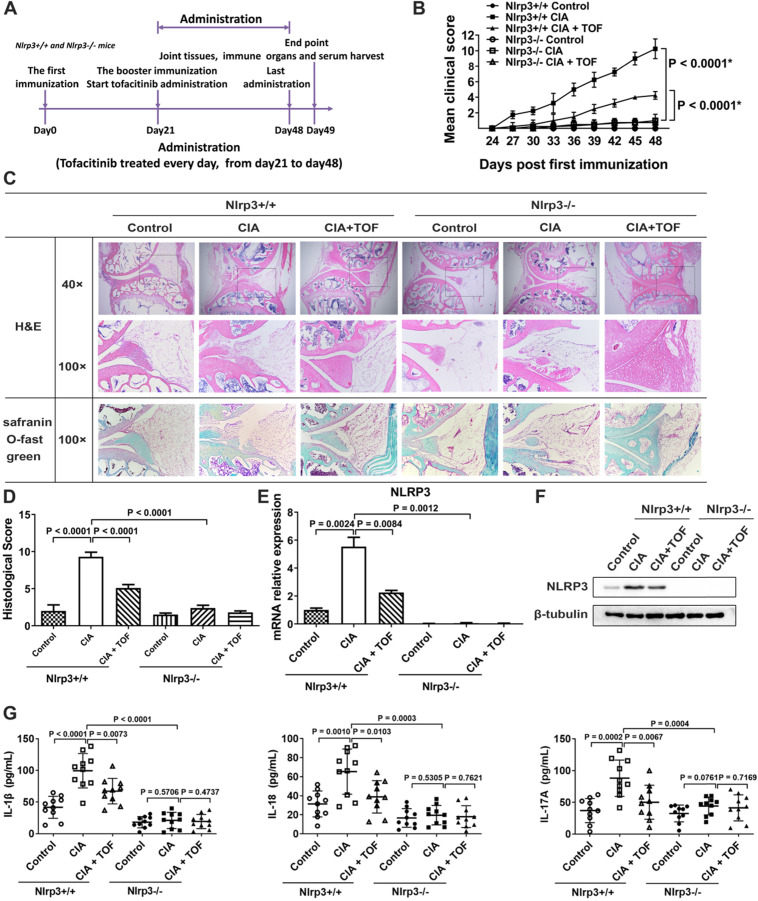
Objective: Tofacitinib (TOF) is a Janus kinase (JAK) inhibitor used in the treatment of rheumatoid arthritis (RA), but the mechanism of its action remains unclear. In this study, we investigated the influence of TOF on gamma delta regulatory T-cell (γδTreg)/γδT17 cell balance in RA and the role of the nucleotide-binding domain (NOD)-like receptor protein 3 (NLRP3) inflammasome in this process. Methods: We detected levels of inflammatory factors in the serum of RA patients before and after administration of TOF using an enzyme-linked immunosorbent assay (ELISA). A collagen-induced arthritis (CIA) model was constructed to investigate the effect of TOF on arthritis symptoms, γδTreg/γδT17 cell balance and the NLRP3 inflammasome. We used bone marrow-derived macrophages (BMDMs) to study the effect of TOF on NLRP3 inflammasome activation. Nlrp3-/- mice were introduced to assess the influence of NLRP3 on γδT17 cell activation in RA. Results: TOF treatment decreased levels of γδT17 cell-related cytokine interleukin-17 (IL-17) in RA patients. In addition, TOF intervention in the CIA model reduced joint inflammation and damage, rebalanced the γδTreg/γδT17 cell ratio and inhibited excessive NLRP3 inflammasome activation in draining lymph nodes and arthritic joints. BMDM intervention experiments demonstrated that TOF decreased the level of secreted IL-1β via downregulation of NLRP3. Furthermore, experiments using Nlrp3-/- mice verified that the NLRP3 inflammasome mediated the effect of TOF on γδT17 cell activation. Conclusions: Recovery of γδTreg/γδT17 cell balance was a novel mechanism by which TOF alleviated RA. Meanwhile, NLRP3 played a pivotal role in the process of TOF-mediated γδT17 cell activation.

Keywords: Inflammation; NLRP3 inflammasome; Rheumatoid arthritis; Tofacitinib; γδT cells.


Conflict of interest statement

Competing Interests: The authors have declared that no competing interest exists.

Figures

Figure 1
Figure 1
TOF treatment inhibited NLRP3 inflammasome levels and balanced the γδlang/γδT17 ratio in rheumatoid arthritis (RA). (A) Levels of cytokines IL-1β, IL-18, IL-6, TNF-α, IL-17, IL-10 and TGF-β in synovia of RA patients (n = 33) and HCs (n = 33) were detected by ELISA. (B) Immunoblot analysis of NLRP3 in synovial lysates of RA patients (n = 10) and HCs (n = 10). (C) Representative images of paraffin sections from RA patients (n = 10) and HCs (n = 10) that were stained with anti-human TCRγ/δ (green) and anti-human IL-17 (upper, red) or with anti-human TCRγ/δ (green) and anti-human Foxp3 (lower, red) for IF analyses. Arrows in the merged image indicate γδT17 cells (upper) or γδTregs (lower). IL-17-positive but TCRγ/δ-negative staining areas of the merged images represent other IL-17-secreting cells. One of the 10 independent experiments is shown. (D) Levels of IL-1β, IL-18, IL-17, IL-6, TNF-α, IL-10 and TGF-β in serum of RA patients (n = 13) and TOF-treated patients (n = 13) were detected by ELISA.
Figure 2
Figure 2
TOF ameliorated joint inflammatory response and inhibited NLRP3 inflammasome activation in a CIA model. (A) Timeline of TOF intervention experiment in the CIA model. (B) Clinical scores of CIA mice during TOF administration. Two independent observers who were not aware of the animals' treatment inspected the mice every 3 days for the severity of arthritis. Scoring was as follows: 0 = no evidence of erythema or swelling; 1 = erythema and mild swelling confined to the tarsals or ankle joint; 2 = erythema and mild swelling extending from the ankle to the tarsals; 3 = erythema and moderate swelling extending from the ankle to the metatarsal joints; and 4 = erythema and severe swelling encompassing the ankle, paw and digits, or ankylosis of the limb. Statistical significance was determined by ANOVA of repeated measurements. n = 10 per group. (C) Knee joints of CIA mice (n = 12) were stained with H&E (blue for cell nuclei and membranes; red for cytoplasm and extracellular matrix [ECM]) and Safranin O Fast Green (green for cartilages) and graded on a scale of 0 (normal) to 3 (severe). Expression of IL-1β in paraffin sections of synovia of CIA mice was detected by IHC analysis. (D) Histological score for CIA mice was F(3, 36) = 9.358. n = 10 per group. (E) Using RT-qPCR, expression of NLRP3 mRNA was primarily detected in cultured lymphocytes from lymph nodes of CIA mice treated with TOF (F, = 13.97). n = 10 per group. (F) Concentrations of IL-1β, IL-18, IL-6, TNF-α, IL-17, IL-10 and TGF-β in serum from CIA mice were detected by ELISA. Results were F(3, 36) = 12.19 for IL-1β, F(3, 36) = 6.971 for IL-18, F(3, 36) = 7.258 for IL-6, F(3, 36) = 19.26 for TNF-α, F(3, 36) = 6.016 for IL-17, F(3, 36) = 8.083 for IL-10 and F(3, 36) = 19.98 for TGF-β. n = 10 per group. (G) Protein levels of NLRP3 in lymph nodes of CIA mice treated with TOF were detected by Western blot. (H) LPS-primed BMDMs were stimulated with nigericin and then treated with TOF for 2 h. Expression of NLRP3 mRNA in BMDMs was detected by RT-qPCR (F, = 31.37). n = 10 per group. (I) Immunoblot analysis of IL-1β and cleaved CASP-1 (p20) in culture supernatants (SNs); immunoblot analysis of NLRP3, precursors of IL-1β (pro-IL-1β) and precursors of CASP-1 (pro-CASP-1) in lysates of BMDMs. n = 10 per group.
Figure 3
Figure 3
TOF influenced γδTreg/γδT17 balance in CIA mice. (A, C) Representative FCM images indicated percentages of γδT17 cells in lymph nodes of CIA mice treated with TOF (F, = 23.68). n = 10 per group. (B, D) Percentage of γδTregs was detected in lymph nodes of CIA mice treated with TOF (F(3, 36) = 13.16). n = 10 per group. (E) γδTreg/γδT17 cell ratio in lymph nodes of CIA mice treated with TOF (F, = 69.96). (F-I) Representative images of paraffin sections were stained with anti-mouse TCRγ/δ (green) and anti-mouse IL-17 (upper, red), or with anti-mouse TCRγ/δ (green) and anti-mouse Foxp3 (lower, red) for IF analyses. Arrows in the merged image indicate γδT17 cells (upper) or γδTregs (lower). One of the 10 independent experiments is shown.
Figure 4
Figure 4
TOF alleviated joint injury and inflammatory response via suppression of the NLRP3 inflammasome. (A) Timeline of TOF treatment in Nlrp3+/+ and Nlrp3-/- mice in CIA models. (B) Clinical scores of CIA models established from Nlrp3+/+ and Nlrp3-/- mice. Two independent observers who were not aware of the animals' treatment inspected the mice every 3 days for the severity of arthritis. Statistical significance was determined by ANOVA of repeated measurements. *Data are compared with Nlrp3+/+ control group. n = 10 per group. (C) Knee joints of mice were stained with H&E and Safranin O Fast Green. (D) Histological score of Nlrp3+/+ and Nlrp3-/- mice was F(5, 54) = 62.96. n = 10 per group. (E) Using RT-qPCR, expression of NLRP3 mRNA was primarily detected in cultured lymphocytes from lymph nodes of Nlrp3+/+ and Nlrp3-/- mice (F[5, 54 = 60.10). n = 10 per group. (F) Protein levels of NLRP3 were detected in lymph nodes of Nlrp3+/+ and Nlrp3-/- mice using Western blot. n = 10 per group. (G) Concentrations of IL-1β, IL-18 and IL-17 in serum from Nlrp3+/+ and Nlrp3-/- mice were detected by ELISA. Results were F(5, 54) = 35.92 for IL-1β, F(5, 54) = 15.26 for IL-18 and F(5, 54) = 9.061 for IL-17. n = 10 per group.
Figure 5
Figure 5
TOF influenced the activation of γδT17 cells via suppression of the NLRP3 inflammasome. (A, B) Representative FCM pictures indicated percentages of γδT17 cells in the lymph nodes of Nlrp3+/+ and Nlrp3-/- mice (F[5,54] = 39.68). n = 10 per group. (C, D) Representative images of paraffin sections from Nlrp3+/+ and Nlrp3-/- mice were stained with anti-mouse TCRγ/δ (green) and anti-mouse IL-17 (red) for IF analyses. Arrows in the merged image indicate γδT17 cells. One of the 10 independent experiments is shown (F, = 11.87).

References

    1. Wang JG, Xu WD, Zhai WT, Li Y, Hu JW, Hu B. et al. Disorders in angiogenesis and redox pathways are main factors contributing to the progression of rheumatoid arthritis: a comparative proteomics study. Arthritis Rheum. 2012;64:993–1004. - PubMed
    1. Scott DL, Wolfe F, Huizinga TW. Rheumatoid arthritis. Lancet. 2010;376:1094–108. - PubMed
    1. Firestein GS, McInnes IB. Immunopathogenesis of Rheumatoid Arthritis. Immunity. 2017;46:183–96. - PMC - PubMed
    1. Miossec P, Kolls JK. Targeting IL-17 and TH17 cells in chronic inflammation. Nat Rev Drug Discov. 2012;11:763–76. - PubMed
    1. Lubberts E, Koenders MI, Oppers-Walgreen B, van den Bersselaar L, Coenen-de Roo CJ, Joosten LA. et al. Treatment with a neutralizing anti-murine interleukin-17 antibody after the onset of collagen-induced arthritis reduces joint inflammation, cartilage destruction, and bone erosion. Arthritis Rheum. 2004;50:650–9. - PubMed

Publication types