. 2020 Dec;10(2):e12050.
doi: 10.1002/jev2.12050. Epub 2020 Dec 28.

Extracellular vesicles containing ACE2 efficiently prevent infection by SARS-CoV-2 Spike protein-containing virus

Affiliations

Extracellular vesicles containing ACE2 efficiently prevent infection by SARS-CoV-2 Spike protein-containing virus

Federico Cocozza et al. J Extracell Vesicles. 2020 Dec.

Abstract

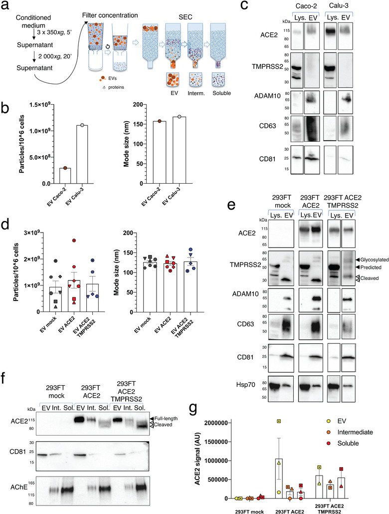
SARS-CoV-2 entry is mediated by binding of the spike protein (S) to the surface receptor ACE2 and subsequent priming by host TMPRSS2 allowing membrane fusion. Here, we produced extracellular vesicles (EVs) exposing ACE2 and demonstrate that ACE2-EVs are efficient decoys for SARS-CoV-2 S protein-containing lentivirus. Reduction of infectivity positively correlates with the level of ACE2, is much more efficient than with soluble ACE2 and further enhanced by the inclusion of TMPRSS2.

Keywords: ACE2; EV therapy; SARS‐CoV‐2; TMPRSS2.


Figures

FIGURE 1
FIGURE 1
Isolation and characterization of EVs containing ACE2 and TMPRSS2. (a) Scheme of EV isolation and separation from soluble components by SEC. (b) NTA quantification and mode size of the particles in EV‐containing SEC fractions obtained from Caco‐2 and Calu‐3 cells. (c) WB analysis of ACE2, TMPRSS2 and different EV markers in lysates from 4 × 105  cells and 1 × 1010  2020 (Caco‐2) or 0.5 × 1010 2020 (Calu‐3) particles obtained from EV SEC fractions. One experiment. (d) NTA quantification and mode size of the particles in EV‐containing fractions obtained from 293FT‐mock, 293FT‐ACE2 and 293FT‐ACE2‐TMPRSS2 cell lines. Different symbols correspond to independent experiments. Error bars indicate SEM. (e) WB analysis of ACE2, TMPRSS2 and different EV markers in lysates from 4 × 105 cells and 1 × 1010 2020 particles from EV SEC fractions obtained from the three 293FT cell lines. (f‐g) WB analysis of ACE2, CD81 and AChE on EV, intermediate and soluble fractions from the three 293FT cell lines. 1 × 109  particles from the EV fraction or material recovered from the same corresponding volume of CCM, for the intermediate or soluble fractions, were loaded on the gel. (f) Representative WB. (g) Quantification of ACE2 signal (pooled full‐length and cleaved forms) in 2–3 independent WB
FIGURE 2
FIGURE 2
Inhibition of SARS‐CoV‐2‐S‐pseudotyped virus infection with ACE2 EVs. (a) Infection of 293FT‐ACE2, Caco‐2 and Calu‐3 cells with different dilutions of a SARS‐CoV‐2‐S‐pseudotyped lentivirus encoding for eGFP. The number of infected cells was calculated by multiplying the percentage of GFP‐positive cells by the initial number of cells. (b) Scheme of the infectivity assay with different treatments. (c) Dot plots showing the percentage of infected (= eGFP+) 293FT‐ACE2 cells obtained after incubation with viruses alone (0.05 dilution), in combination with 1 × 1010 2020 EVs from the different 293FT cell lines or in combination with rACE2 (25 μg/ml). This rACE2 level represents 250 and 2960 times more ACE2 than the one contained in ACE2‐EVs and ACE2‐TMPRSS2‐EVs, respectively, measured by ELISA. (d) Quantification of the number of infected 293FT‐ACE2 cells in the presence of EVs. The percentage of eGFP+ cells was measured by FACS and normalized to infection with the virus alone (100%). Results from three independent experiments are shown. All replicates from each experiment are included. *P < 0.05; **P < 0.01; ***P < 0.001 (Dunnett's test). (e) Caco‐2 infection in the presence of ACE2‐EVs and ACE2‐TMPRSS2‐EVs. *P < 0.05 (Dunnett's test). (f) ACE2 quantification by ELISA in EV and soluble fractions obtained from the three different 293FT cell lines. Results are expressed as ng per million of secreting cells. *P < 0.05; ***P < 0.001 (non‐parametric ANOVA with Kruskal‐Wallis test for comparison among all groups). ***P < 0.001 (Mann‐Whitney test for comparison among EV vs soluble for each cell line). (g) Comparison of the effect on 293T‐ACE2 infection of 1 × 1010 2020 EVs and soluble fractions from an equivalent volume of CCM of 293FT‐ACE2‐TMPRSS2 cells. **P < 0.01 (t‐test) (h) Percentage of infectivity from Figure 2d related to the amount of EV‐associated ACE2, quantified by ELISA in Figure 2f. As a comparison, cell infection rates in the presence of increasing amount of recombinant ACE2 (rACE2) were also determined (n = 4). Lines represent results of linear regression analysis. Comparison of slopes and intercepts using Prism indicated that the three regression lines are distinct but parallels. ACE2‐TMPRSS2 is different from ACE2 (P = 0.0017) and rACE2 is different from ACE2‐TMPRSS2 (P < 0.0001) and from ACE2 (P < 0.0001)

References

    1. Afar, D. E. H. , Vivanco, I , Hubert, R. S. , Kuo, J , Chen, E , Saffran, D. C. , Raitano, A. B. , & Jakobovits, A et al. (2001). Catalytic Cleavage of the Androgen‐regulated TMPRSS2 Protease Results in Its Secretion by Prostate and Prostate Cancer Epithelia. Cancer Research, 59, 6015–6022. - PubMed
    1. Börger, V. , Weiss, D. J , Anderson, J. D , Borràs, F. E , Bussolati, B. , Carter, D. R. F , Dominici, M. , Falcón‐Pérez, J. M. , Gimona, M. , Hill, A. F. , Hoffman, A. M. , de Kleijn, D. , Levine, B. L. , Lim, R. , Lötvall, J. , Mitsialis, S. A. , Monguió‐Tortajada, M. , Muraca, M. , Nieuwland, R. … Giebel, B. (2020). International Society for Extracellular Vesicles and International Society for Cell and Gene Therapy statement on extracellular vesicles from mesenchymal stromal cells and other cells: Considerations for potential therapeutic agents to suppress coronavirus disease‐19. Cytotherapy, 22, 482–485 - PMC - PubMed
    1. Cerboni, S. , Jeremiah, N. , Gentili, M. , Gehrmann, U. , Conrad, C. , Stolzenberg, M. C. , Picard, C. , Neven, B. , Fischer, A. , Amigorena, S. , Rieux‐Laucat, F. , & Manel, N. (2017). Intrinsic antiproliferative activity of the innate sensor STING in T lymphocytes. Journal of Experimental Medicine, 214, 1769–1785 - PMC - PubMed
    1. De Carvalho, J. V. , De Castro, R. O. , Da Silva, E Z. M. , Silveira, P. P. , Da Silva‐Januário, M. E. , Arruda, E. , Jamur, M. C. , Oliver, C. , Aguiar, R. S. , & Dasilva, L. L. P. (2014). Nef Neutralizes the Ability of Exosomes from CD4+ T Cells to Act as Decoys during HIV‐1 Infection. Plos One, 9, e113691 - PMC - PubMed
    1. Grzelak, L. , Temmam, S. , Planchais, C. , Demeret, C. , Huon, C. , Guivel‐Benhassine, F. , Staropoli, I. , Chazal, M. , Dufloo, J. , Planas, D. , Buchrieser, J. , Michael Rajah, M. , Robinot, R. , Porrot, F. , Albert, M. , Chen, K.‐Y. , Crescenzo, B. , Donati, F. , Anna, F. , … van der Werf, S. (2020). SARS‐CoV‐2 serological analysis of COVID‐19 hospitalized patients, pauci‐symptomatic individuals and blood donors. medRxiv 2020.04.21.20068858. 10.1101/2020.04.21.20068858 - DOI

Publication types